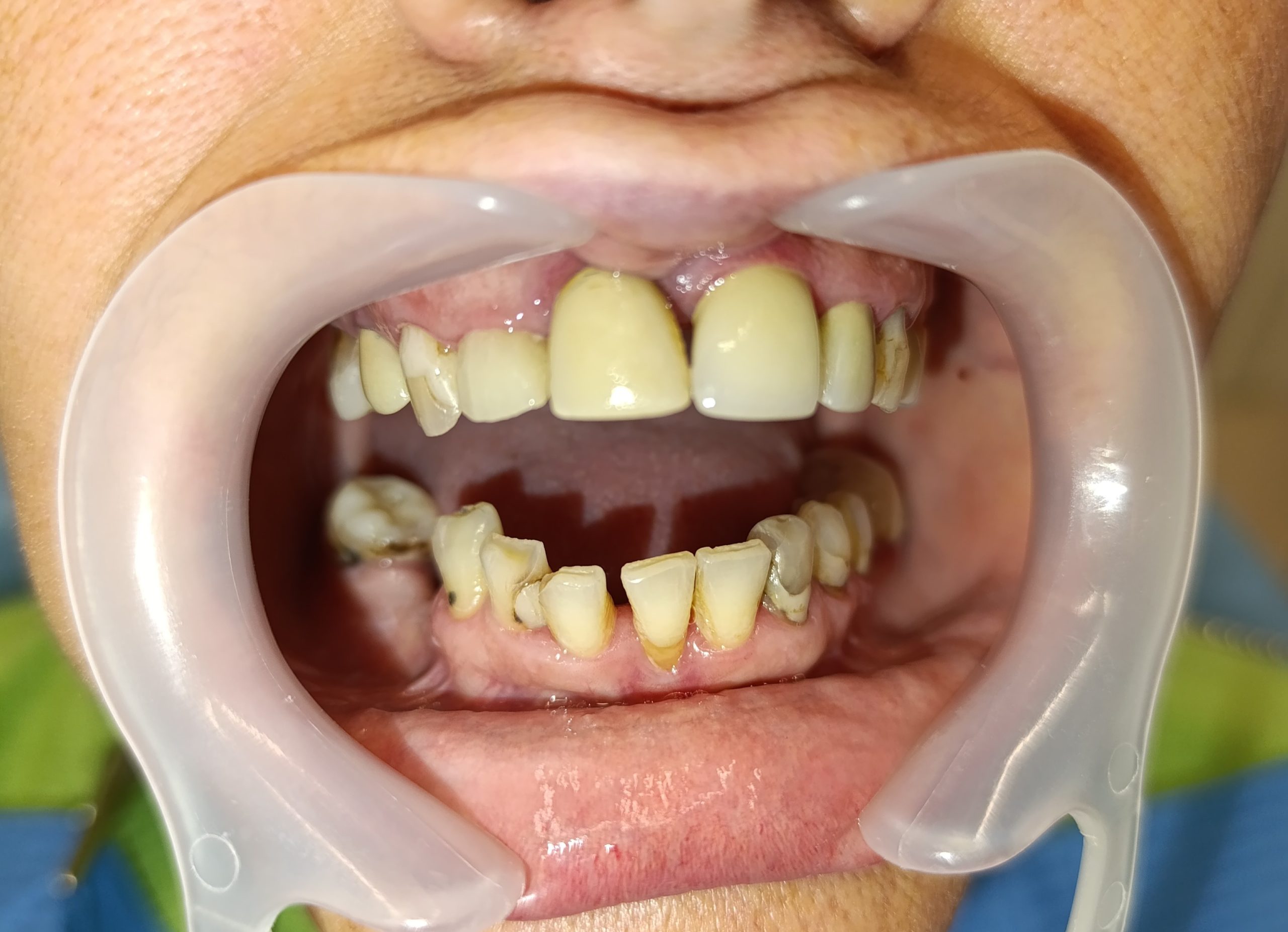
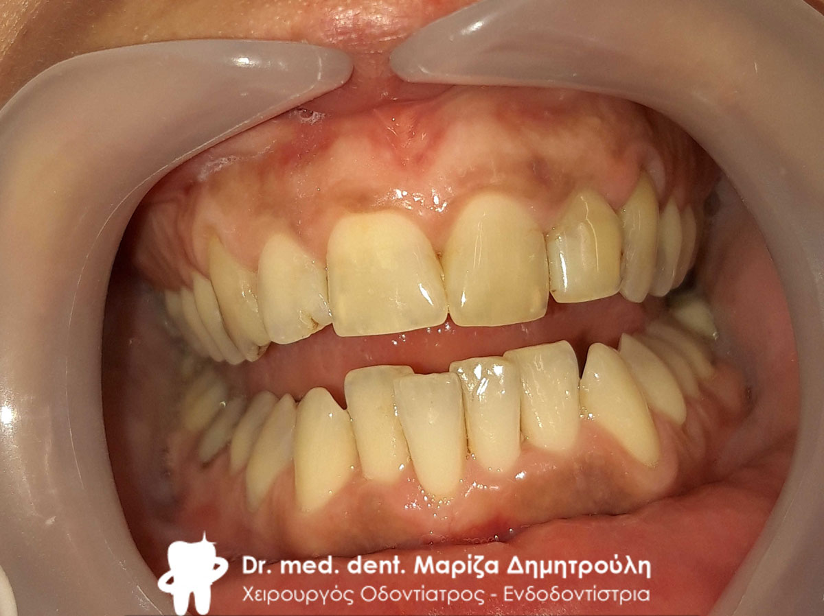
Case – Restoration of lower anterior teeth.
The patient wanted the restoration of the lower anterior teeth, which had strong mobility. She also reports that her left canine, lower right molar and some other teeth hurt. In order to create a perfect prosthetic work, it was decided to extract the lower front teeth, which had strong mobility. Afterwards, those teeth that had severe pain on impact and cold were denervated. Afterwards, the teeth were reconstructed, ground and an impression was taken for the dental technician. An all-ceramic zirconium plate was fabricated and bonded to the mandible.
The patient is very happy with the prosthetic work on the lower jaw.
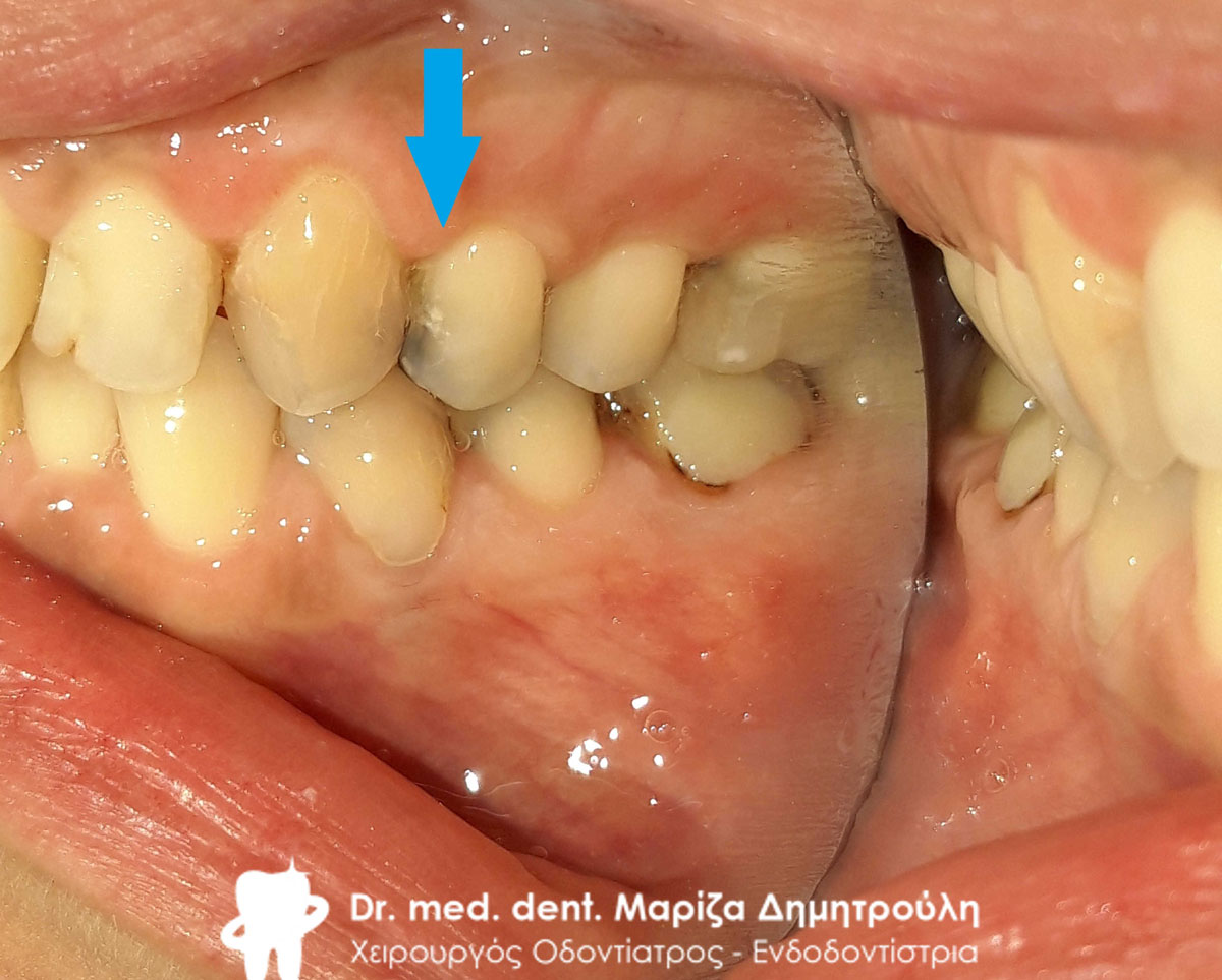
Later he developed severe pain in the second premolar on the left side of the upper jaw. Denervation was performed and the tooth was reconstructed with a white fiberglass shaft and white resin. In the second phase, an all-ceramic zirconium case will be made to cover the tooth.
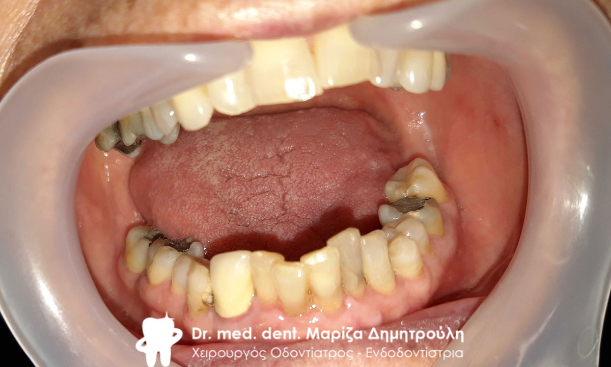
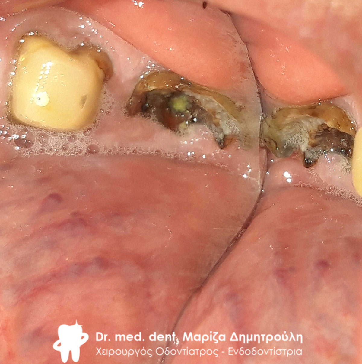
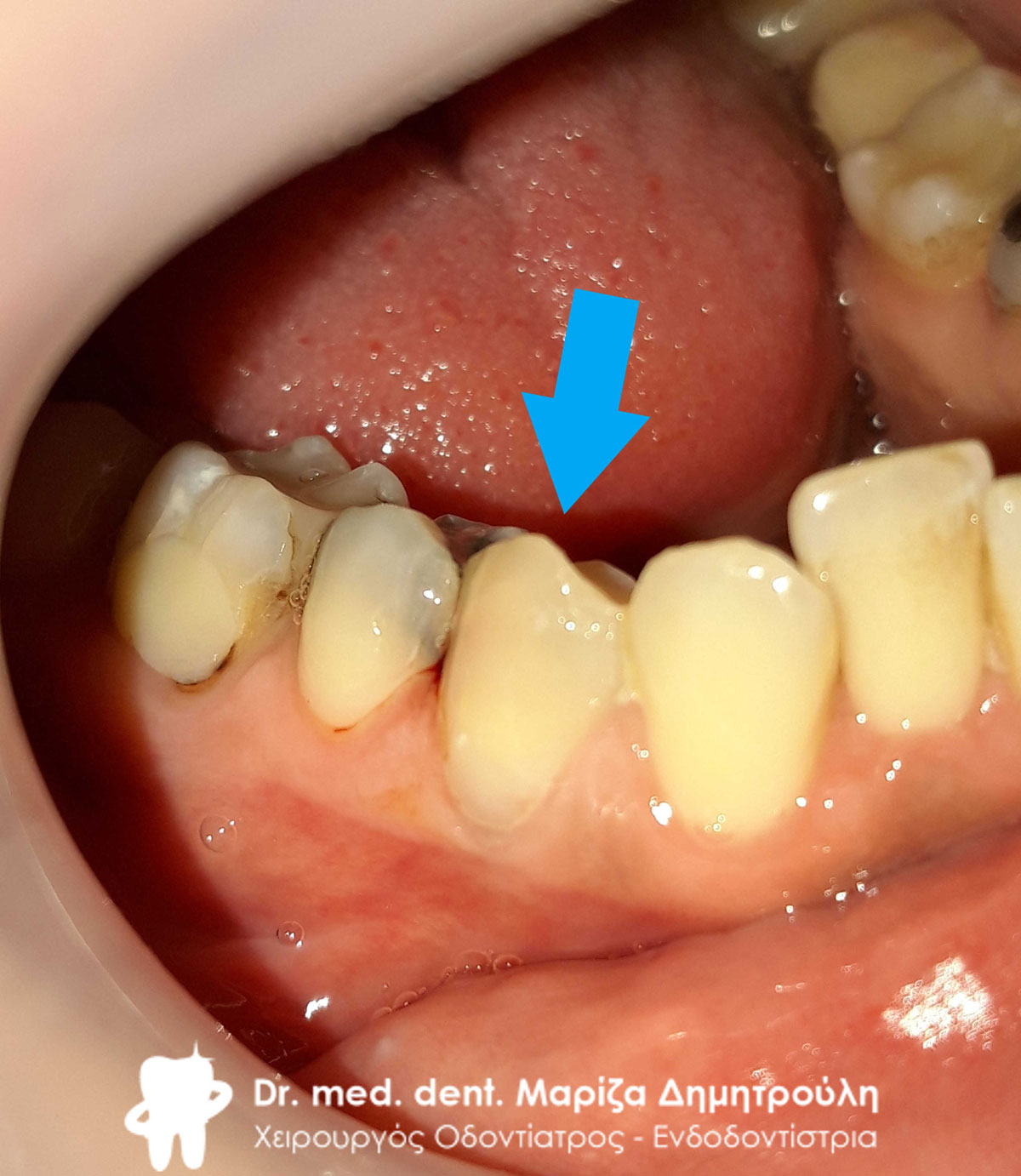
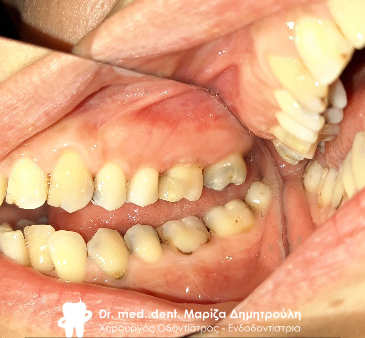
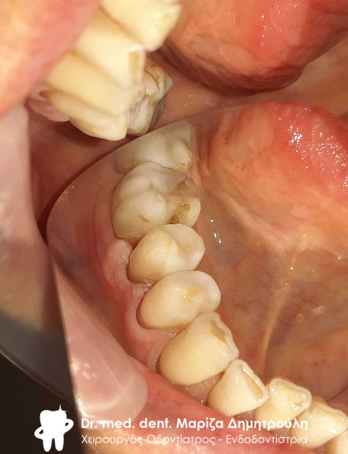
Initial clinical image of the lower jaw
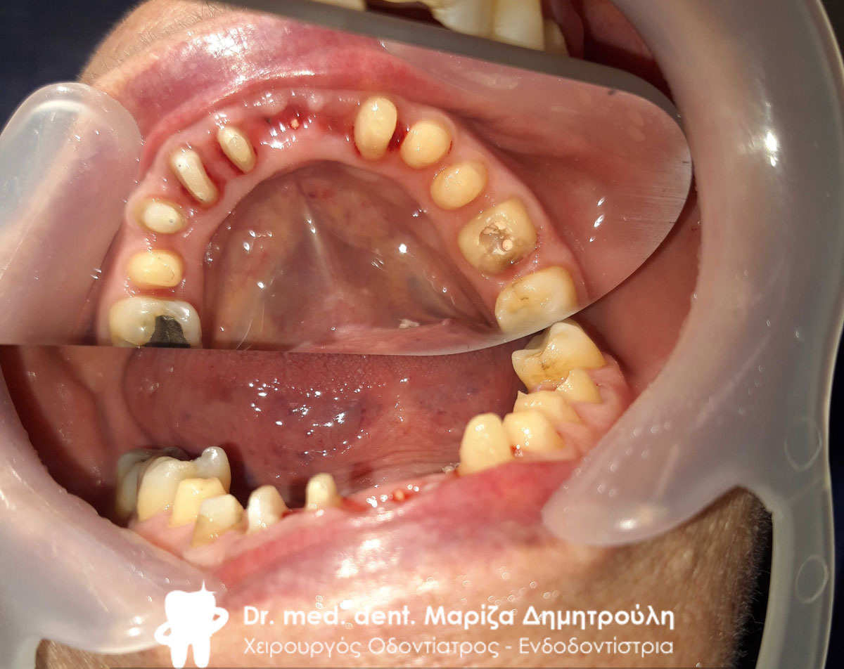
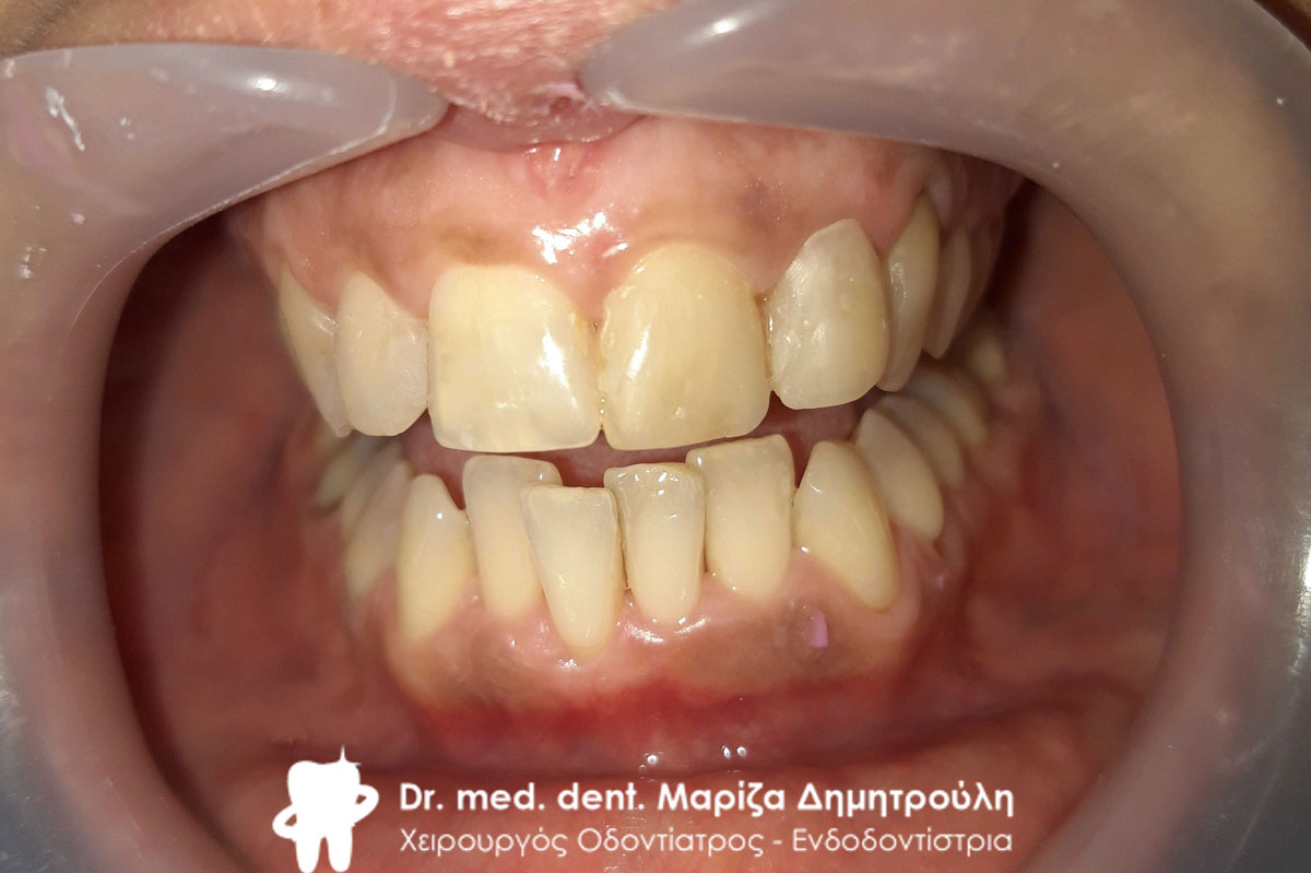
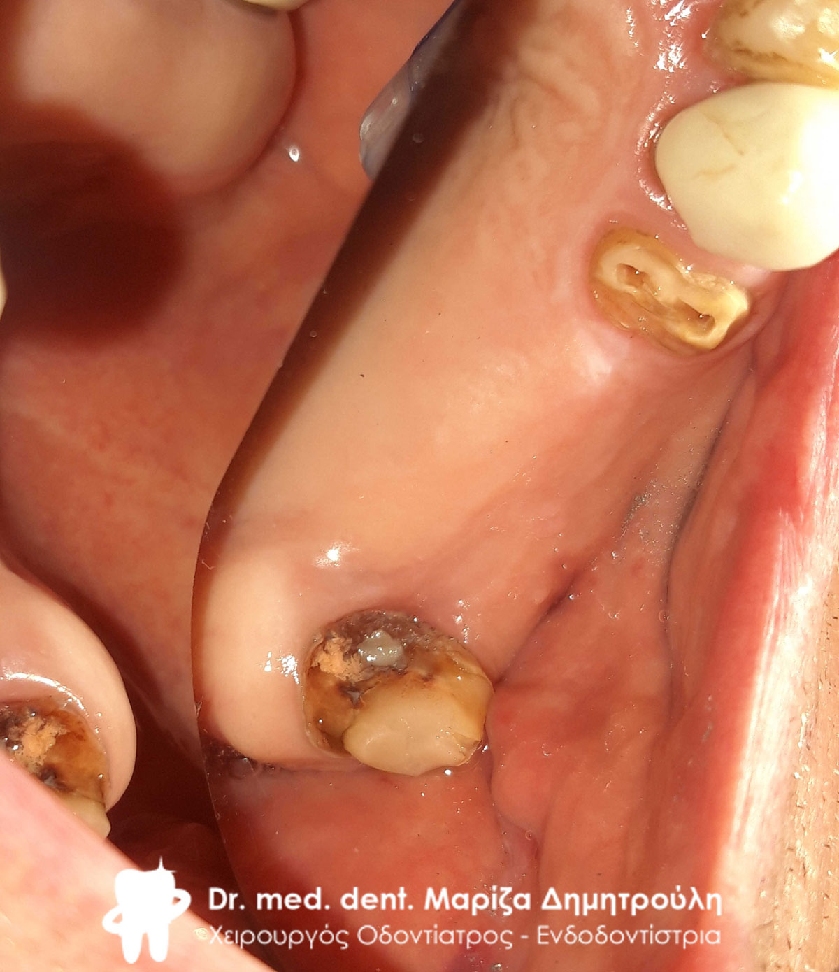
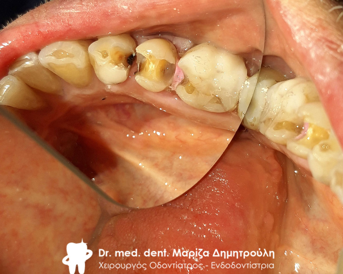
Image of ground teeth with required restorations

Image of ground teeth with required restorations
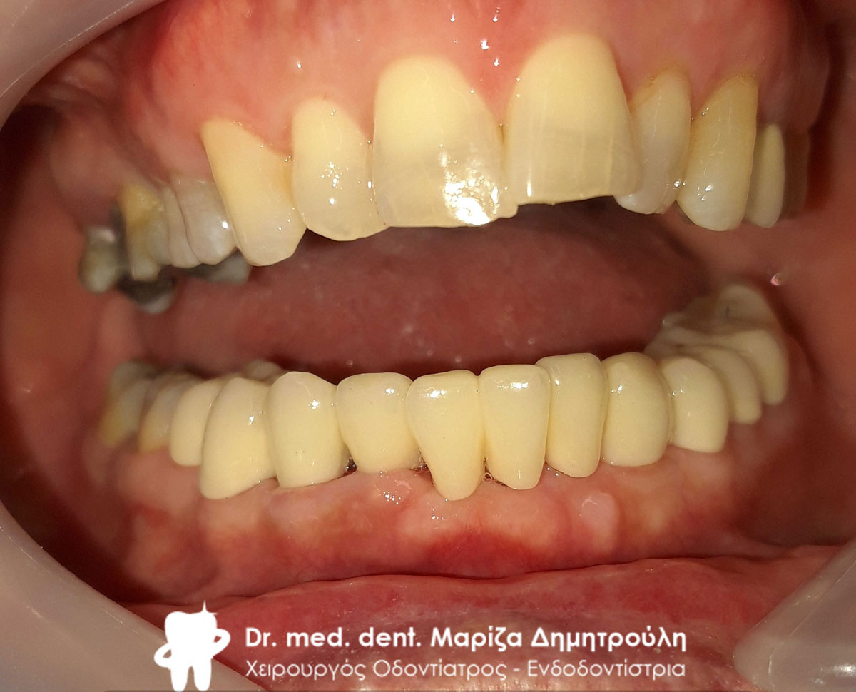
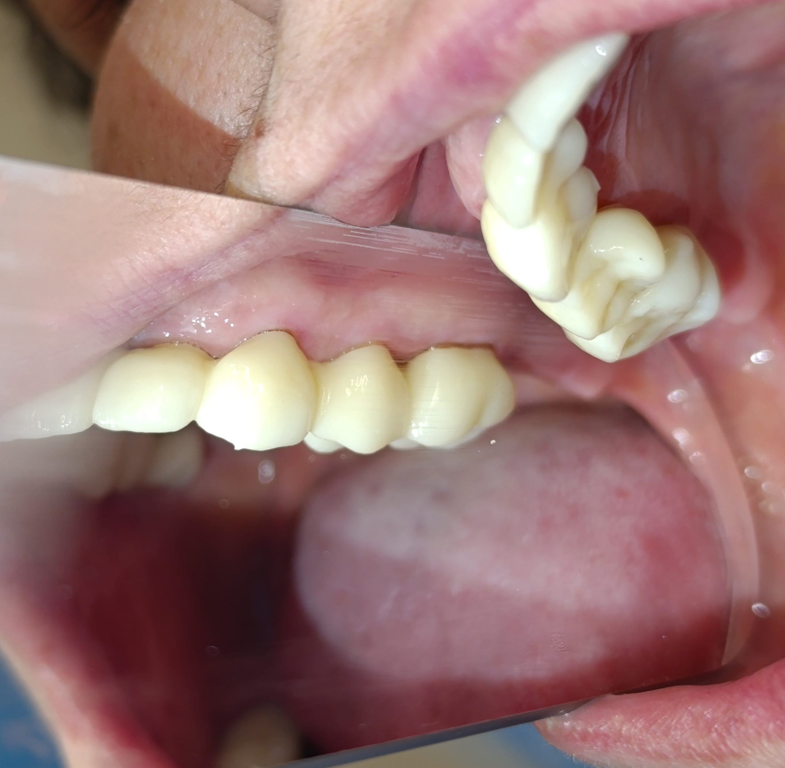
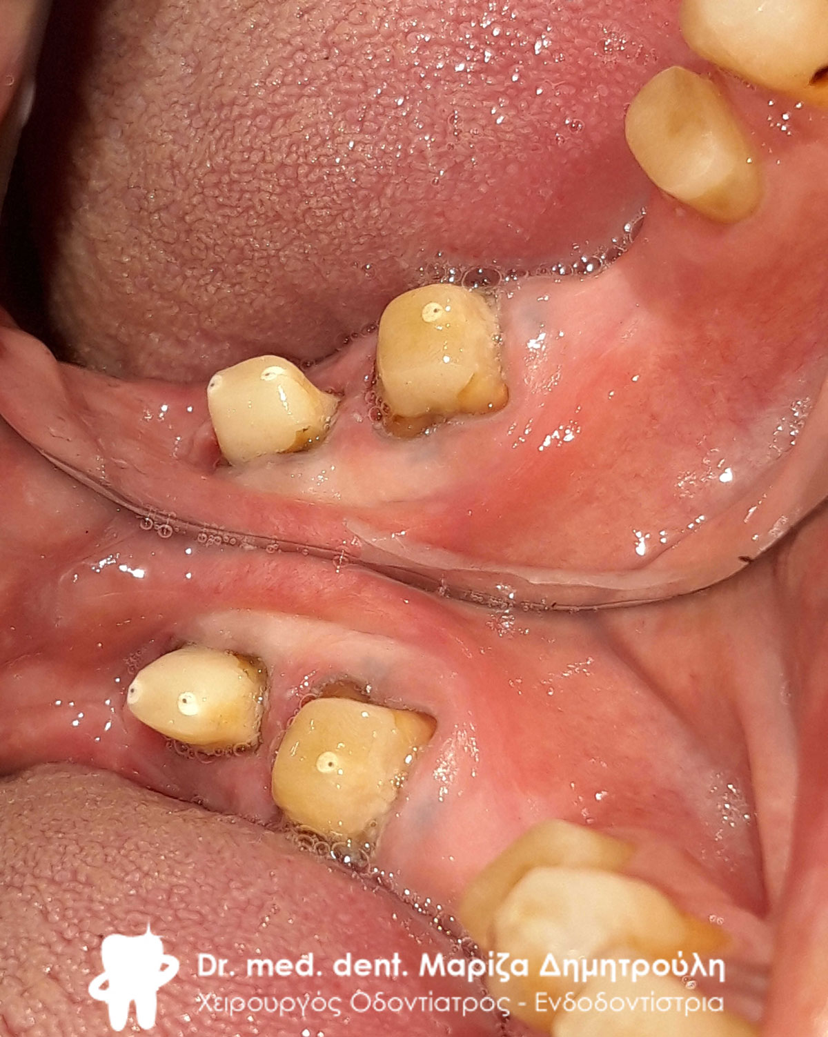
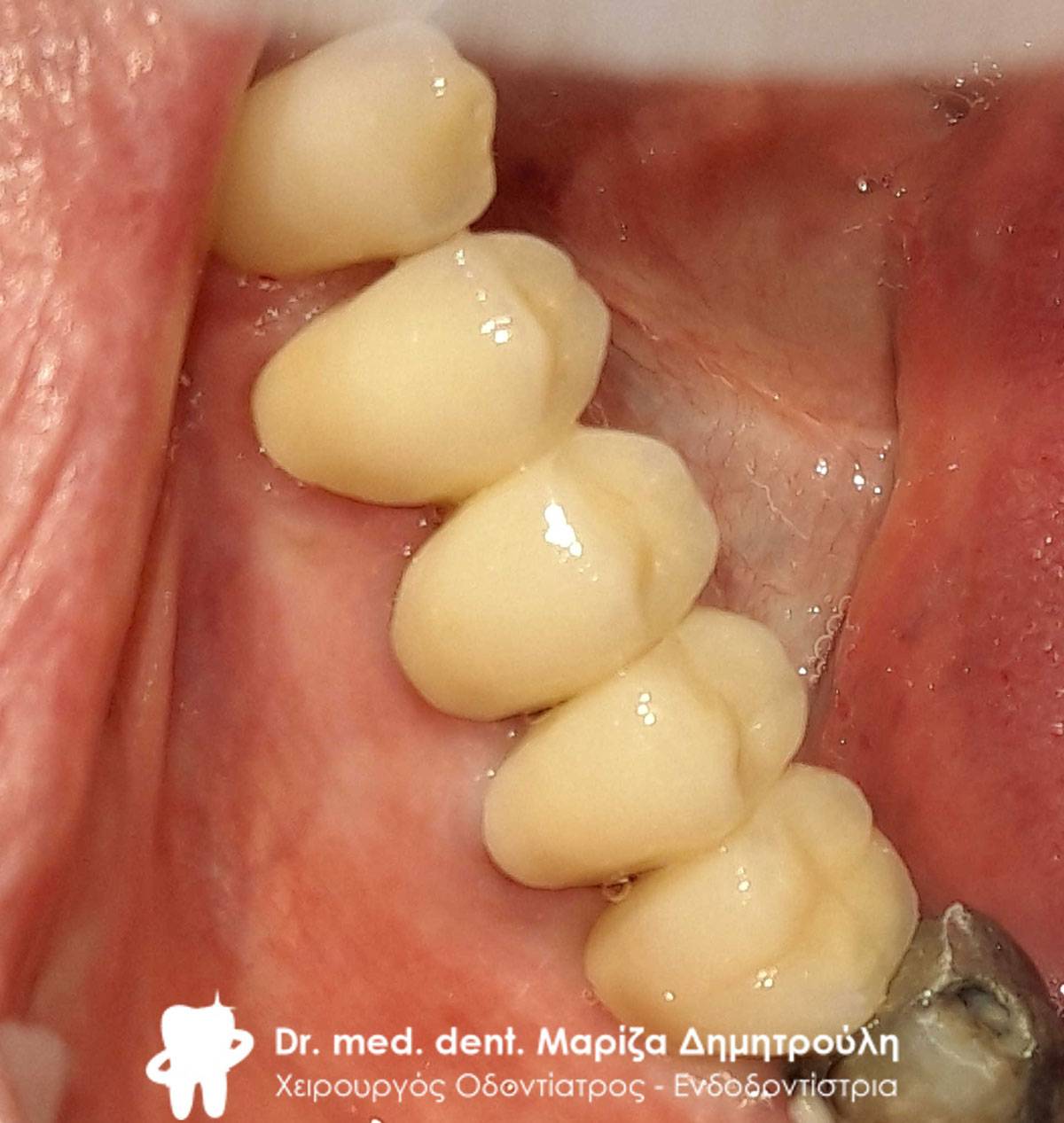
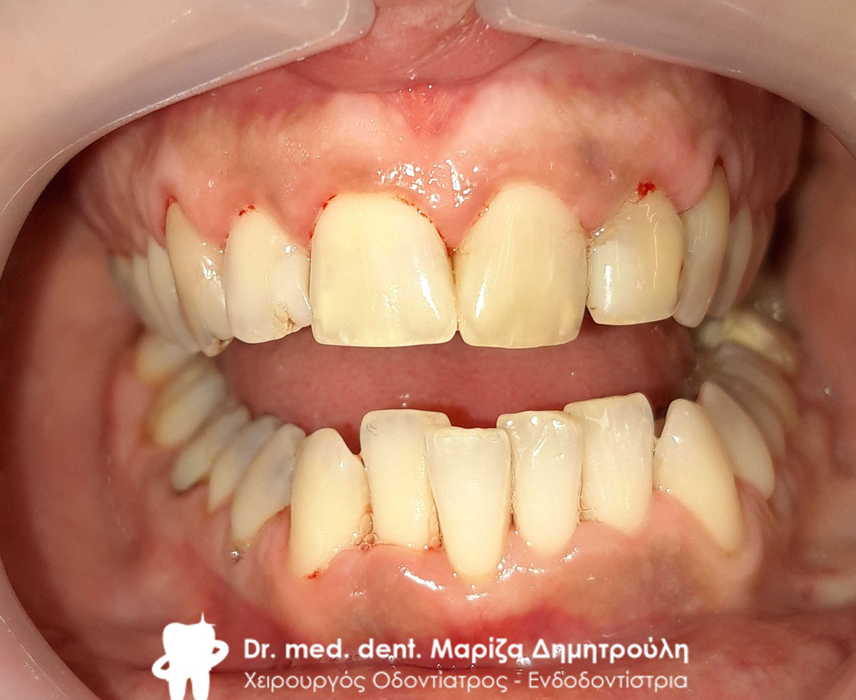
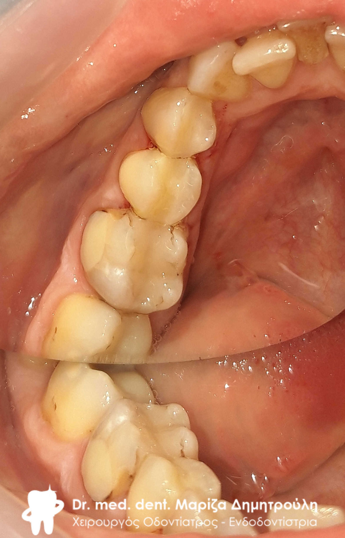
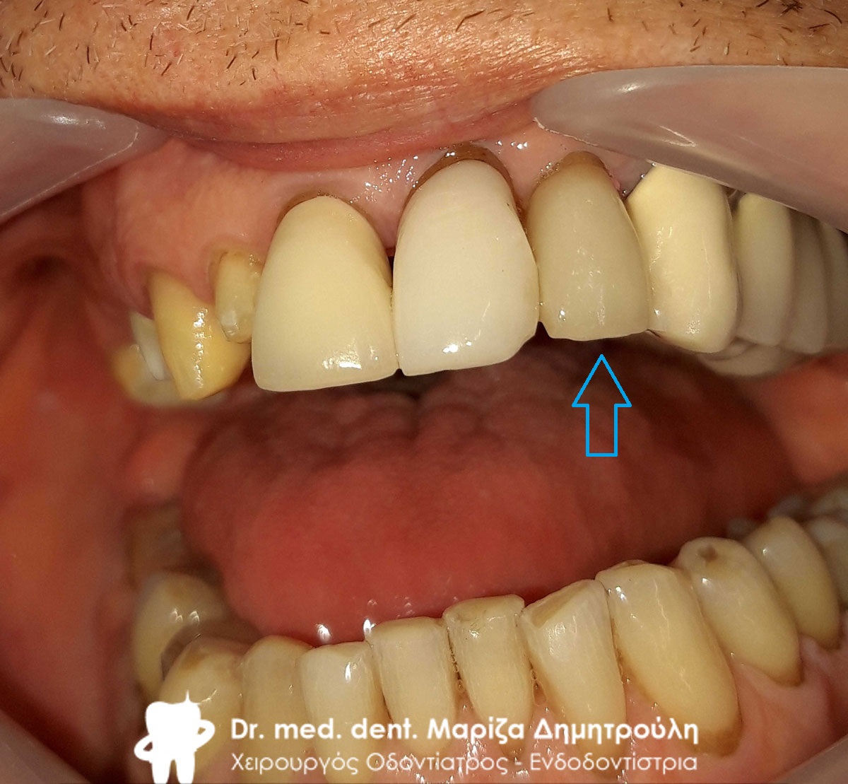
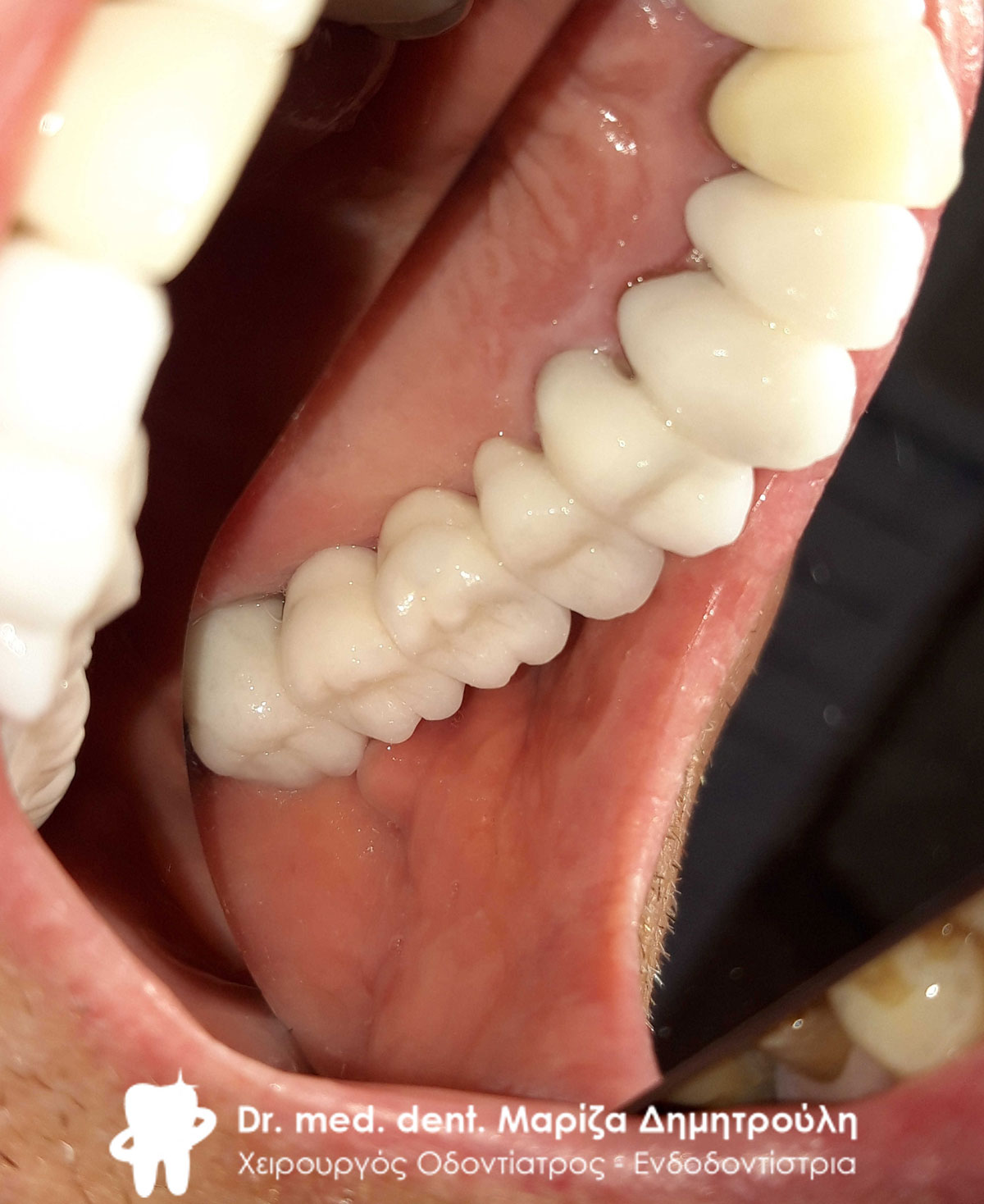
Final image of mandibular all-ceramic zirconium petal
Final image of mandibular all-ceramic zirconium petal
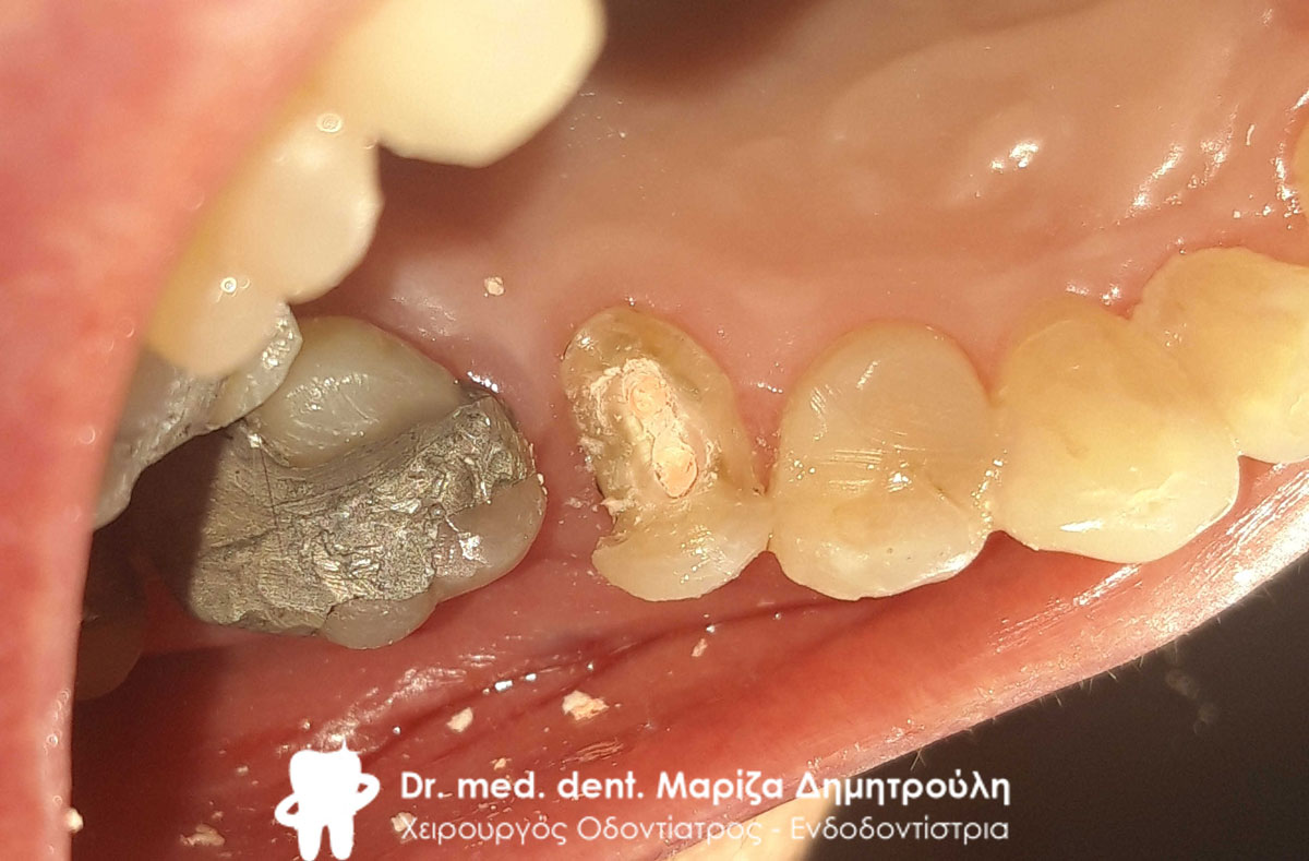
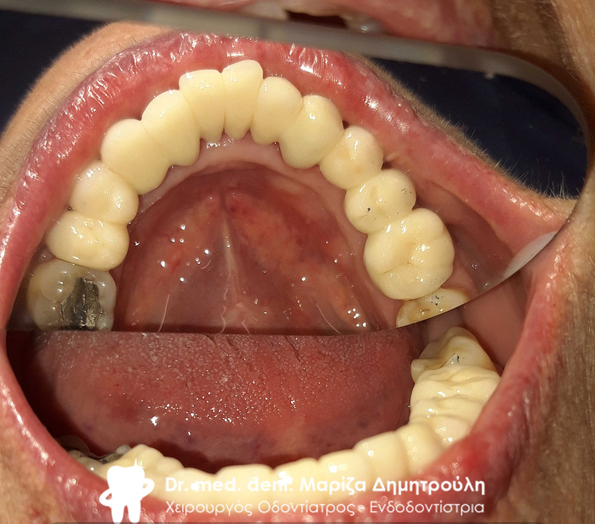
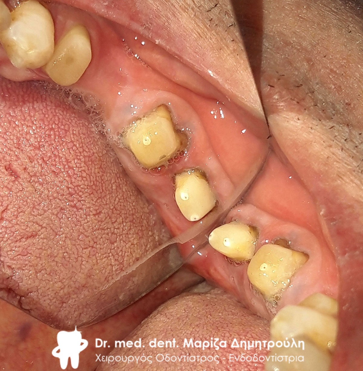
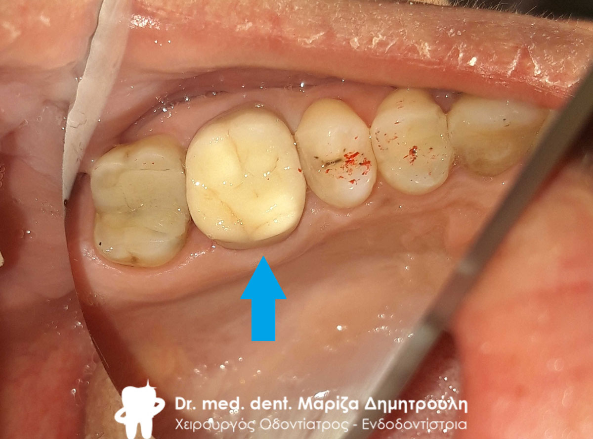
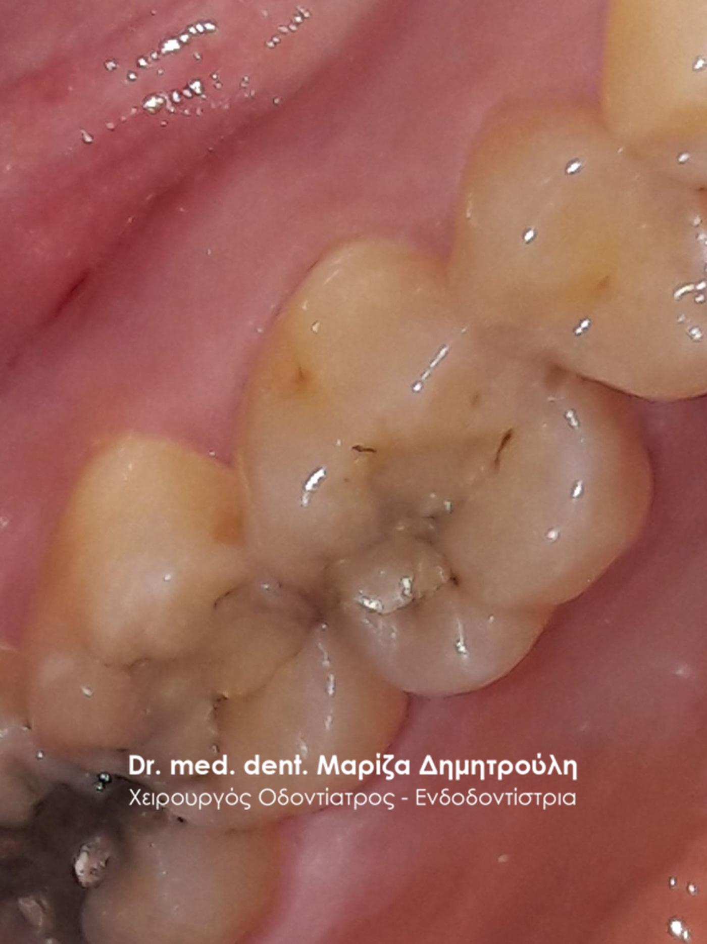
Image of the second premolar after the denervation is complete and before the tooth is reconstructed with a shaft
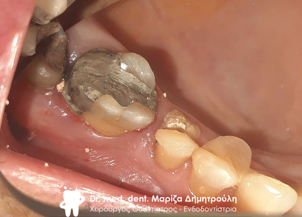
Image of the second premolar after the denervation is complete and before the tooth is reconstructed with a shaft
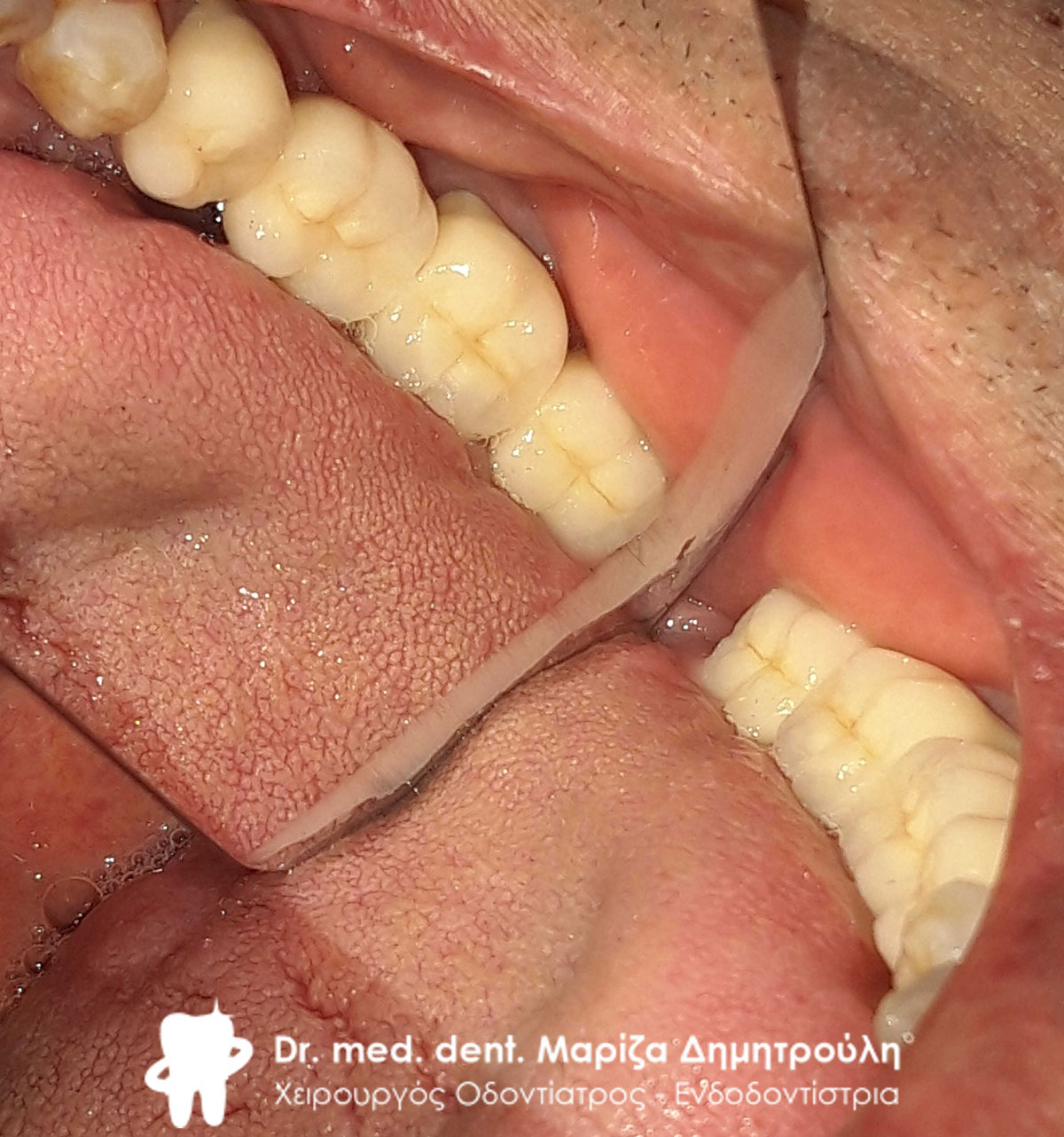
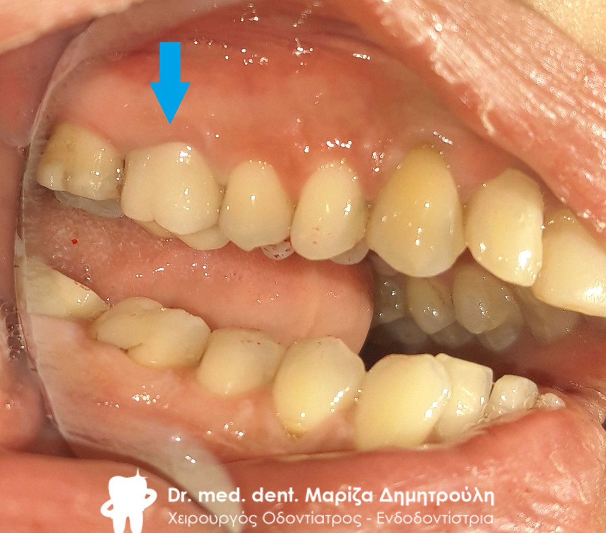
Final image of the premolar with the white fiberglass shaft and the corresponding reconstruction
Final image of the premolar with the white fiberglass shaft and the corresponding reconstruction
Case – Total maxillary restoration
The patient had neglected her teeth for years and made the decision to restore them for both aesthetic and functional reasons.
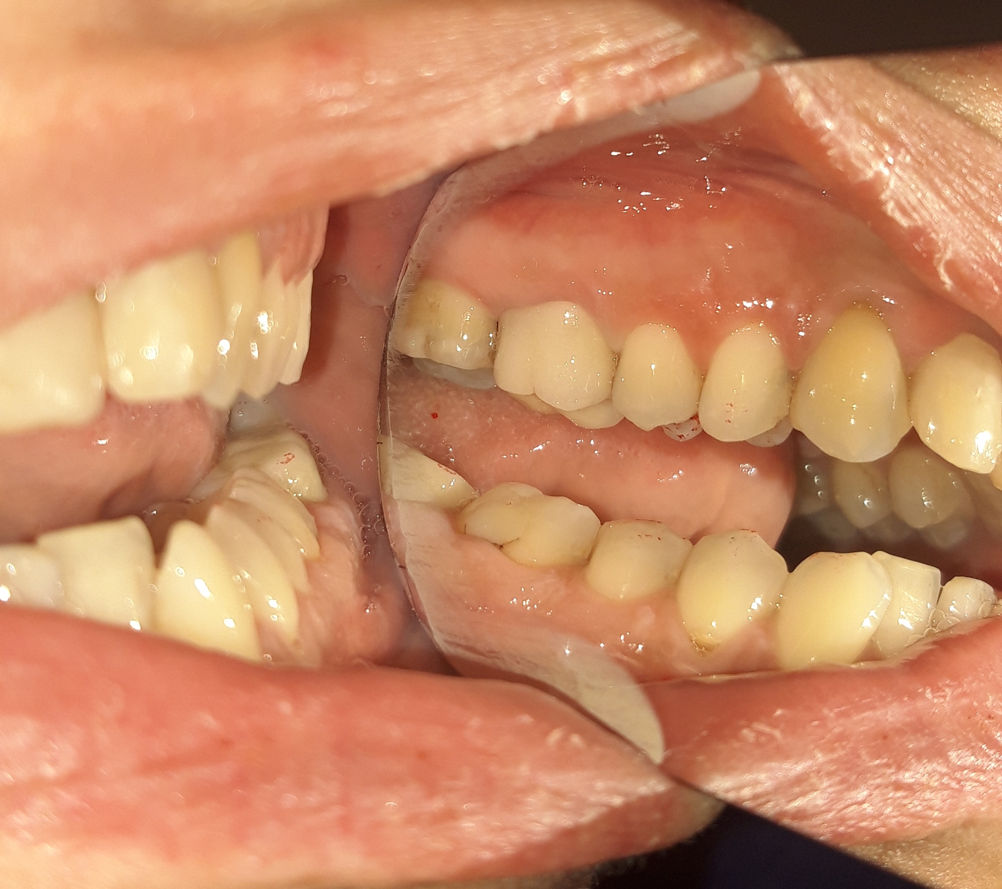
Initial clinical image of the maxilla
Initial clinical image of the left side of the maxilla
Initial clinical image of the right side of the maxilla
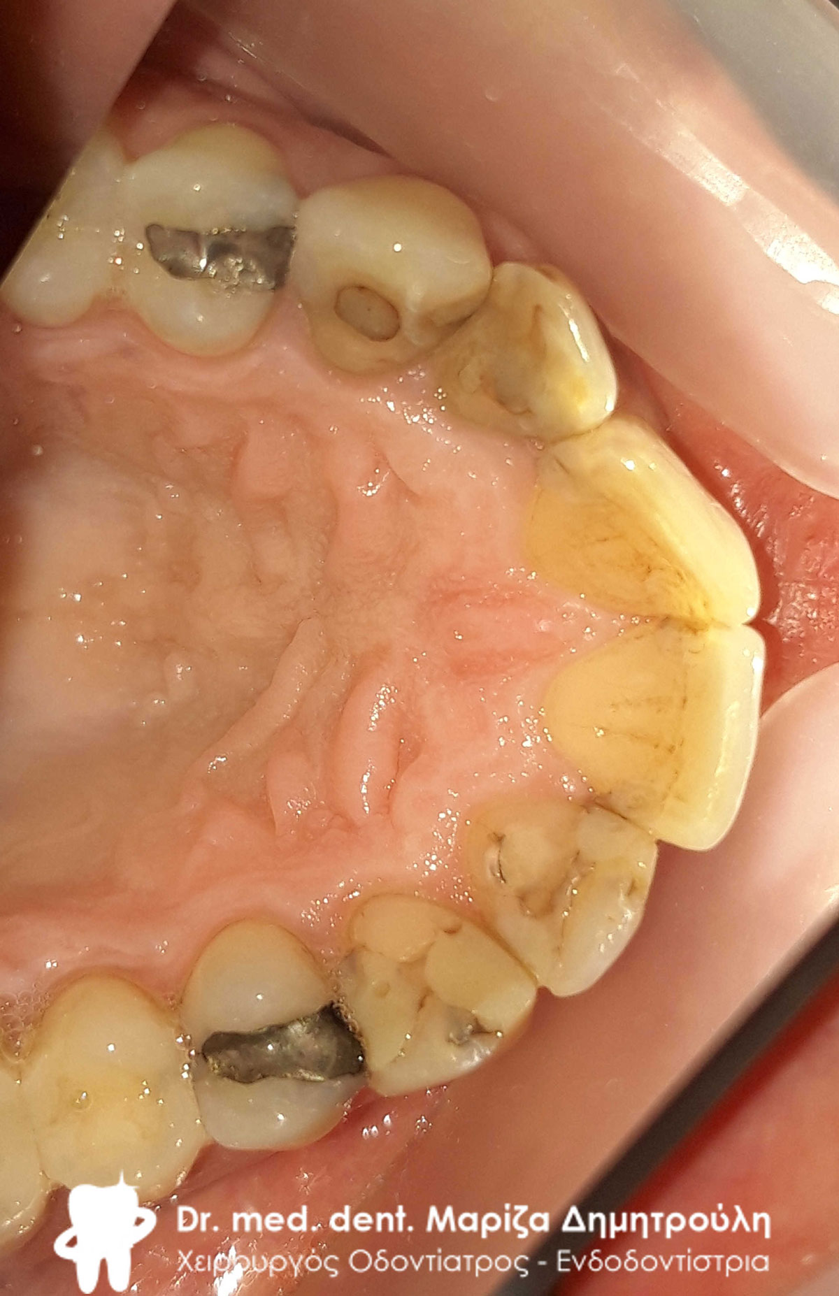
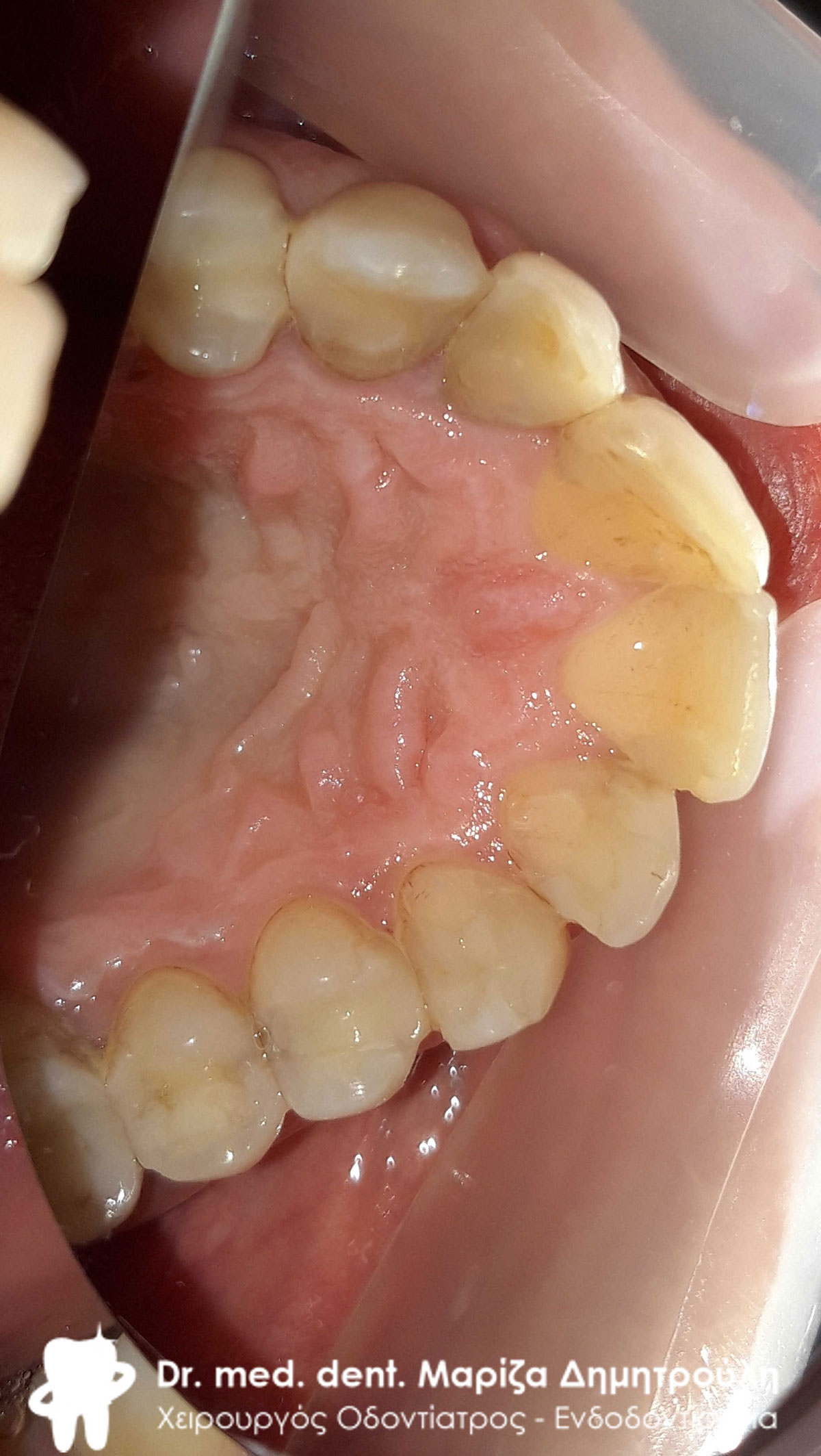
Initial clinical image of the palatal side of the maxilla
Initial clinical image of the palatal side of the maxilla
Final clinical image of the maxilla
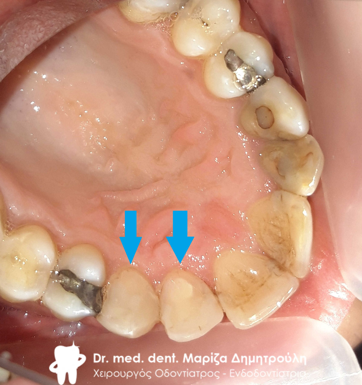
Final clinical image of the palatal side of the maxilla
Final clinical picture of the left side of the maxilla
Final clinical image of the right side of the maxilla
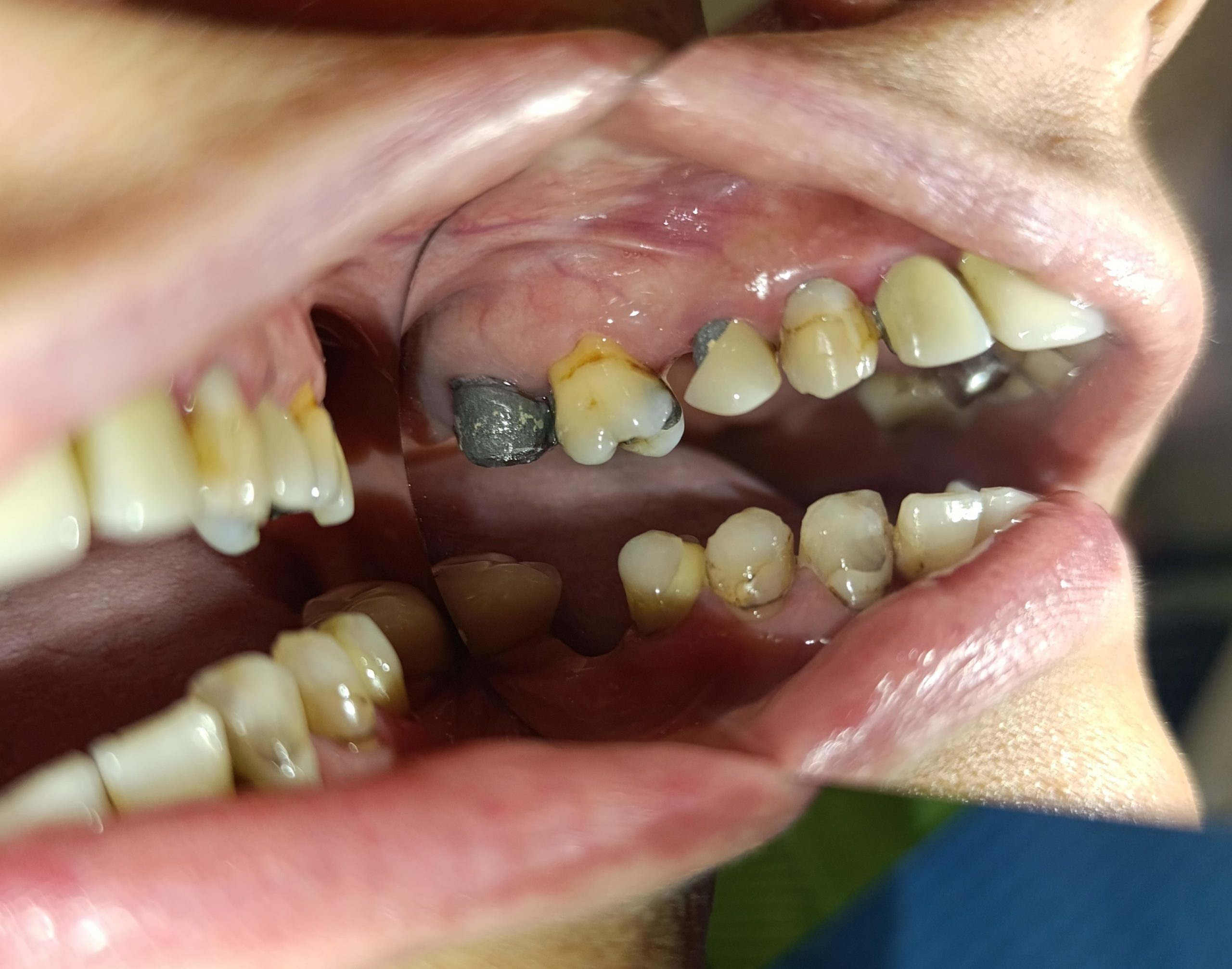
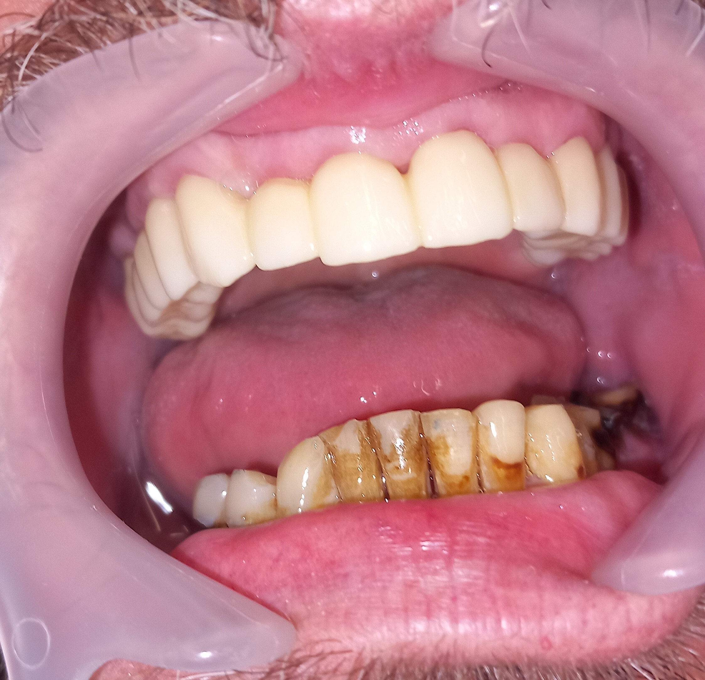
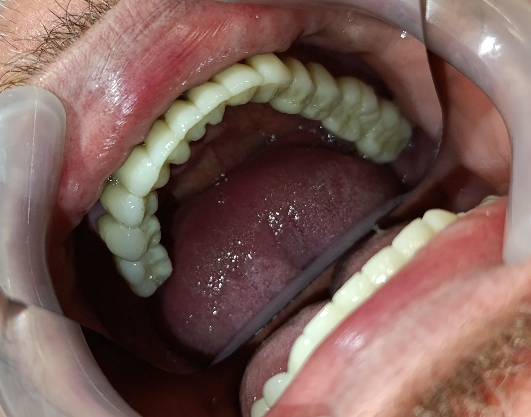
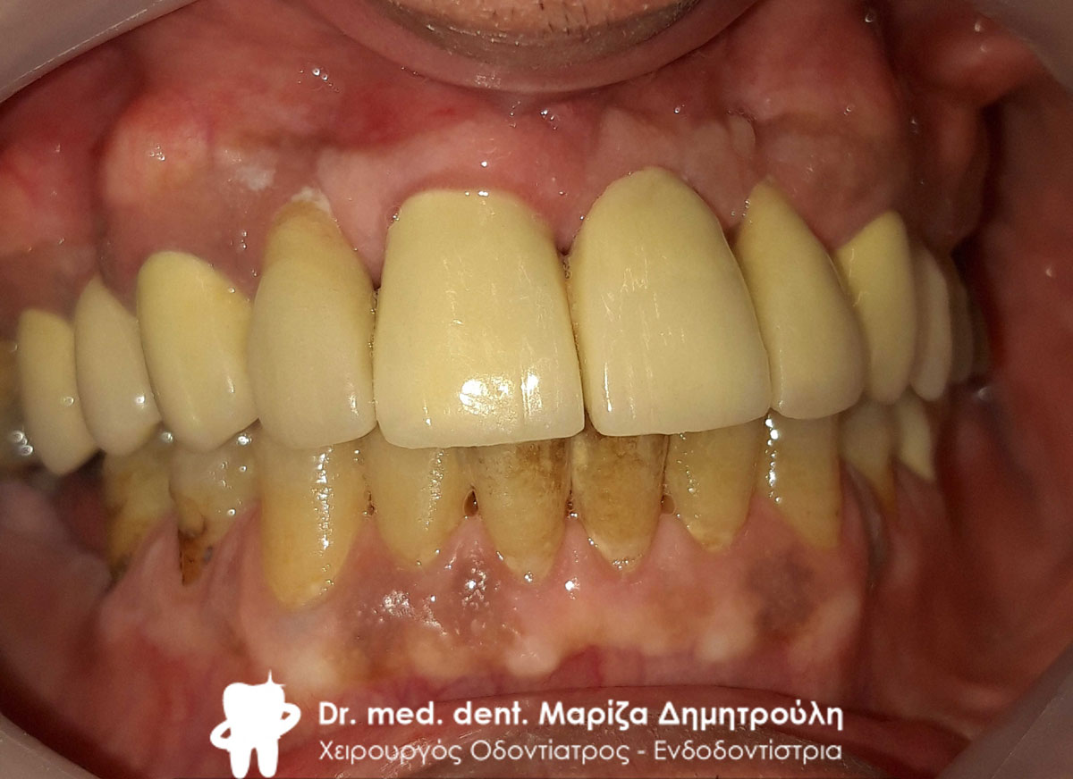
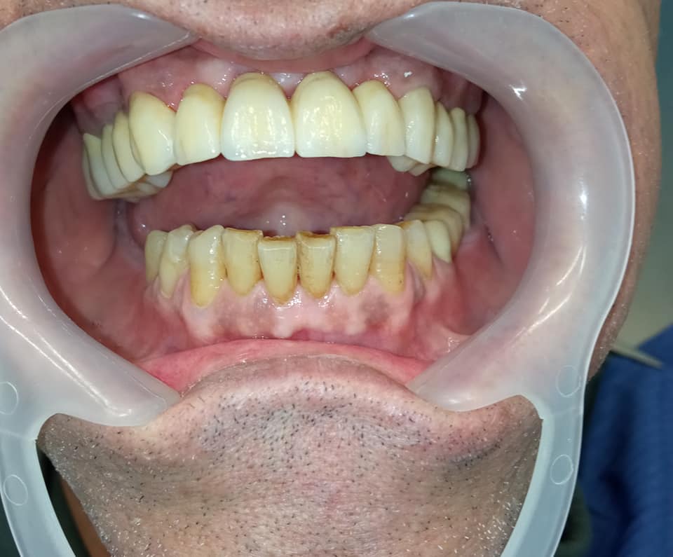
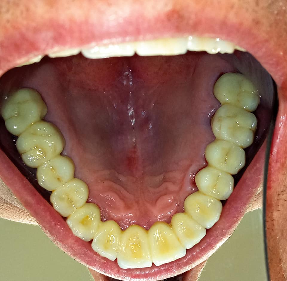
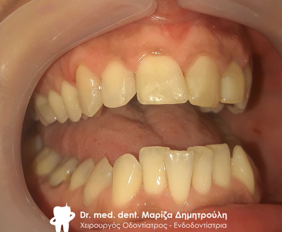
Case – Total restoration of maxilla and mandible
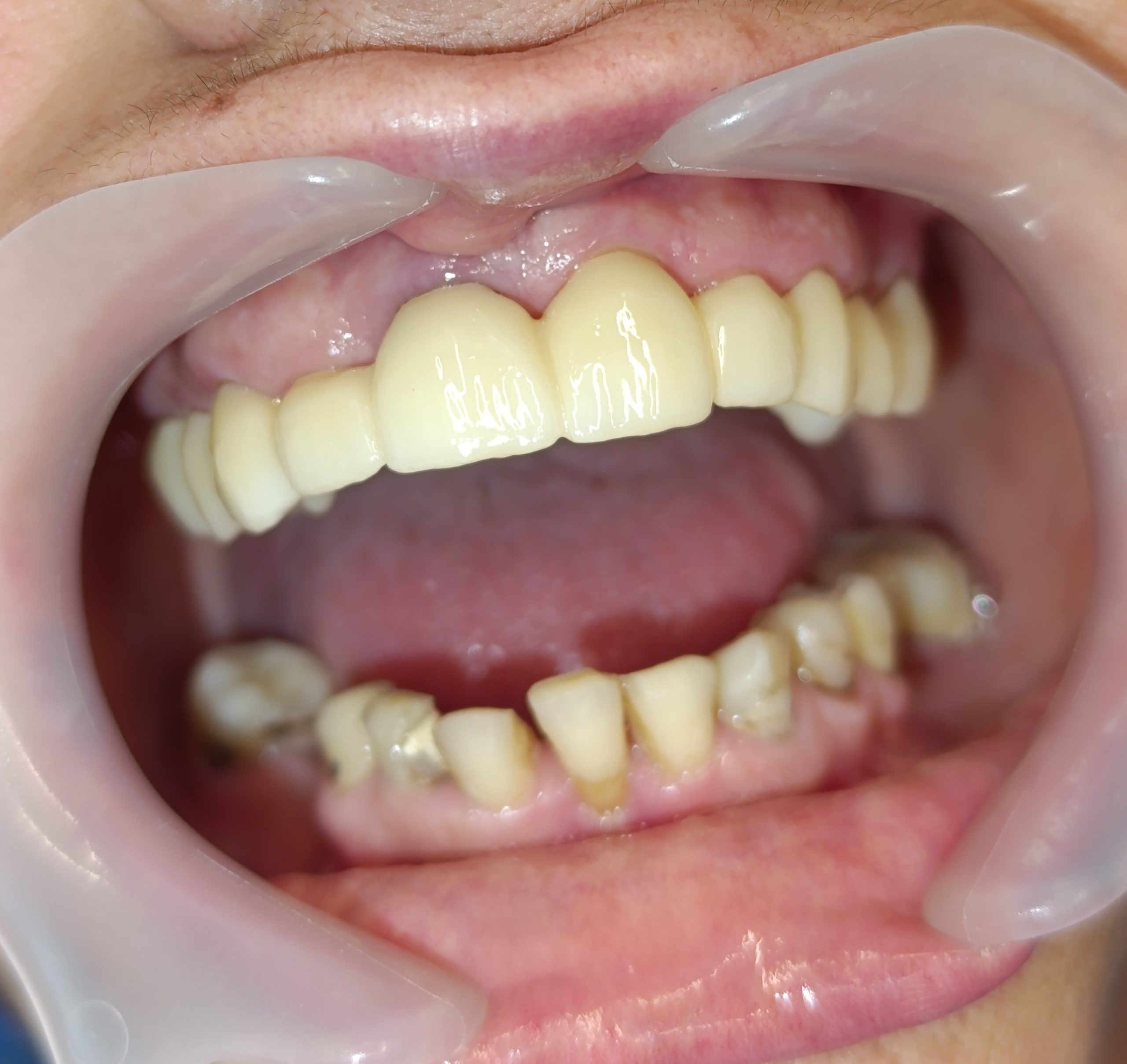
The patient made the decision after many years to restore both maxillary and mandibular teeth. It took several sessions to complete the necessary denervations and tooth reconstructions. Then, once the prosthetic work was ready, the all-ceramic plates were glued to the patient’s mouth by the dental technician.
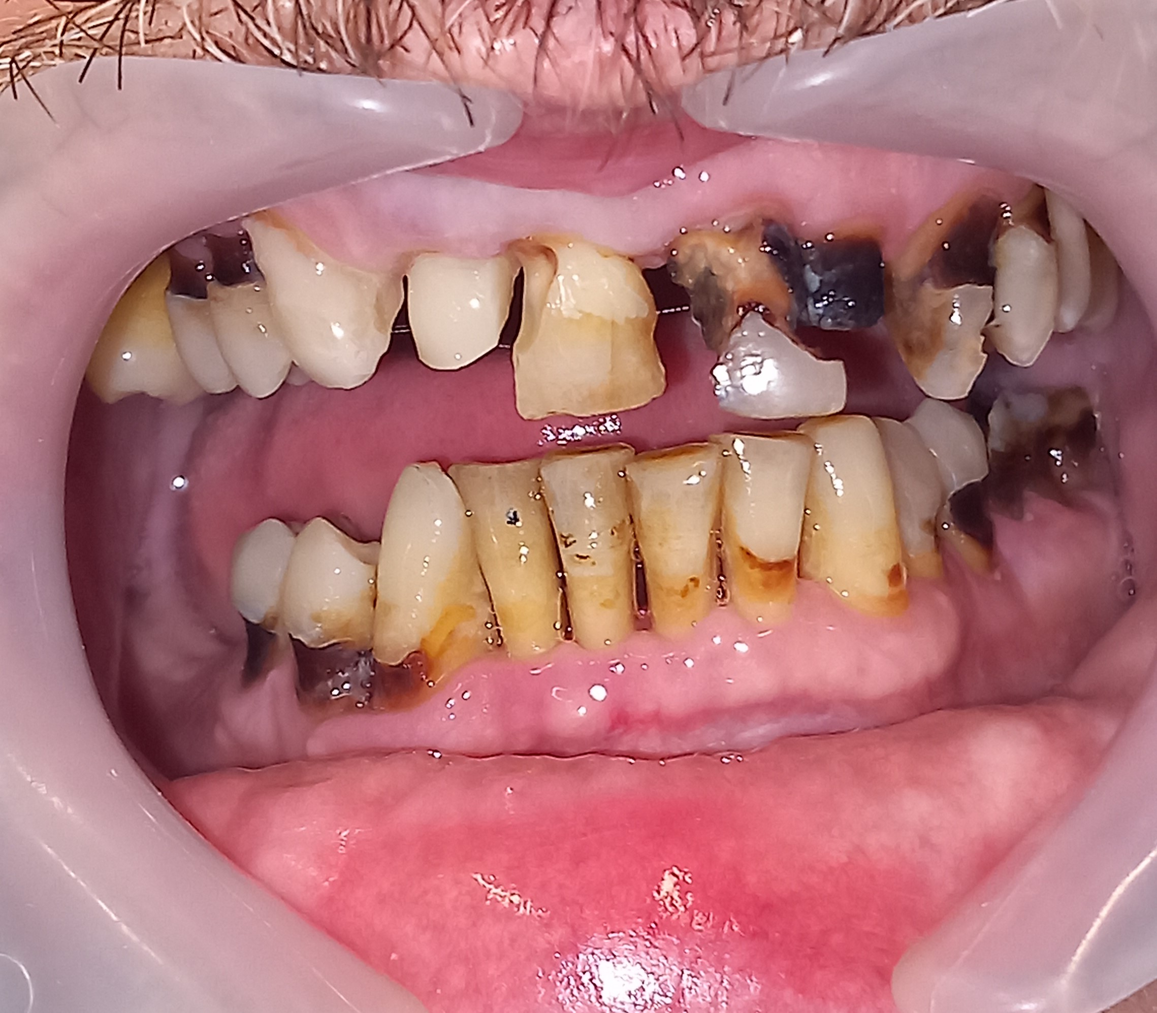
Initial clinical image of the patient’s mouth
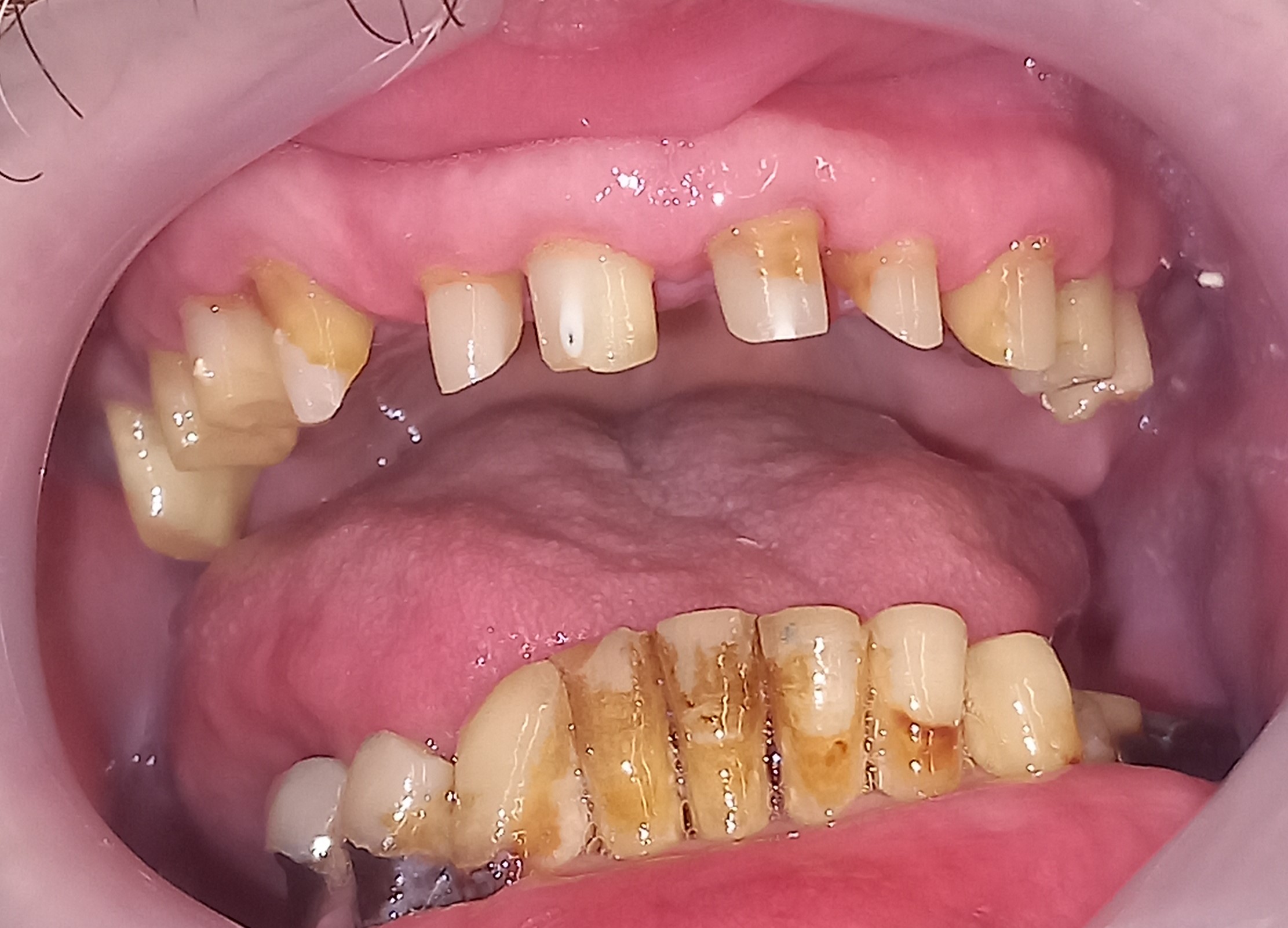
Clinical image of the upper teeth after the necessary operations
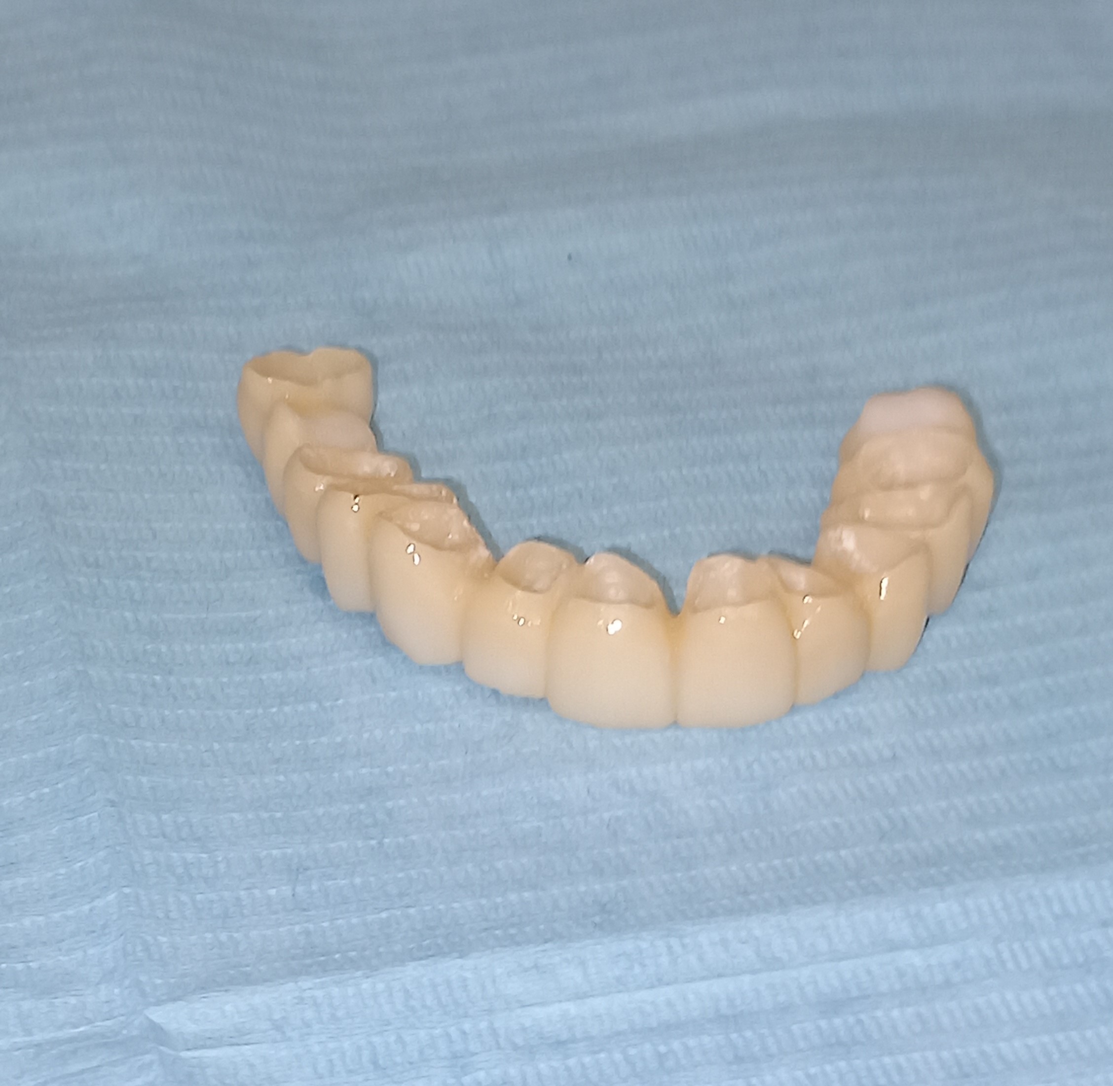
The upper all-ceramic plate before bonding in the patient’s mouth
Final clinical image of the maxilla
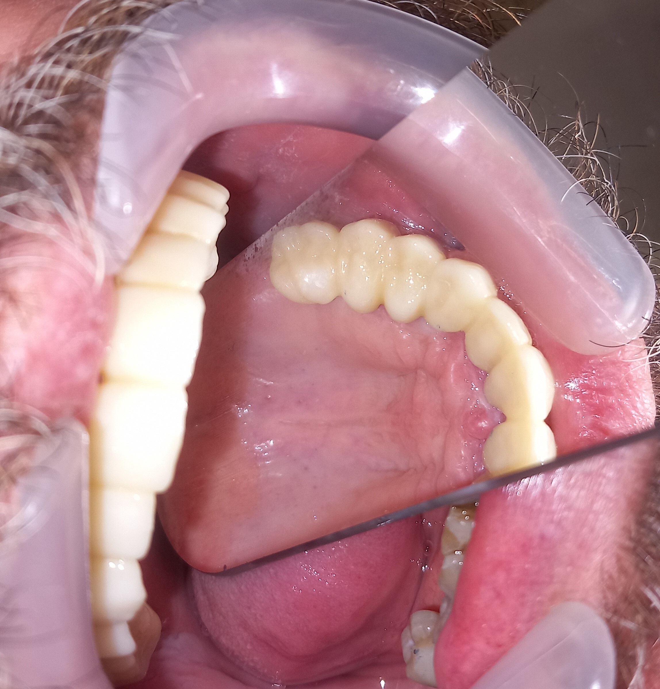
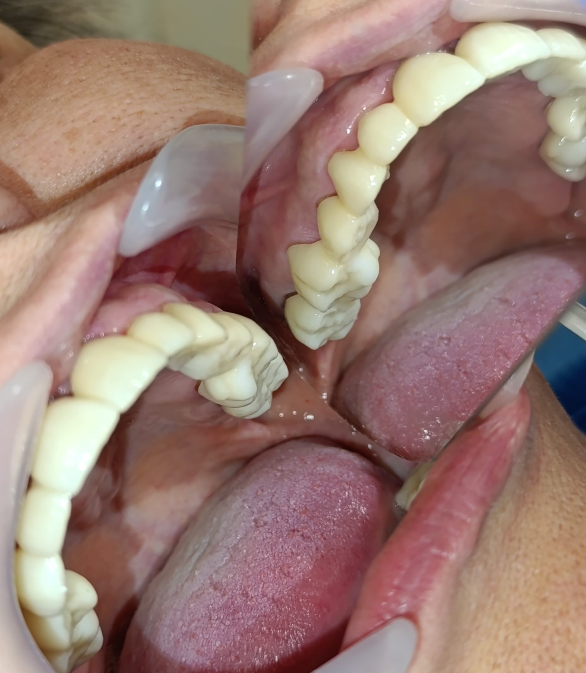
Final clinical image of the palatal side of the maxilla
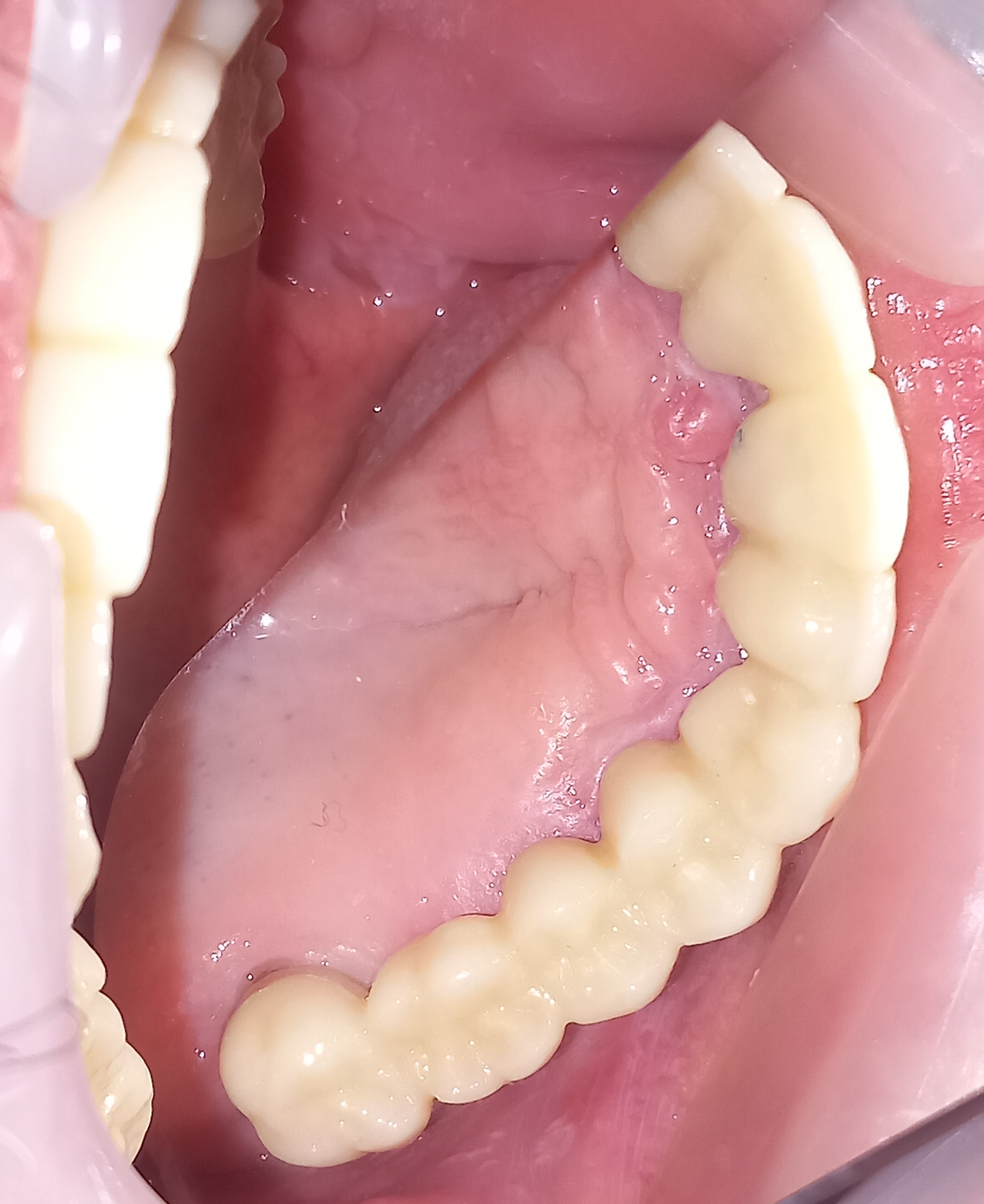
Final clinical image of the palatal side of the maxilla
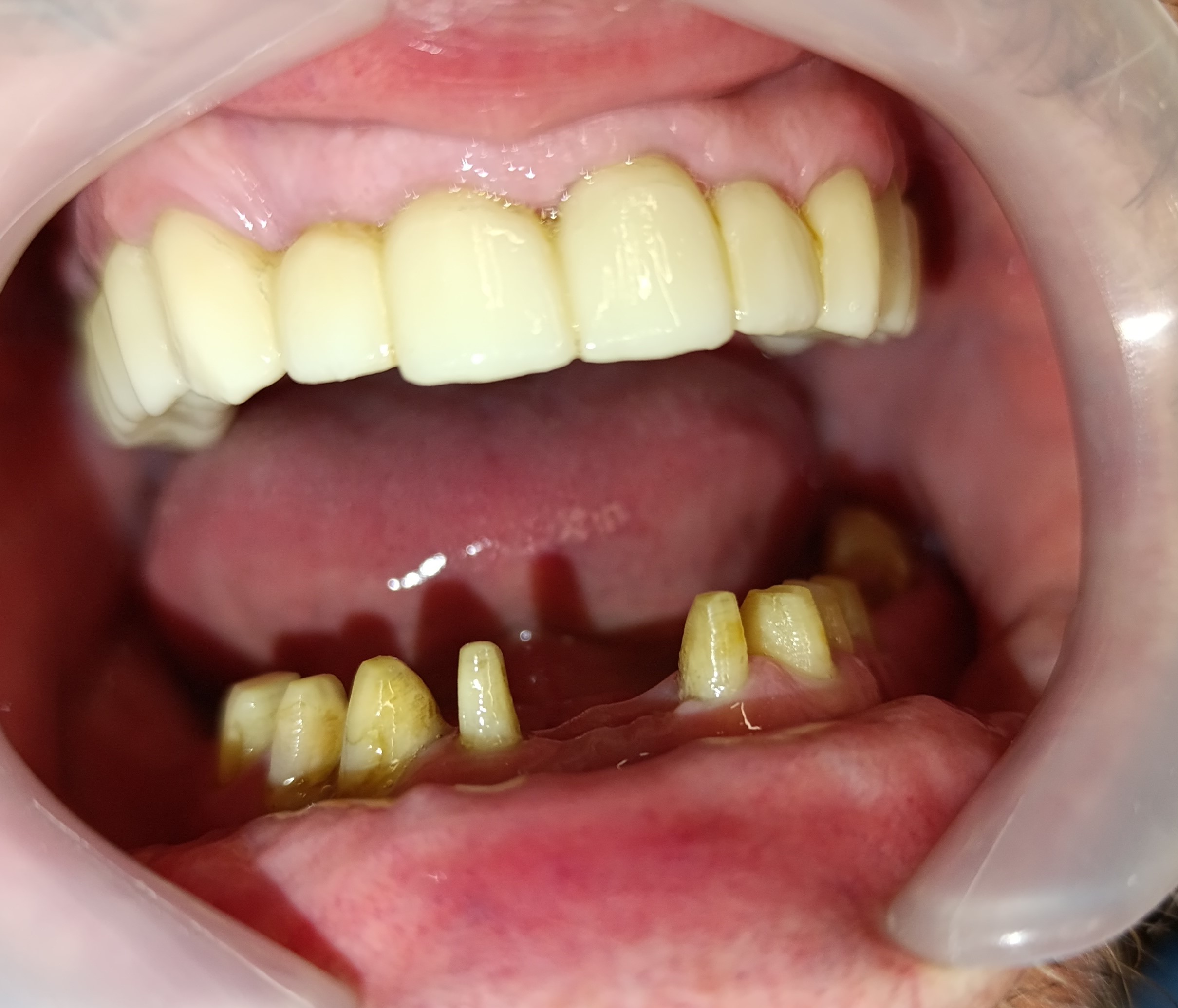
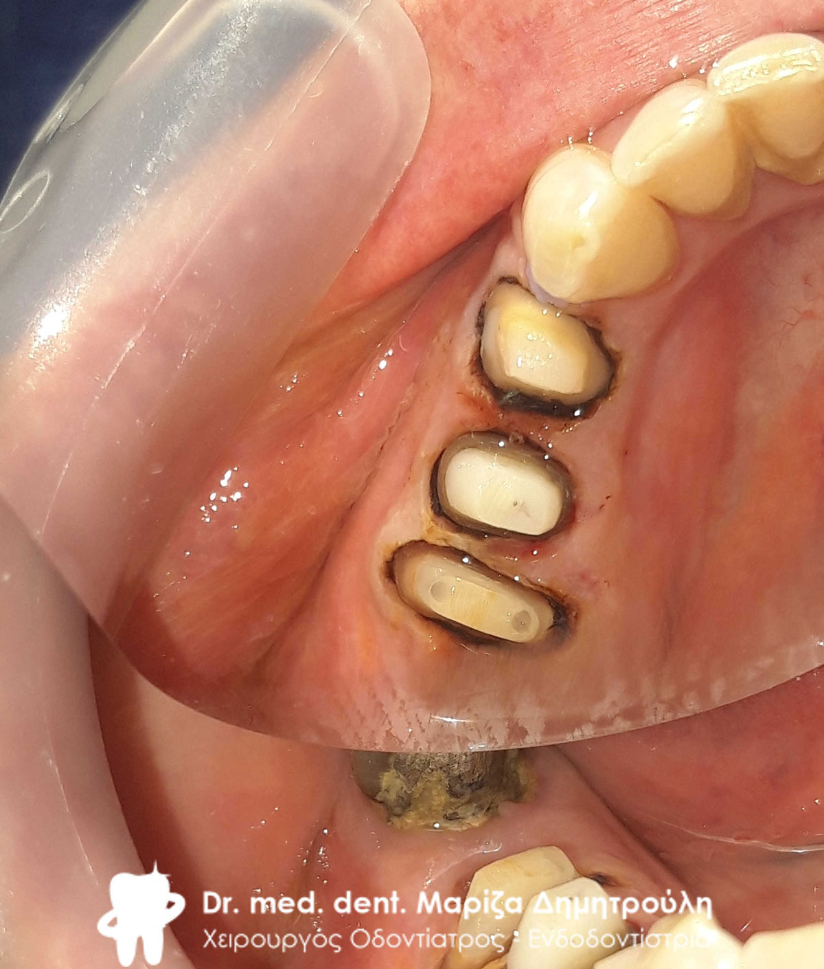
Clinical image of the lower teeth after the necessary operations
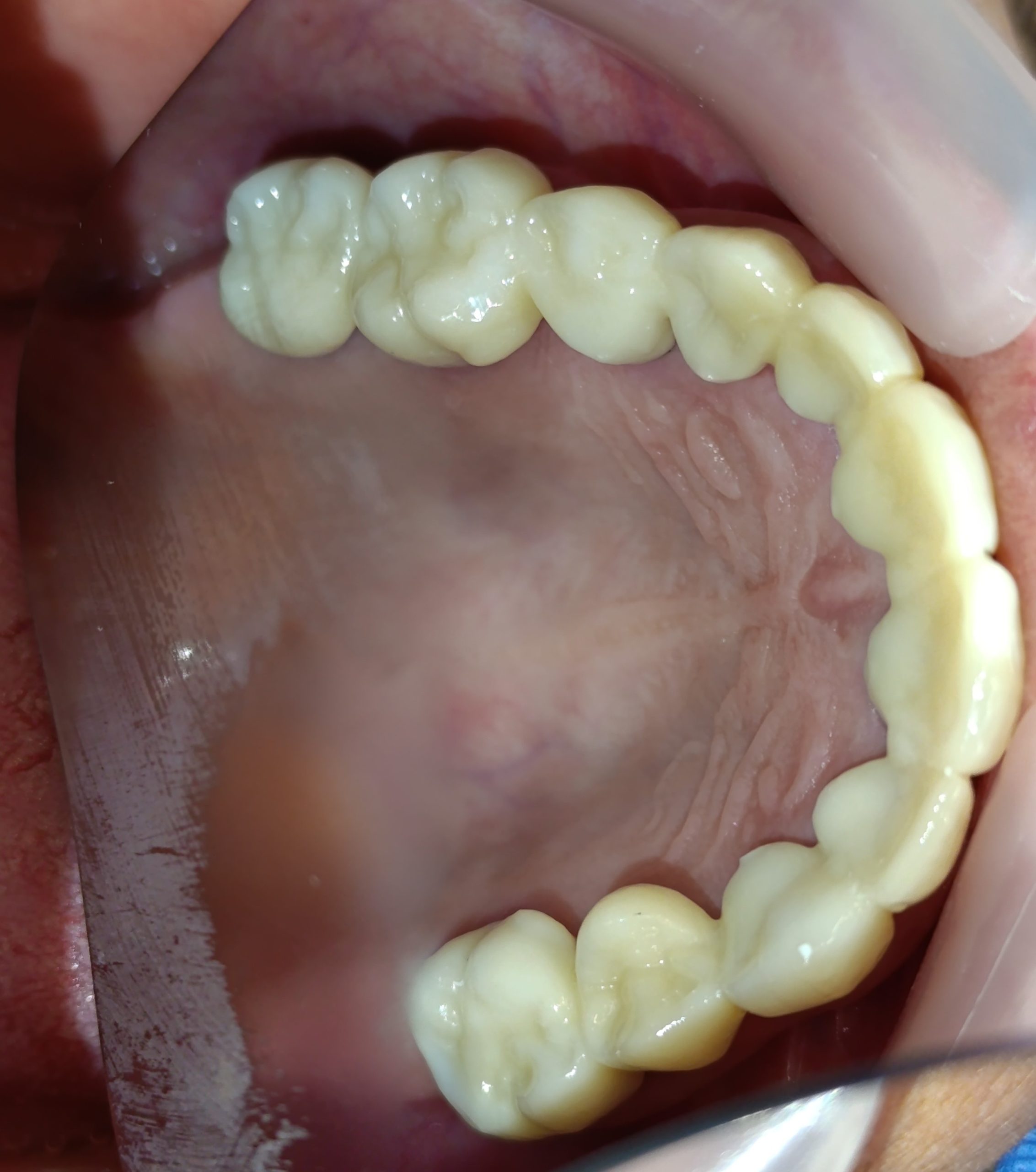
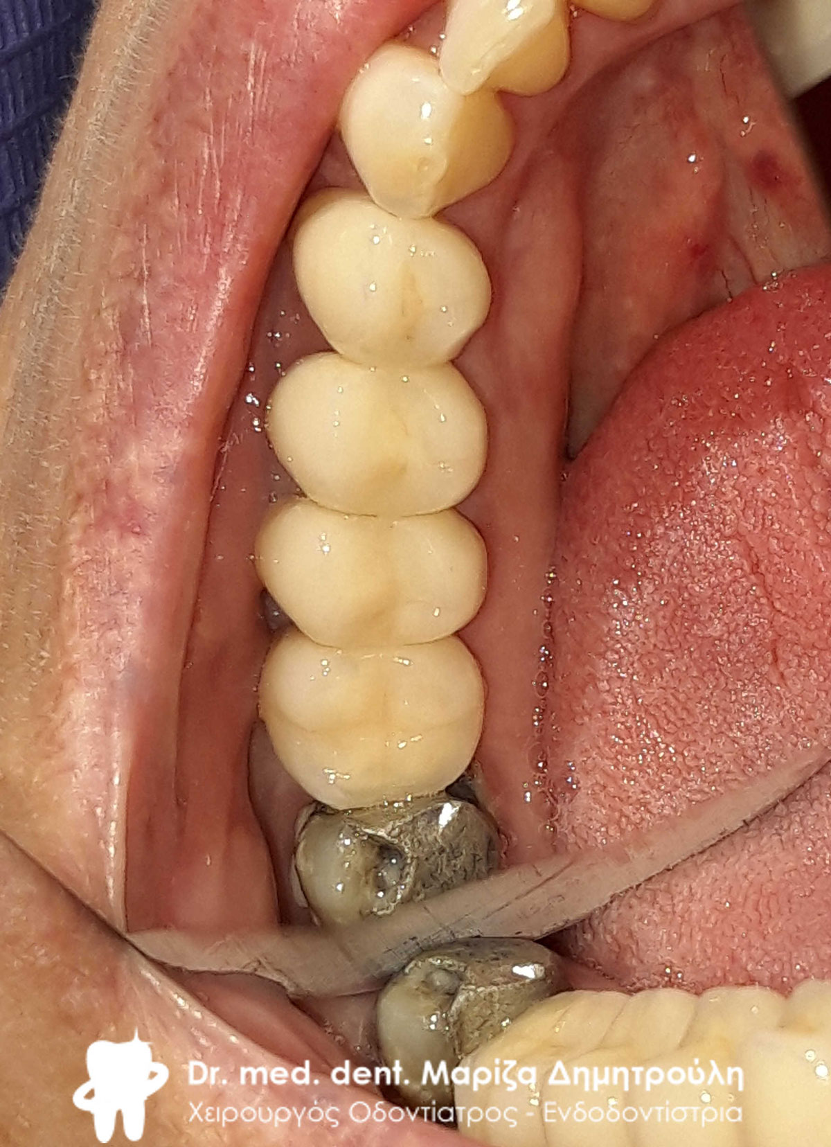
Final clinical image of the occlusal surface of the lower teeth
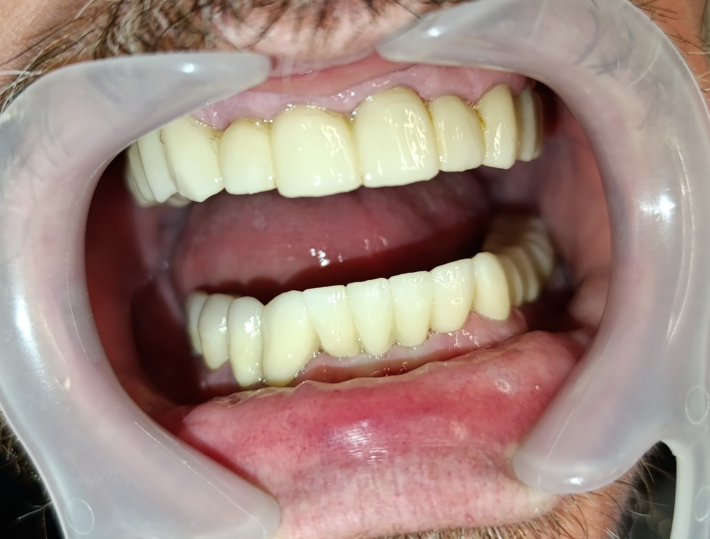
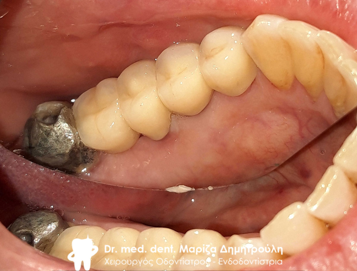
Final clinical image of the patient’s mouth
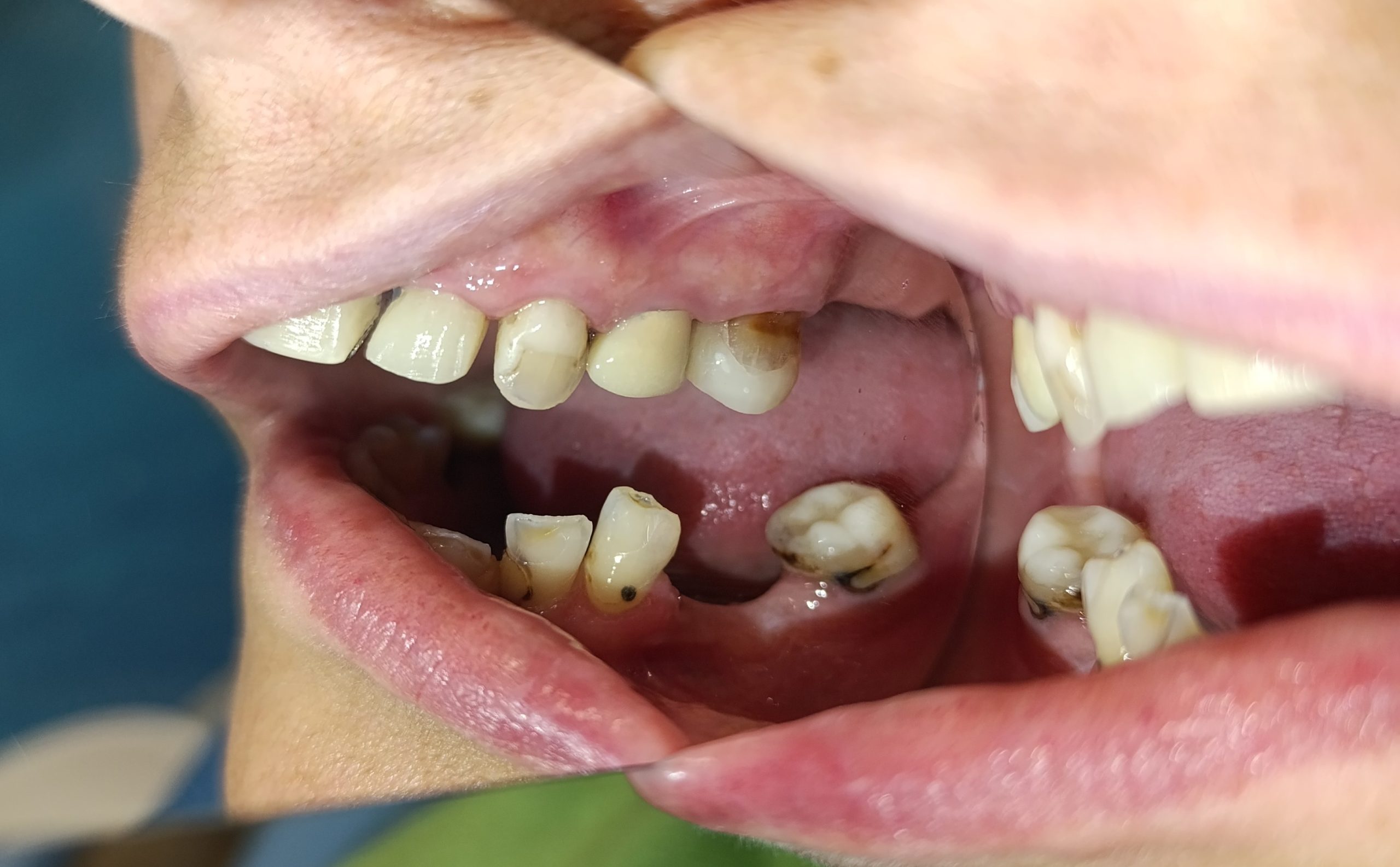
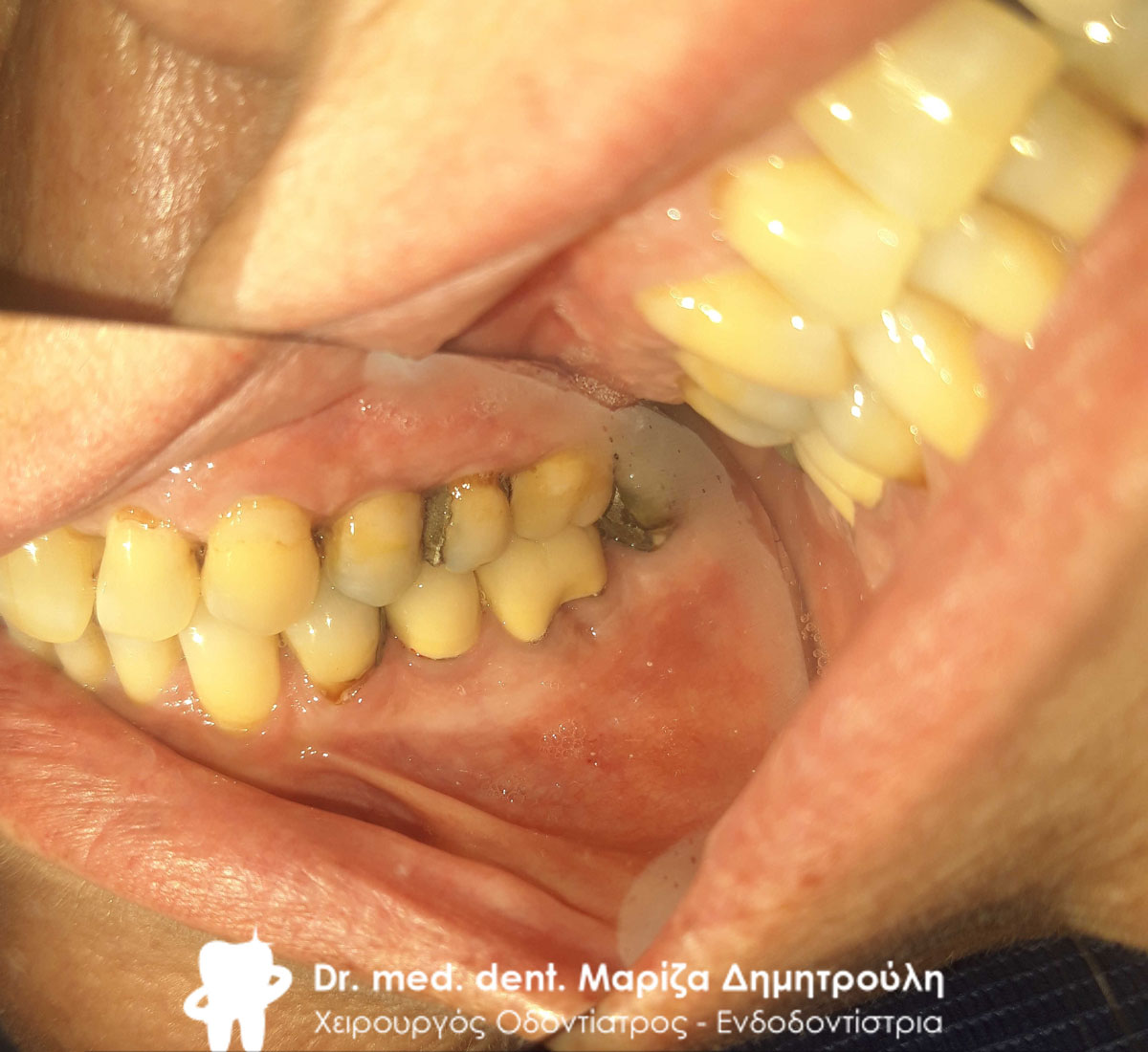
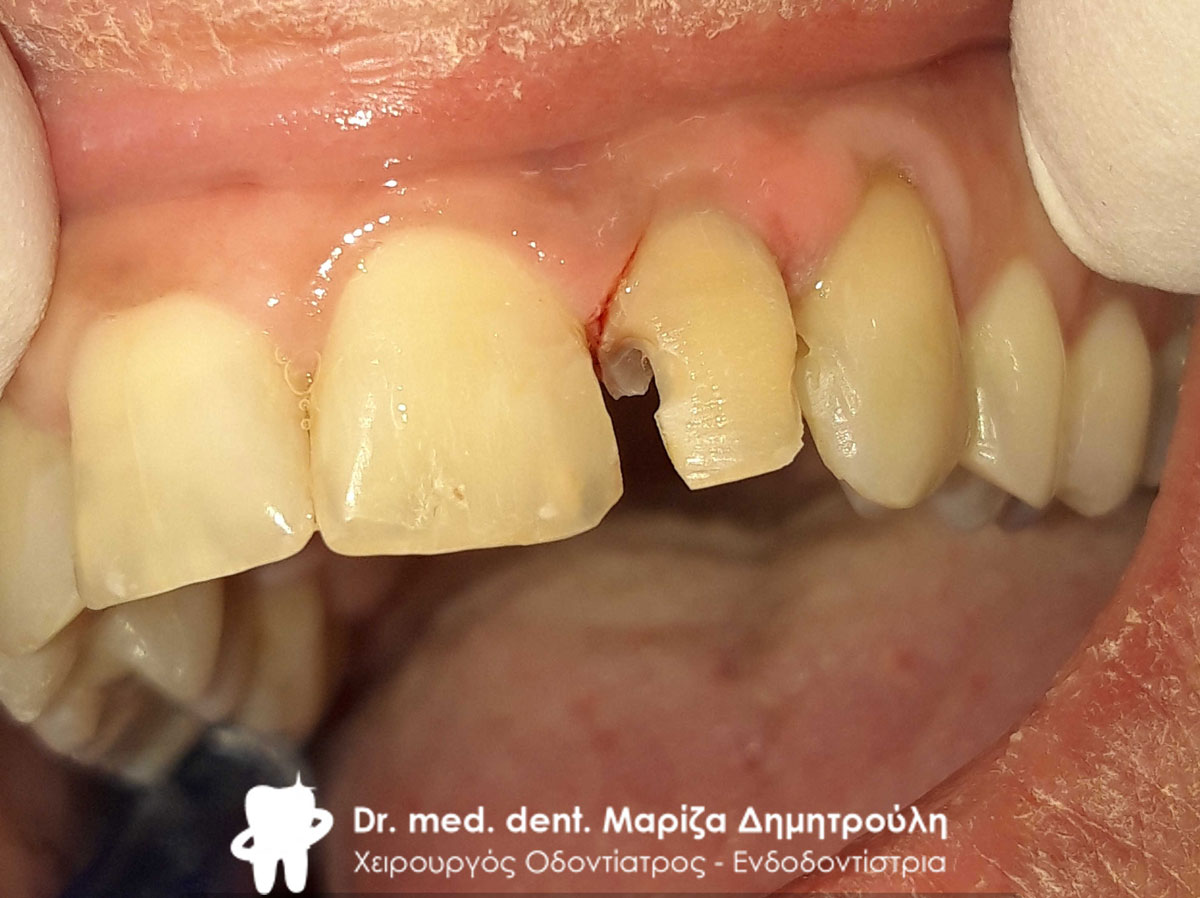
Incident – Replacement of all old re-carious fillings and all-ceramic dental bridge – zirconia
The patient came to the clinic for the restoration of the left side of the lower jaw. The teeth underwent tooth denervation, their reconstruction with white fiberglass shafts. Then they were derailed, impressions taken and sent to the dental technician. Finally, the all-ceramic zirconia bridge was bonded to the teeth.
In the second phase, the patient decided to restore the entire upper jaw. First, the old metal-ceramic bridge was removed and then the metal shaft was removed from the central sector in order to assess whether the tooth can be preserved in the mouth. After the clinical and X-ray examination, it was found that the tooth was perforated and should therefore be extracted. This perforation of the tooth had caused swelling in the area of the central sector. After the extraction of the tooth, the entire upper jaw of the patient was studied and we ended up creating an all-ceramic zirconium petal. This horseshoe would include all the teeth of the upper jaw in order to better stabilize the prosthetic work and restore all the teeth, which had many problems.
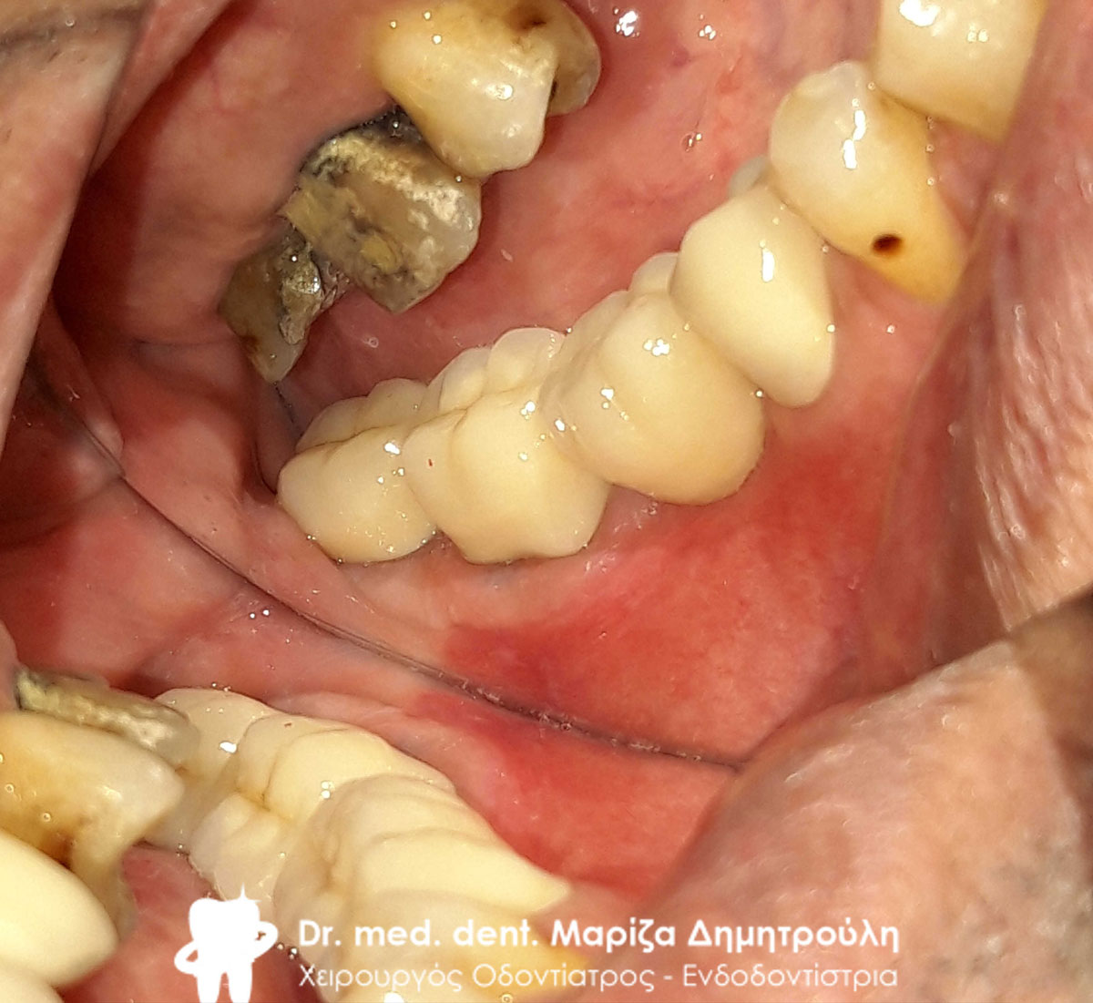
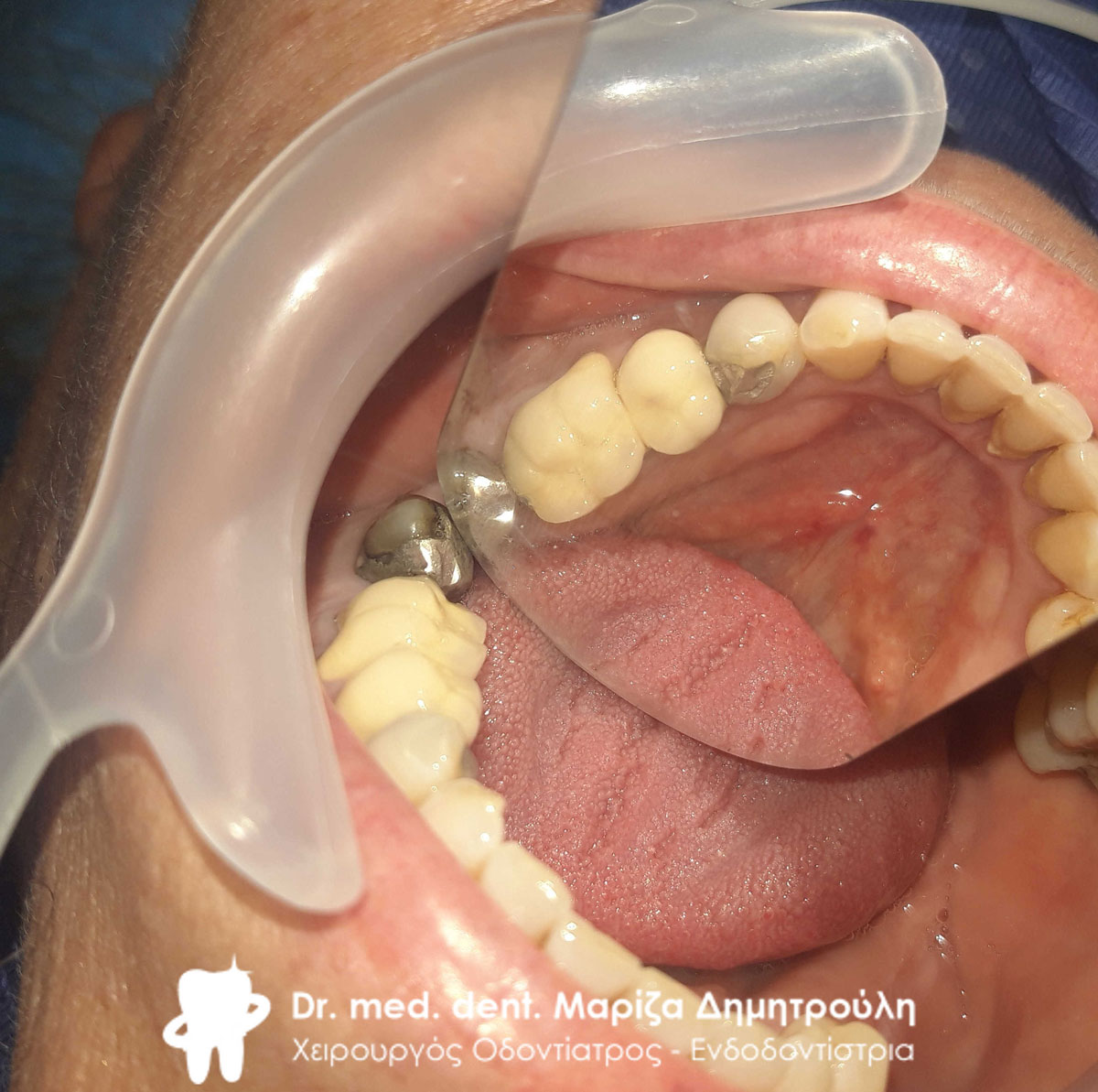
Initial clinical picture of the left side of the lower jaw
Initial clinical picture of the left side of the lower jaw
Initial clinical picture of the left side of the lower jaw
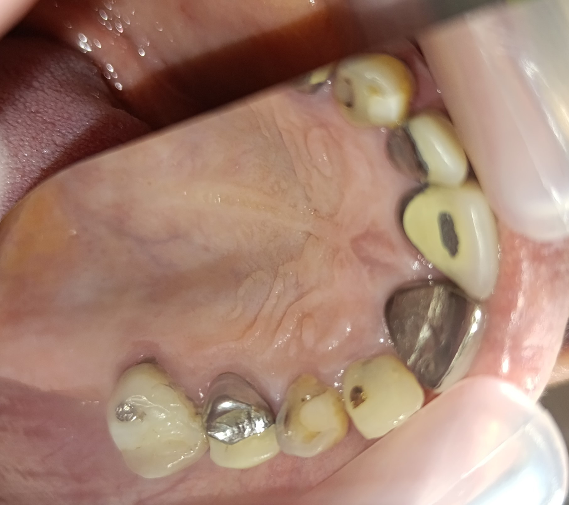
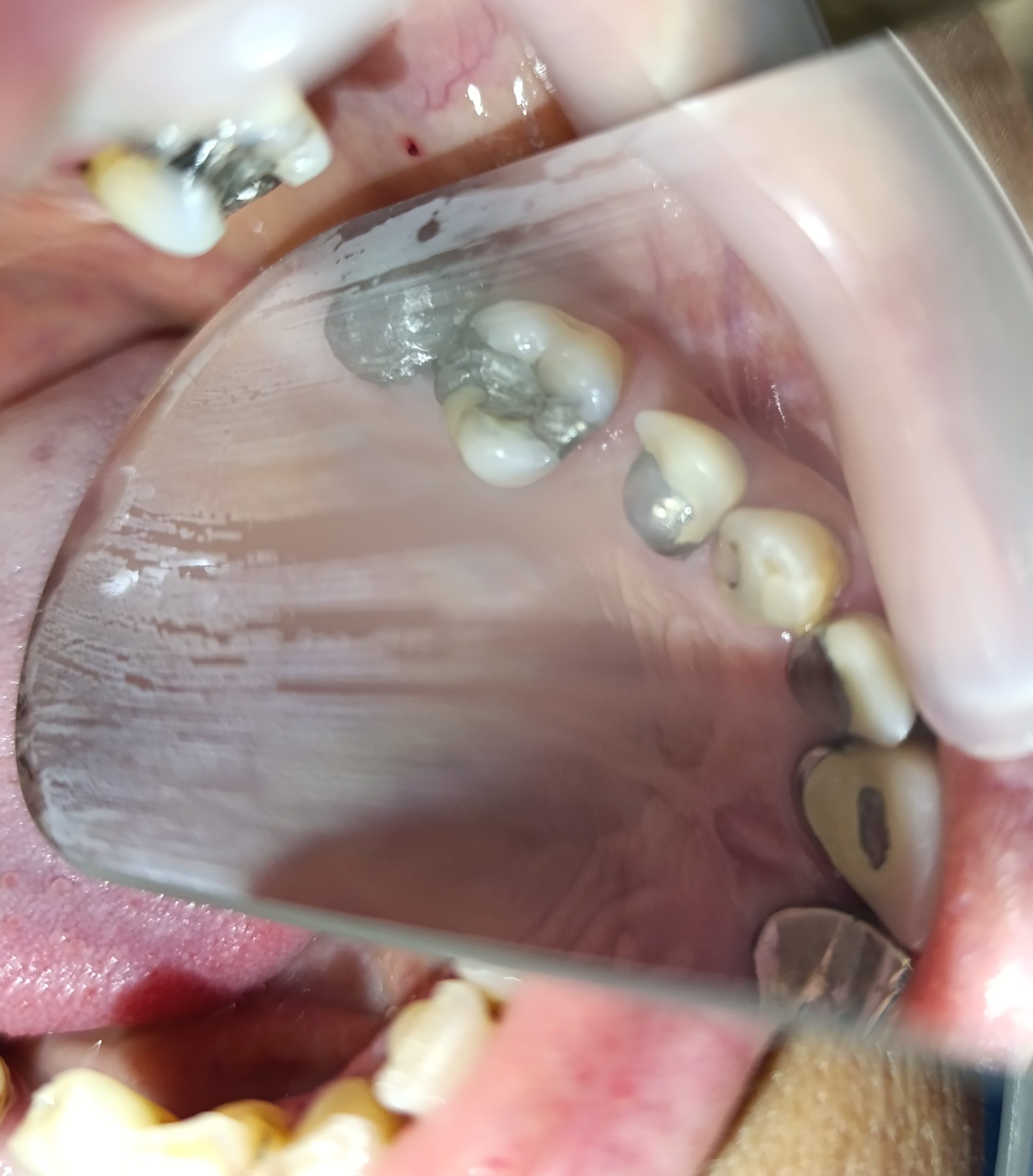
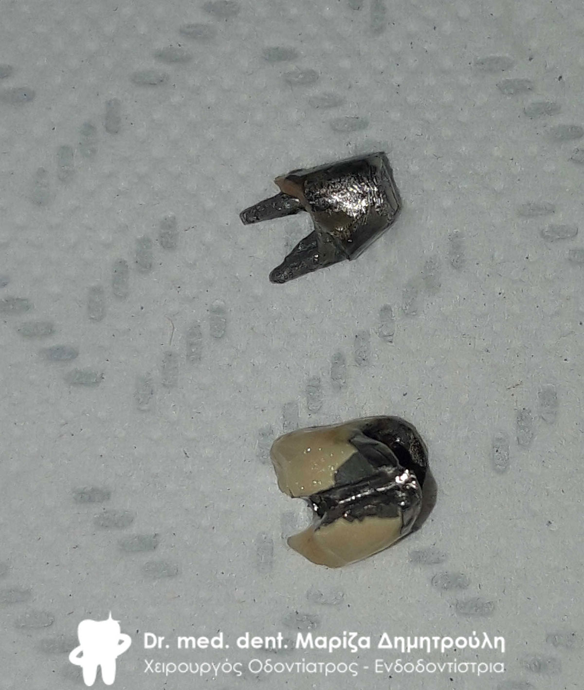
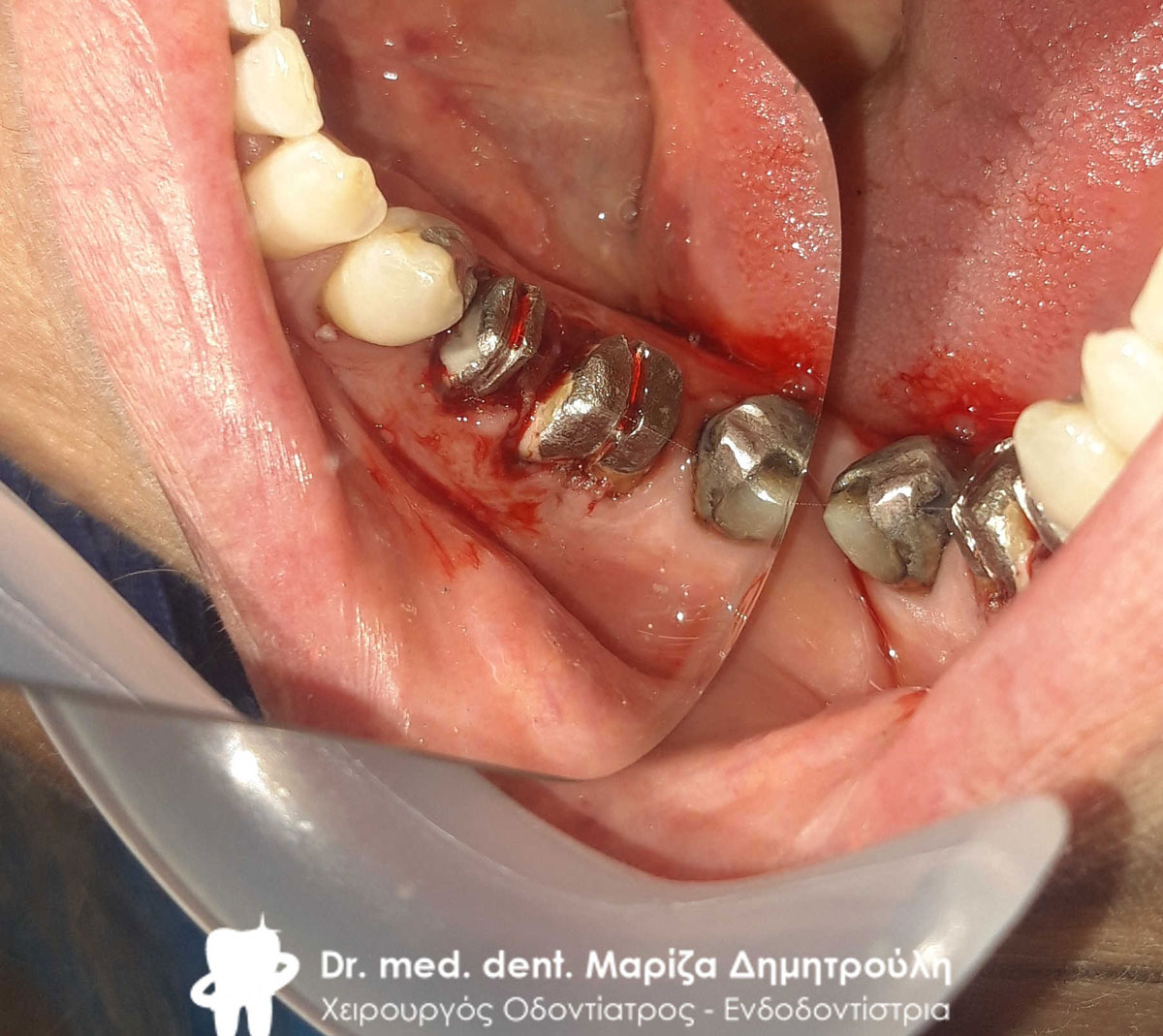
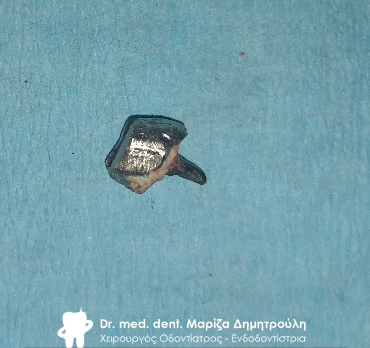
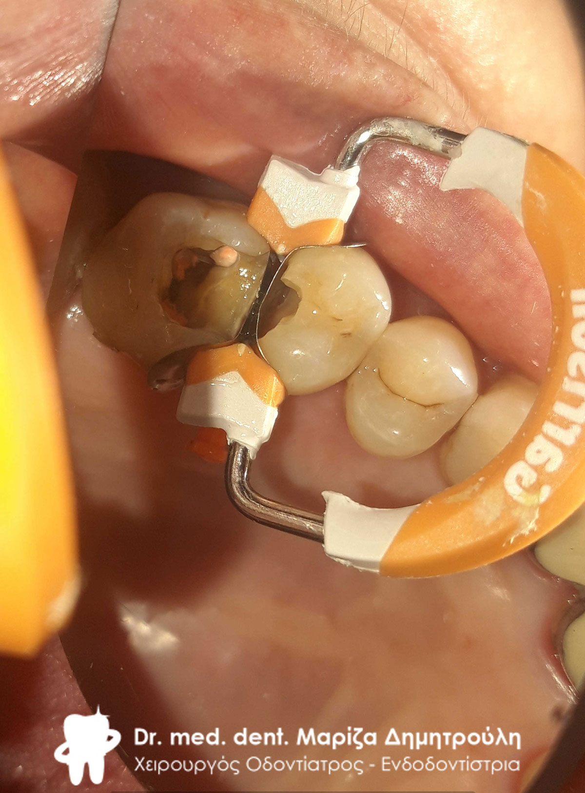
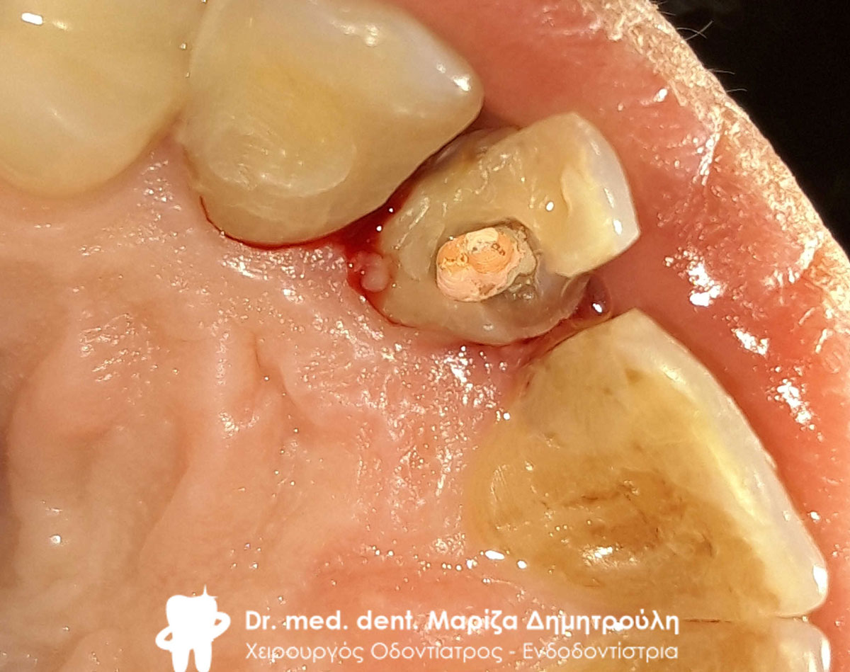
Metal shaft very carefully removed from second molar
Reconstruction of the first premolar and repeat denervation of the second molar
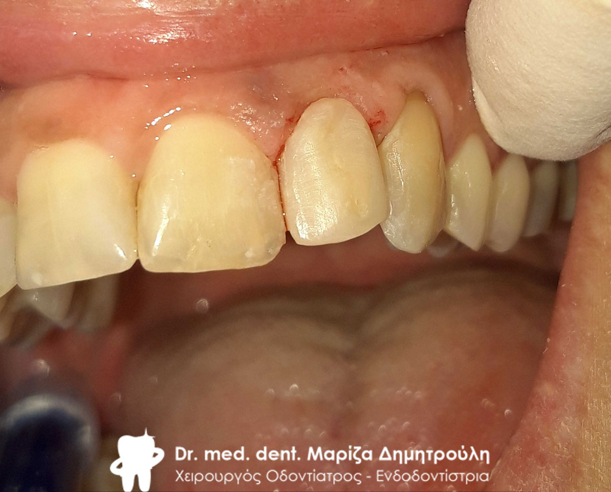
Reconstruction of first premolar and second molar with white fiberglass shafts
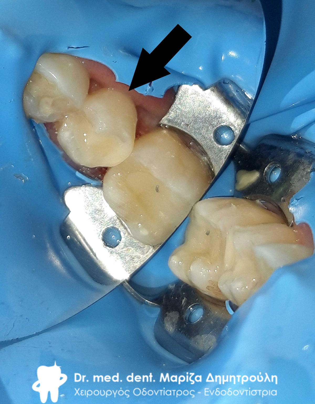
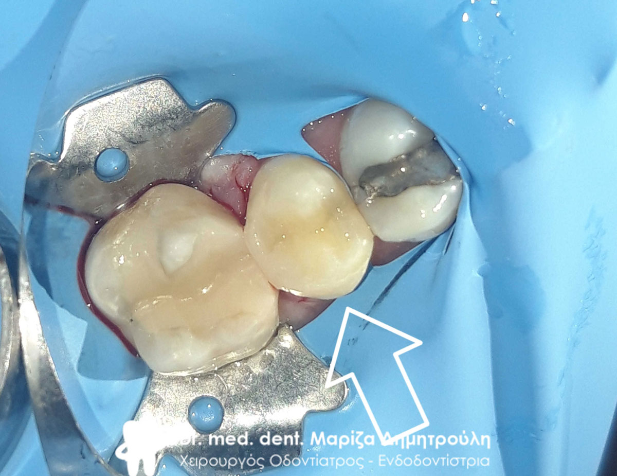
Reconstruction of the second molar and initial clinical picture of the third molar
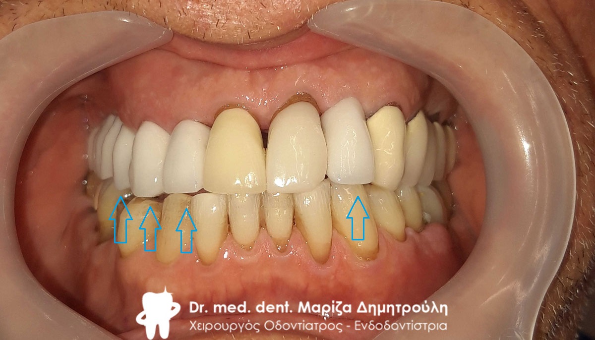
Final image of all teeth before taking impressions
Final image of all teeth before taking impressions
Final image of mandibular all-ceramic zirconia bridge
Final image of mandibular all-ceramic zirconia bridge
Original image of the metal-ceramic holder on the upper left premolar
Removal of the old metal-ceramic case and double metal shaft from the upper left premolar
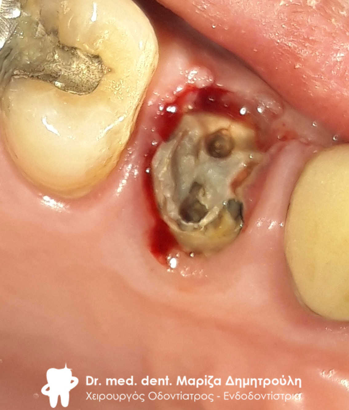
Image of the first premolar immediately after the removal of the double metal shaft
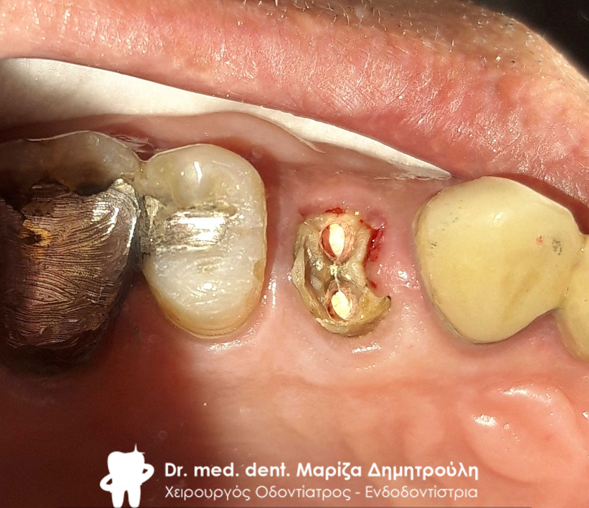
Image of the first premolar immediately after re-denervation
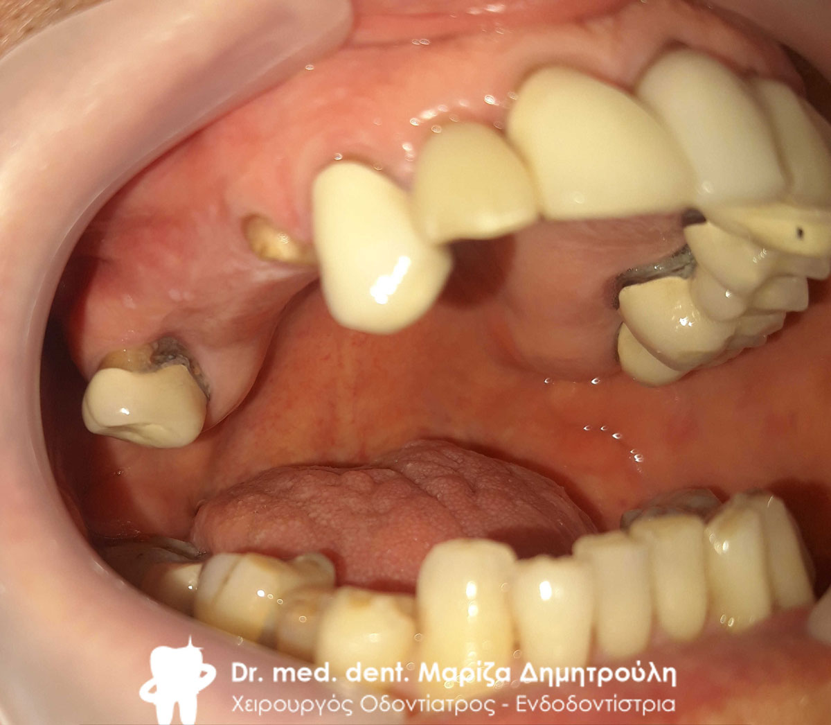
Original image of the old maxillary bridge
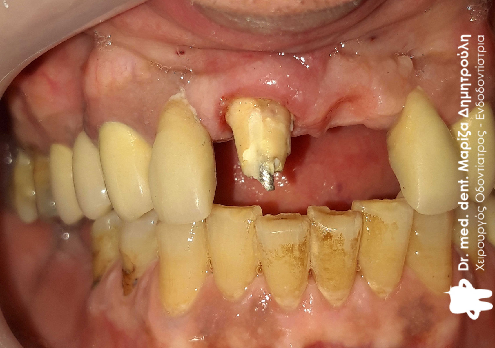
Image of teeth after removal of old bridge
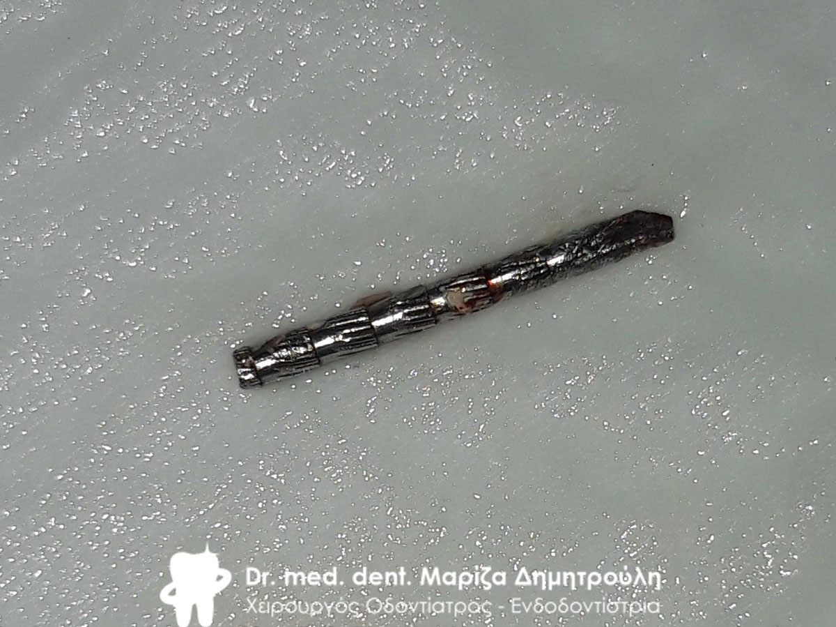
The metal shaft removed from the field to assess the tooth
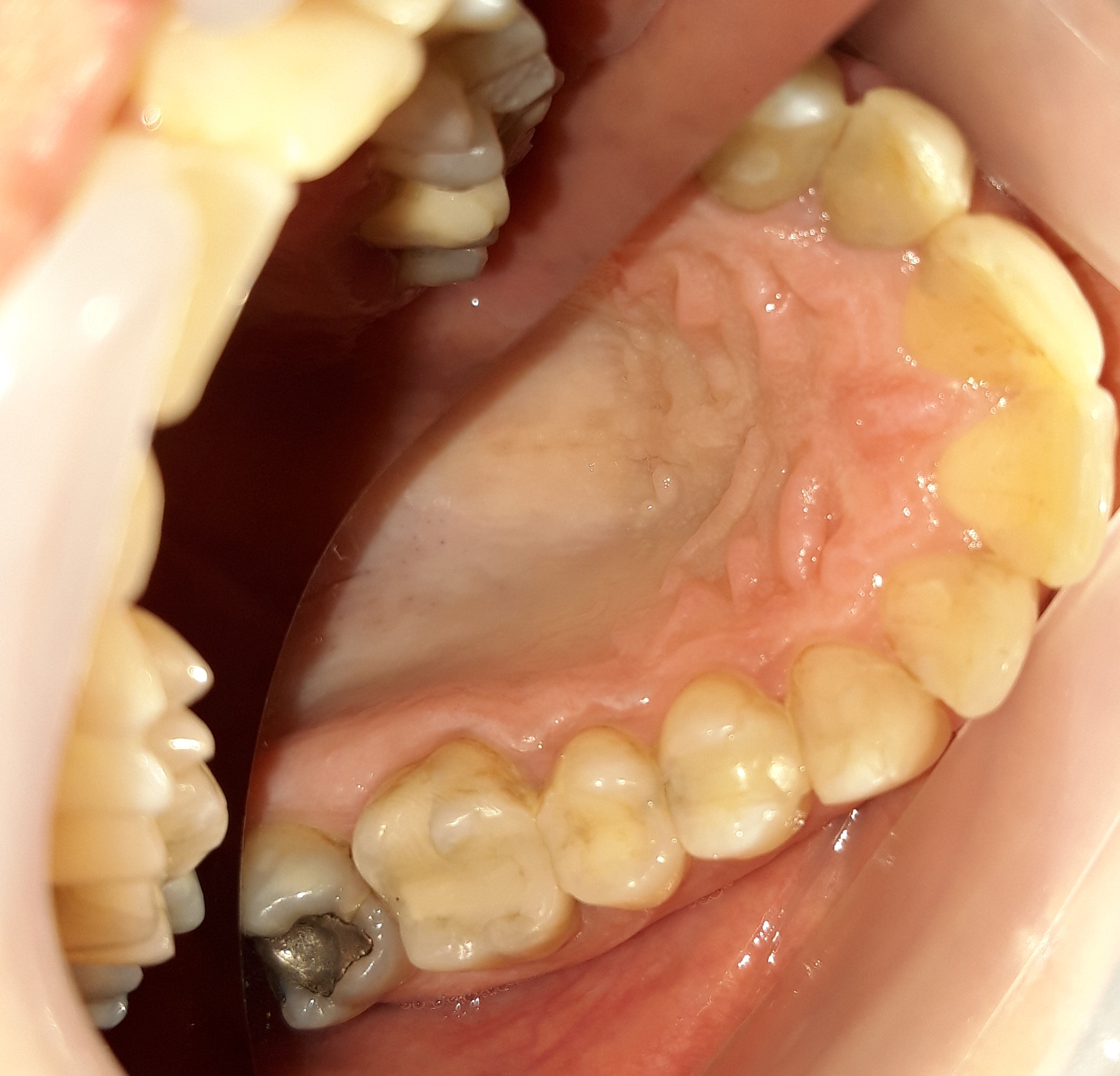
Image of the teeth after the necessary denervation, reconstruction and derailment
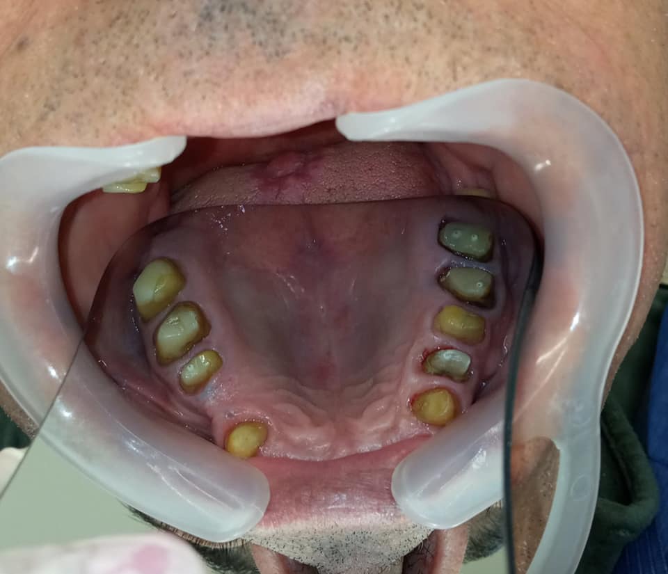
Image of the teeth after the necessary denervation, reconstruction and derailment
Final image of all-ceramic zirconia bridge
Final image of all-ceramic zirconia bridge
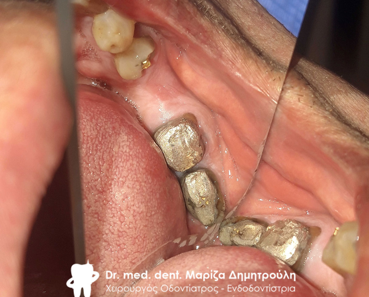
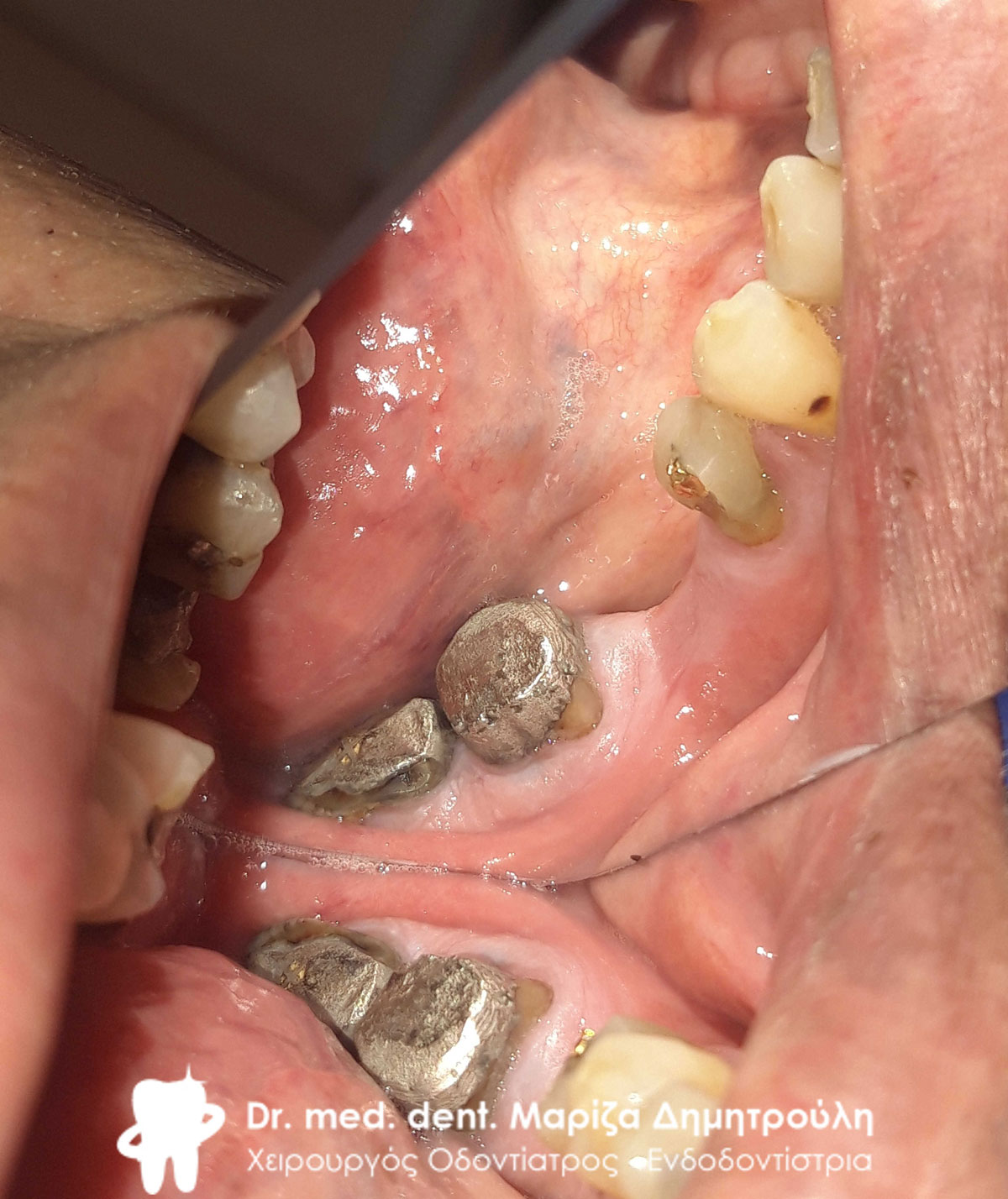
Incident – Replacement of all old re-carious fillings and all-ceramic dental bridge – zirconia
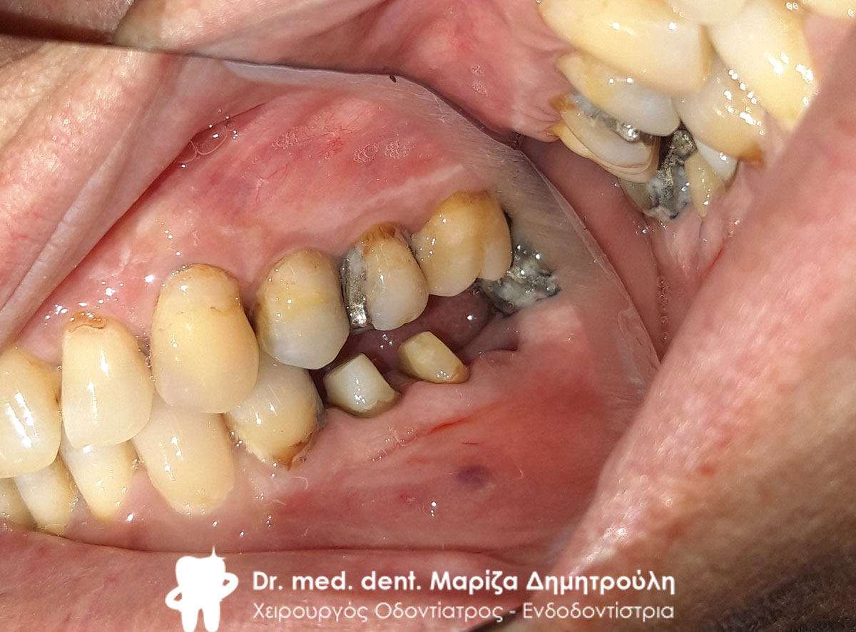
The lady came to the clinic with swelling on the left side of the lower jaw. After the clinical and radiographic examination of the teeth, the old bridge on her teeth was removed. As the photos show, the old metal posts were very carefully removed from inside the roots without causing any damage to the remnants of the tooth roots. The remaining tooth roots were used as „natural implants“ and more specifically an entire tooth abutment was built over the tooth roots using special fiberglass shafts. In the molar, after removing the old case and removing the metal shaft in the same careful way, it was found that one root of the tooth was very damaged, so it was removed. The other half of the root was reconstructed with a fiberglass shaft and prepared accordingly to receive the future zirconia bridge. The first premolar was also prepared accordingly (denervation and tooth reconstruction) to be used as a bridge abutment. when all the necessary preparations were completed, an impression was taken which is sent to the dental technician for the construction of an all-ceramic zirconium bridge. when the bridge was ready it was welded to the patient’s mouth.
In a second phase, the patient decided to restore the left side of the upper jaw, where white cervical resin fillings were performed in the lateral sector and canine. The old carious fillings on the second premolar and first molar on the same side of the upper jaw were also reconstructed.
The patient has no symptoms of pain and is enjoying the new dental work in her mouth.
Initial clinical picture of the left side of the lower jaw
Initial clinical picture of the left side of the lower jaw
Immediately after removing the old metal-ceramic bridge
Removing the metal shaft from the second premolar
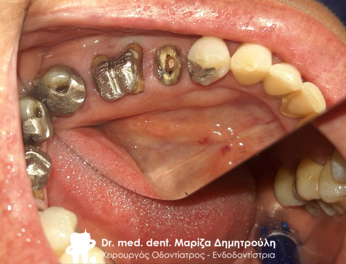
Image of the root of the first premolar after removal of the metal shaft
Removing the double metal shaft from the first molar
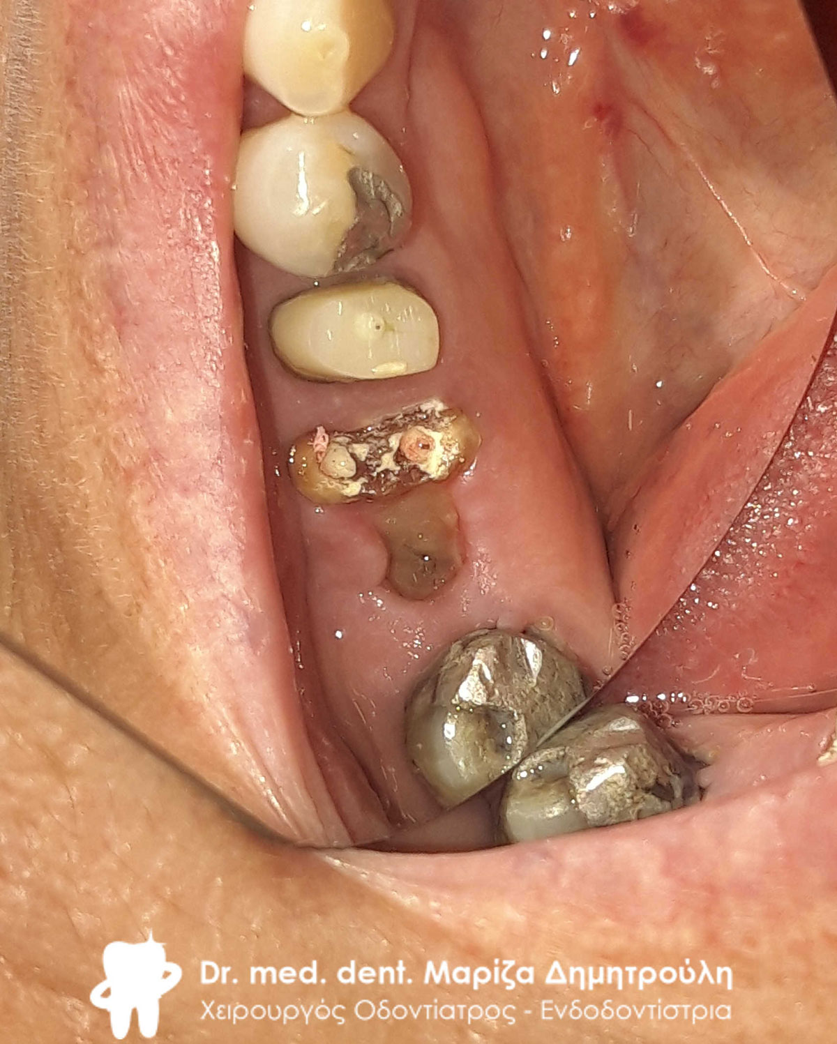
Image of the proximal root of the first molar after repeated denervation andremoval of the distal root
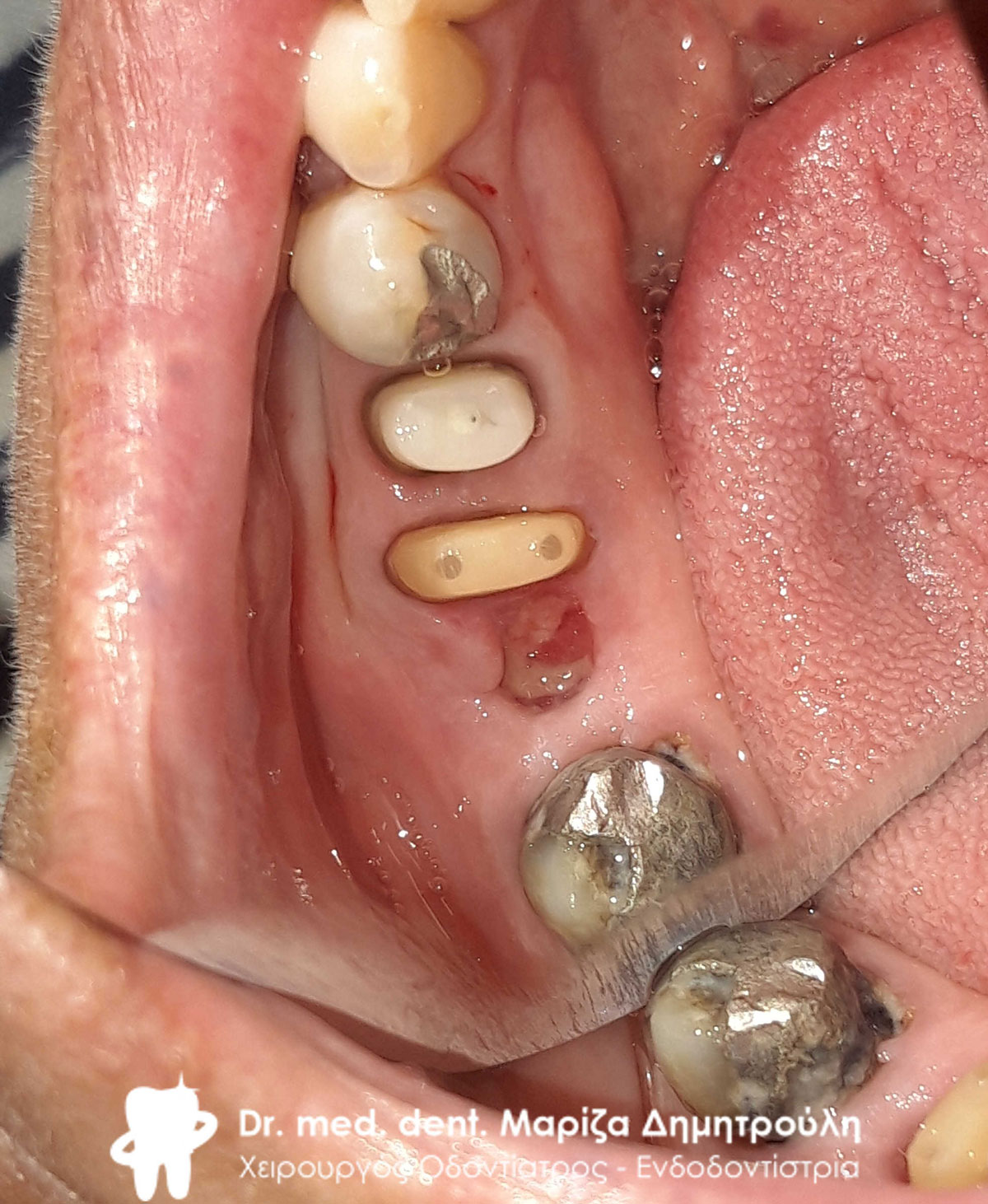
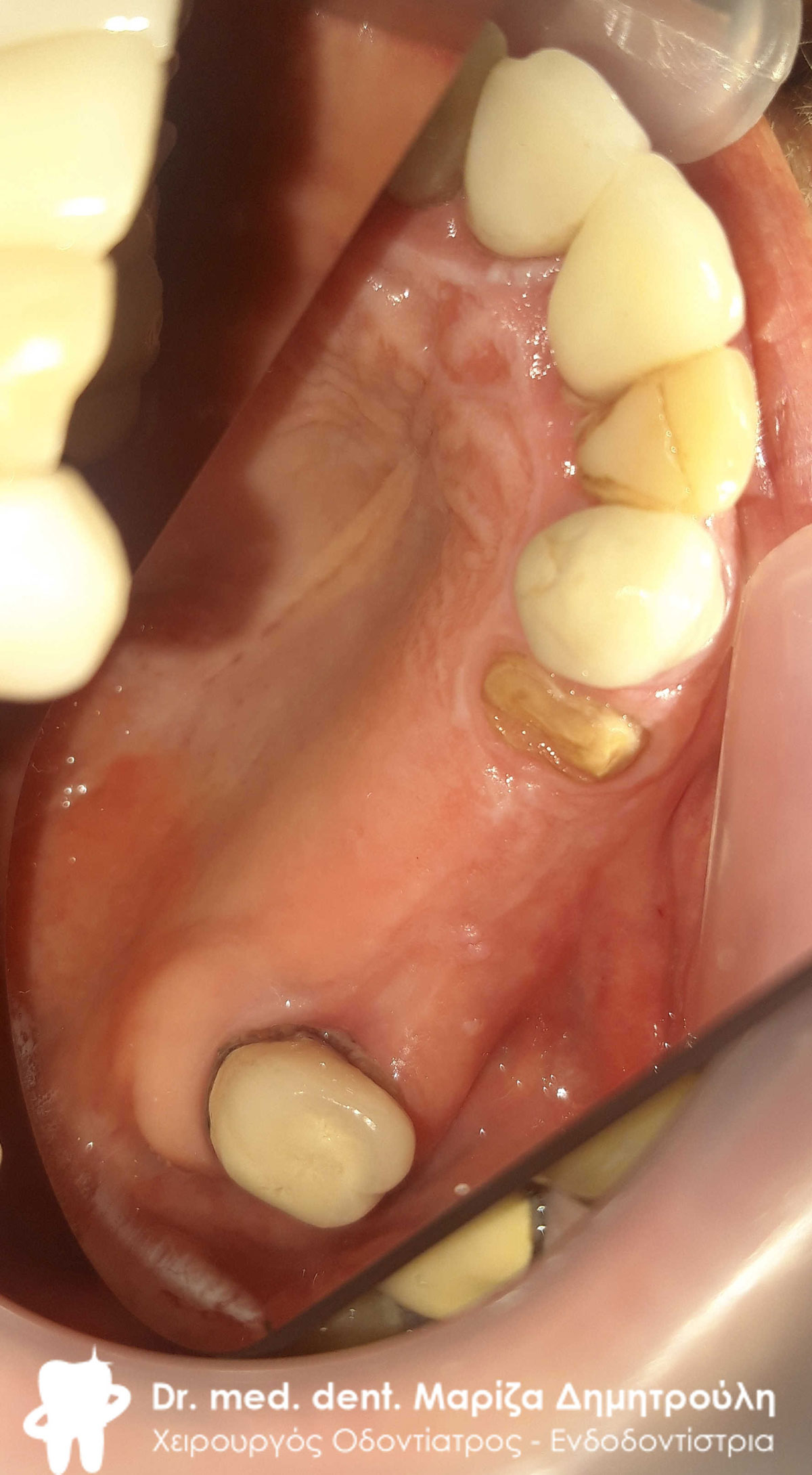
Tooth abutments after reconstruction of the remaining roots with fiberglass posts
Tooth abutments after reconstruction of the remaining roots with fiberglass posts
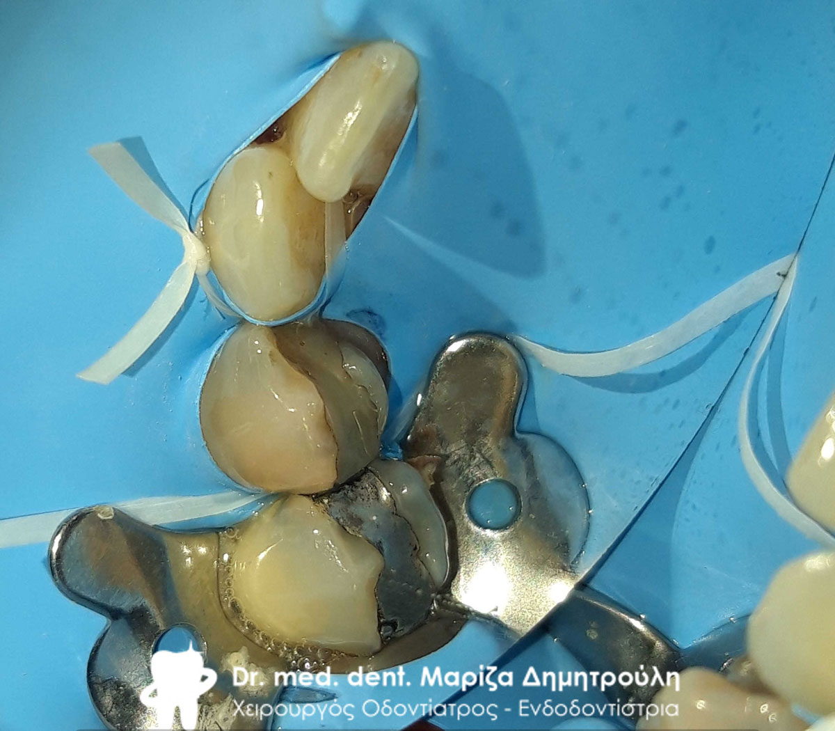
Denervation of the first premolar using an elastic isolator
Final image of teeth before taking impressions
Final image of all-ceramic zirconia bridge
Final image of all-ceramic zirconia bridge
Final image of all-ceramic zirconia bridge
Cervical lesions in the lateral sector and canine
Cervical fillings in the lateral sector and canine
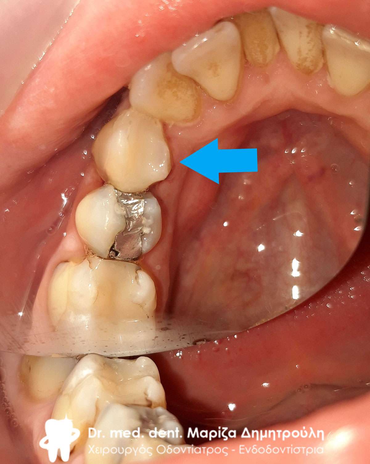
Deficits of upper left first molar and second premolar
White fillings on upper left first molar and second premolar
Incident – Replacement of all old re-carious fillings and all-ceramic tooth trays
The patient visited the clinic for the gradual restoration of her entire mouth.
As the pictures show, all of her old fillings were replaced, while at the same time, an all-ceramic tooth case on the upper and an all-ceramic case on the lower jaw were placed in her mouth.
First, all the old fillings of the upper front area were replaced. The images below depict both the palatal view and the labial view of the old re-carious fillings. Characteristic is the „blackness“ that appeared on the upper front teeth, as the patient typically reported in her first visit. Gingivitis was also treated, which was severe at the lady’s first appointment.
All of the patient’s old fillings were gradually corrected as can be seen in the images below.
The patient is very pleased with the functional as well as aesthetic result of the new dental work and prosthetic restorations.
Anterior Teeth – Original Image
Front teeth – Image after replacement of right side fillings
Anterior Teeth – Final Image
Anterior teeth – Original image
Anterior Teeth – Final Image
Front teeth – Image after gingivitis treatment
Anterior teeth – Original image
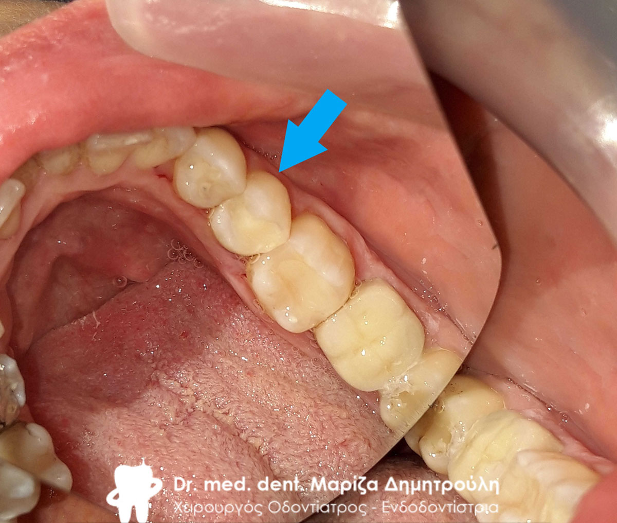
Final right side image
Top left sidebar initial image
Center image of upper left sidebar
Final image of upper left sidebar
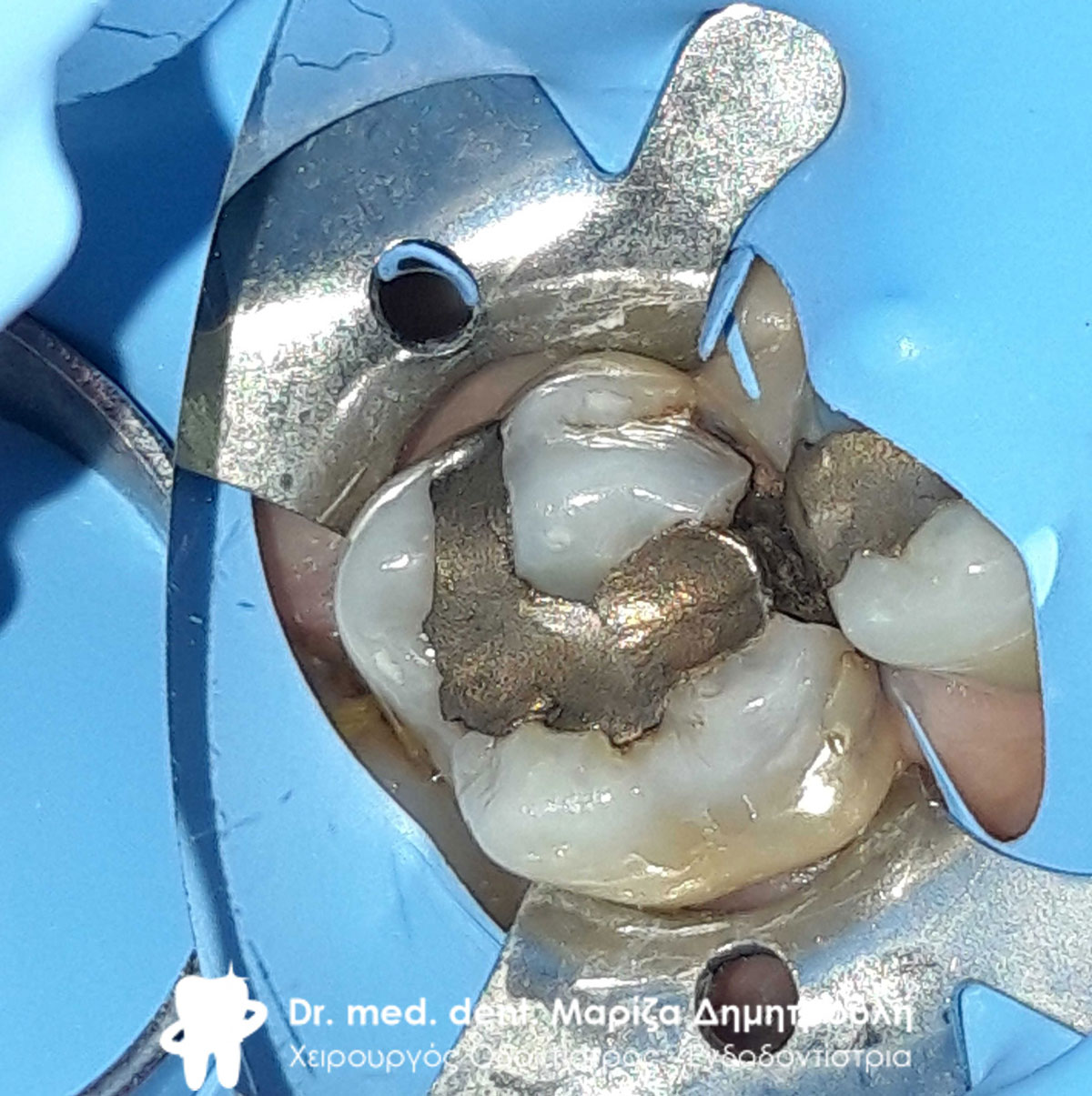
Bottom left – The first molar ground before receiving the all-ceramic zirconia case
Bottom left – The first molar with the all-ceramic zirconium case
Bottom left – The first molar with the all-ceramic zirconium case
Bottom left – Replacing the black filling on the first premolar
Bottom left – Replacing the black filling on the first premolar
Bottom left – Replacing the black filling on the first premolar
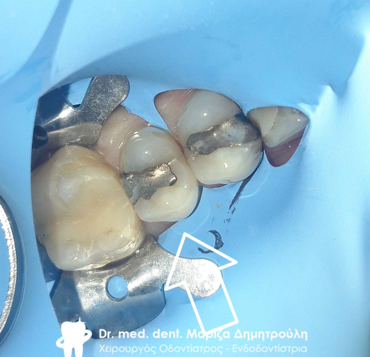
Bottom right- Original image of the two premolars
Bottom right – Final image of first premolar
Bottom right – Final image of first premolar
Bottom right – Final image of the two premolars
All-ceramic socket on left upper first molar
All-ceramic socket on left upper first molar
Left side original image
Final left side image
Top Right – Change Black Amalgam Fillings
Top Right – Change Black Amalgam Fillings
Top Right – Change Black Amalgam Fillings
Top right – Final image after changing black amalgam fillings
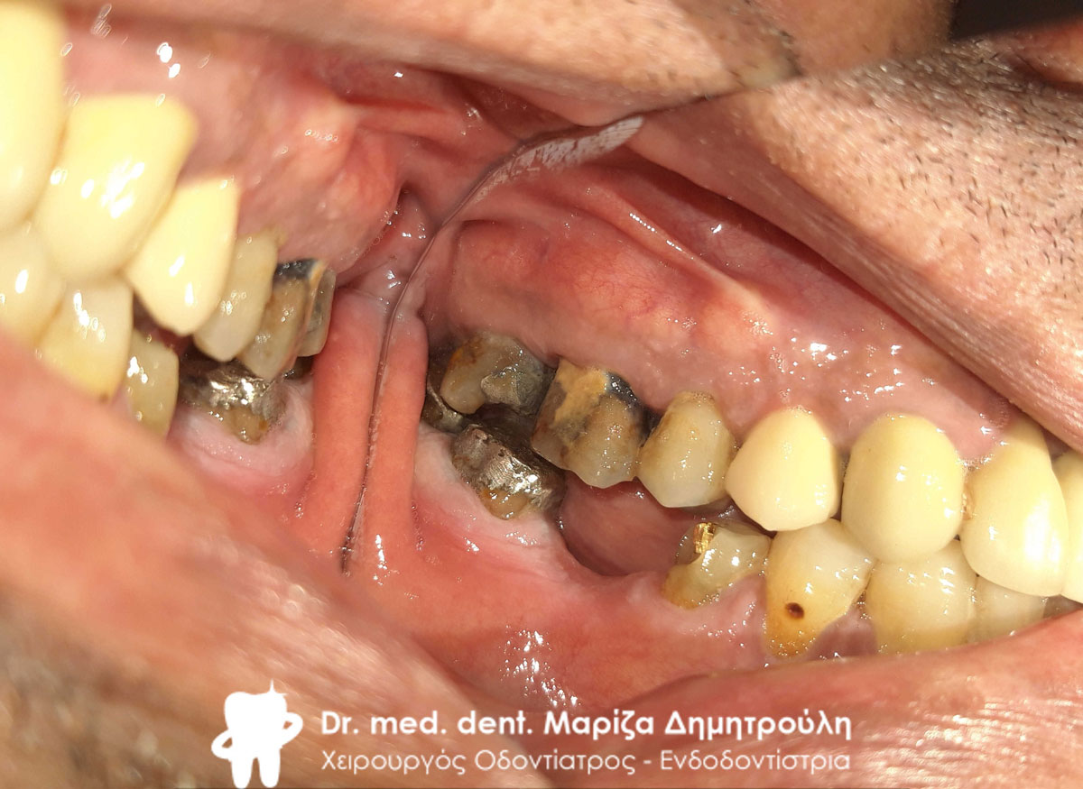
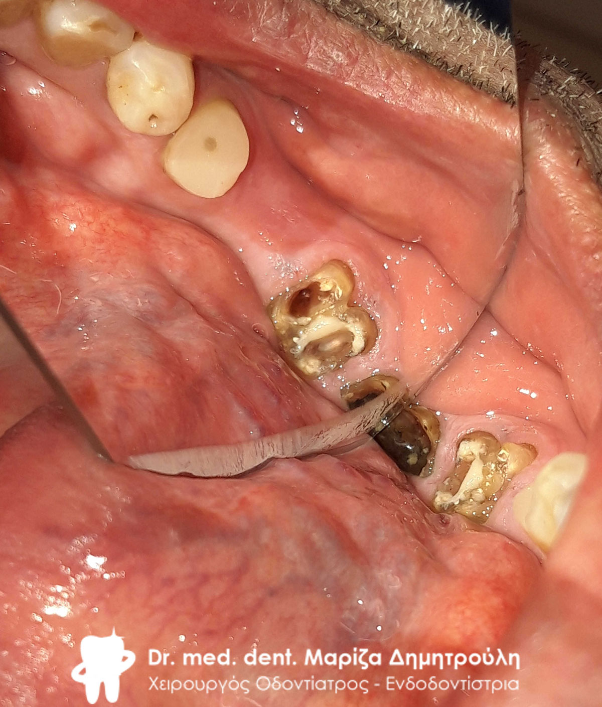
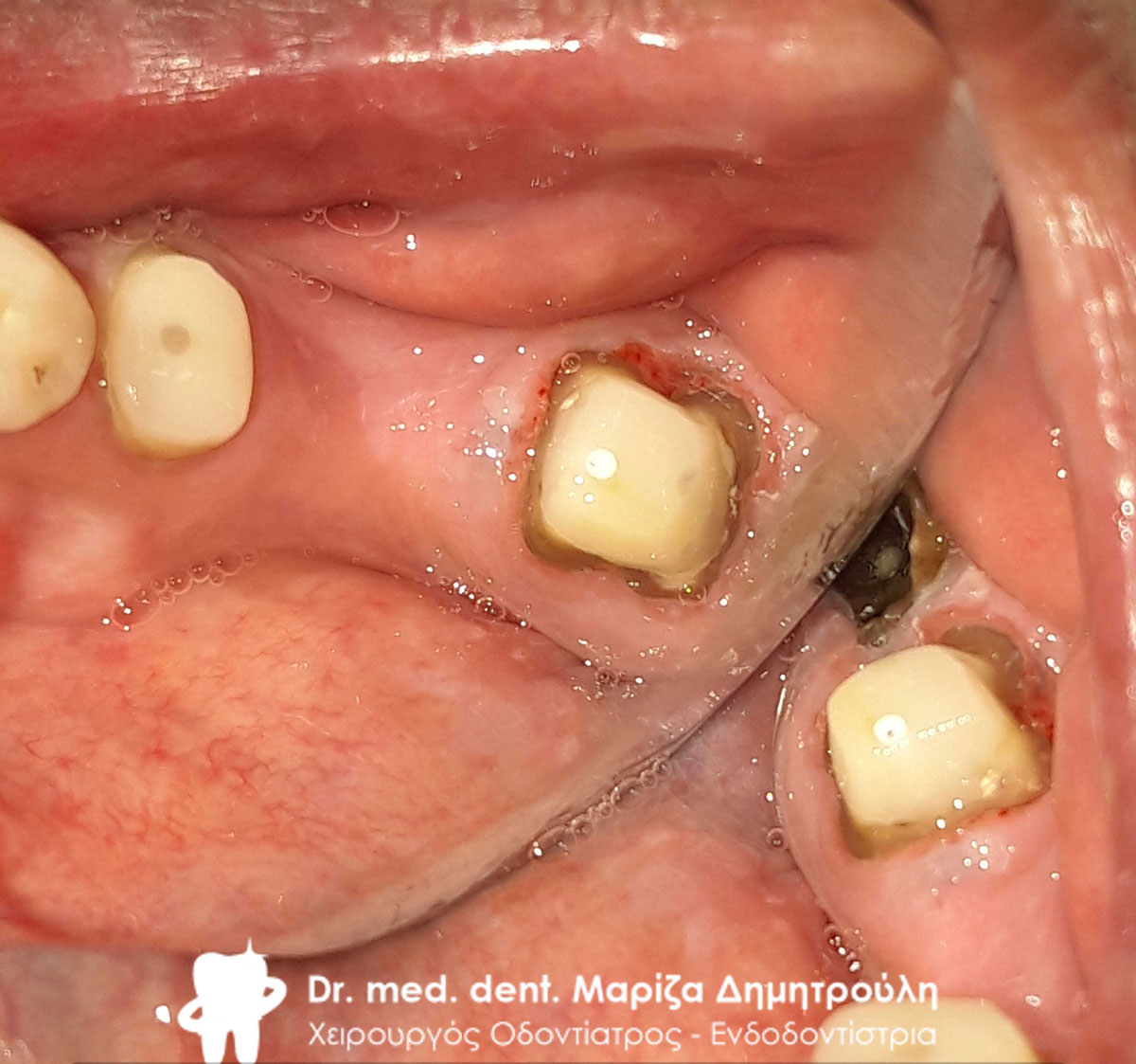
Περιστατικό – Ολοκεραμική γέφυρα ζιρκονίου στη δεξιά πλευρά της άνω γνάθου
Ο ασθενής προσήλθε στο ιατρείο με σκοπό την αποκατάσταση του στόματός του. Το πλάνο θεραπείας αφορούσε το μεγάλο έλλειμμα στη δεξιά πλευρά της άνω γνάθο όπως και την αντικατάσταση μιας παλιάς θήκης στην αριστερή πλευρά της άνω γνάθου. Επίσης επιθυμία του ασθενή ήταν η αντικατάσταση παλιών λευκών σφραγισμάτων στη δεξιά πλευρά της κάτω γνάθου.
Αρχικά αφαιρέθηκαν στη δεξιά πλευρά οι παλιές θήκες των δοντιών. Στη συνέχεια πραγματοποιήθηκαν όπου χρειάστηκε tooth denervation ή επανάληψη απονεύρωσης. Ακολούθησε η ανασύσταση των δοντιών και ο εκτροχισμός τους ώστε να δεχθούν τις νεές προσθετικές δουλειές. Η διαδικασία ολοκληρώθηκε με τη λήψη αποτυπωμάτων και την αποστολή τους στον οδοντοτεχνίτη.
Στο μεσοδιάστημα της προετοιμασίας της ολοκεραμικής γέφυρας και θήκης από τον τεχνίτη αφαιρέθηκαν 2 παλιά επανατερηδονισμένα σφραγίσματα από τους δύο προγομφίους στη δεξιά πλευρά της κάτω γνάθου. Εφόσον αφαιρέθηκε η τερηδόνα πραγματοποιήθηκαν νέα λευκά σφραγίσμτα ρητίνης στα δύο δόντια.
Το χρώμα της νέας προσθετικής εργασίας ζητήθηκε από τον ασθενή να είναι έντονο λευκό με την προϋπόθεση στο μέλλοντα χρόνο να αντικαταστήσει όλες τις παλιές στεφάνες των πρόσθιων δοντιών.
Original image of the left side of the maxilla
Original image of the left side of the maxilla
Clinical image immediately after removal of the old metal-ceramic case
Original image of the old right lateral maxillary case
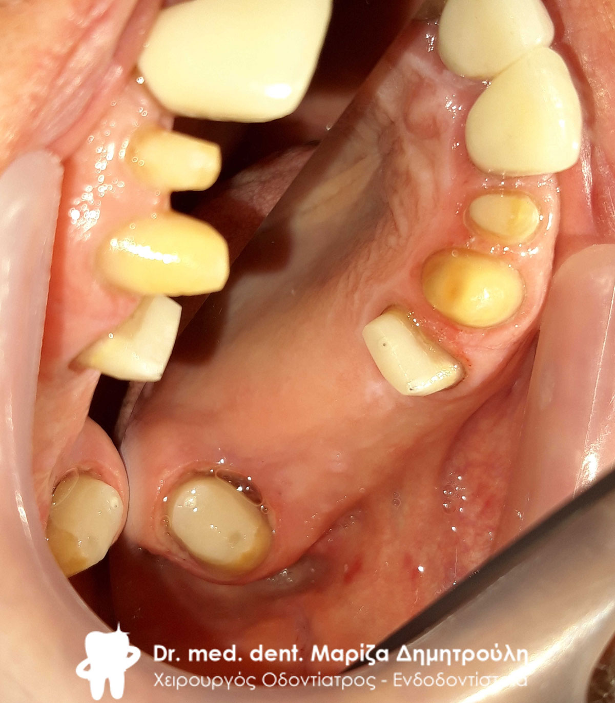
Image after the completion of tooth ablation and reconstruction
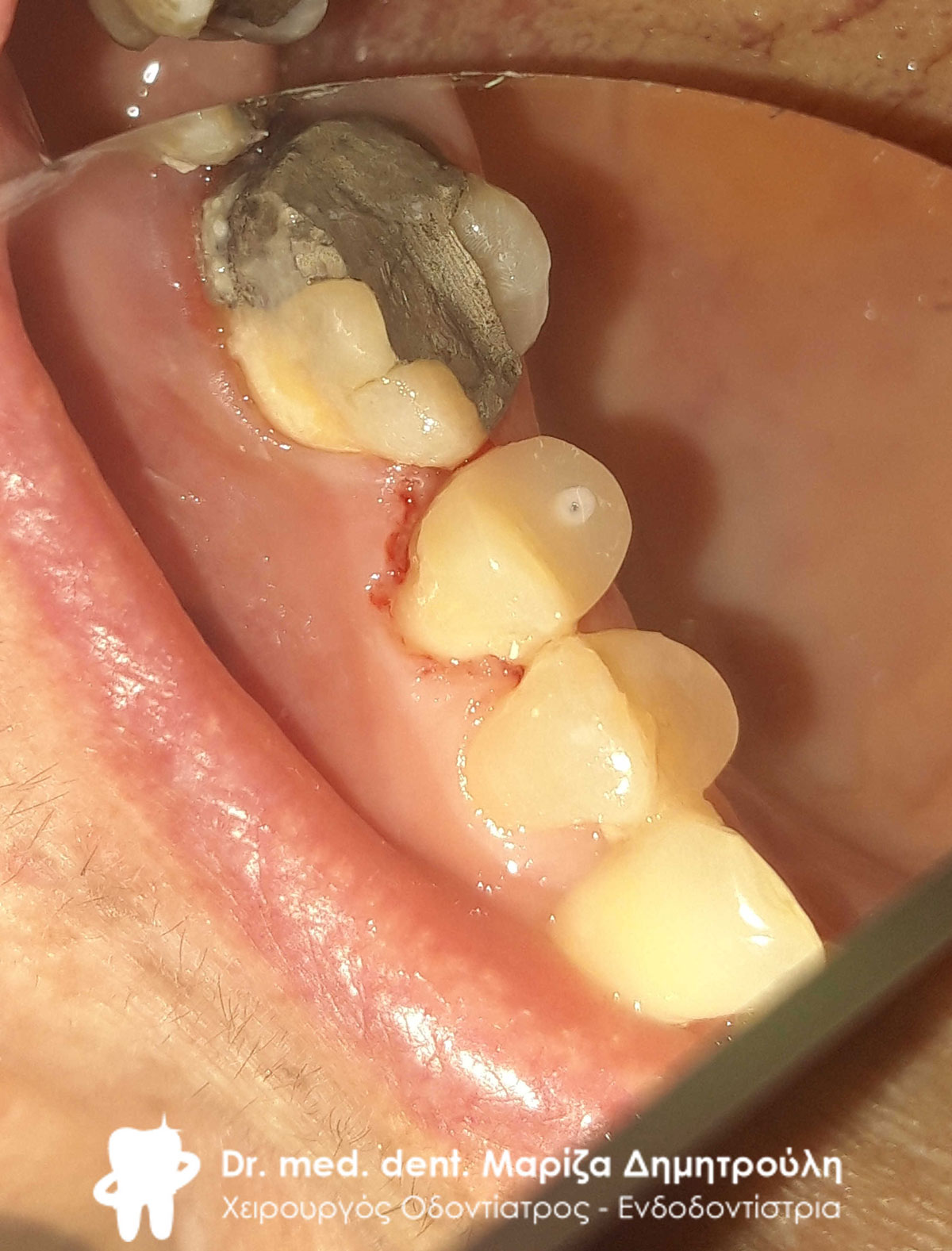
Final image of all-ceramic zirconia bridge
Final image of all-ceramic zirconia bridge
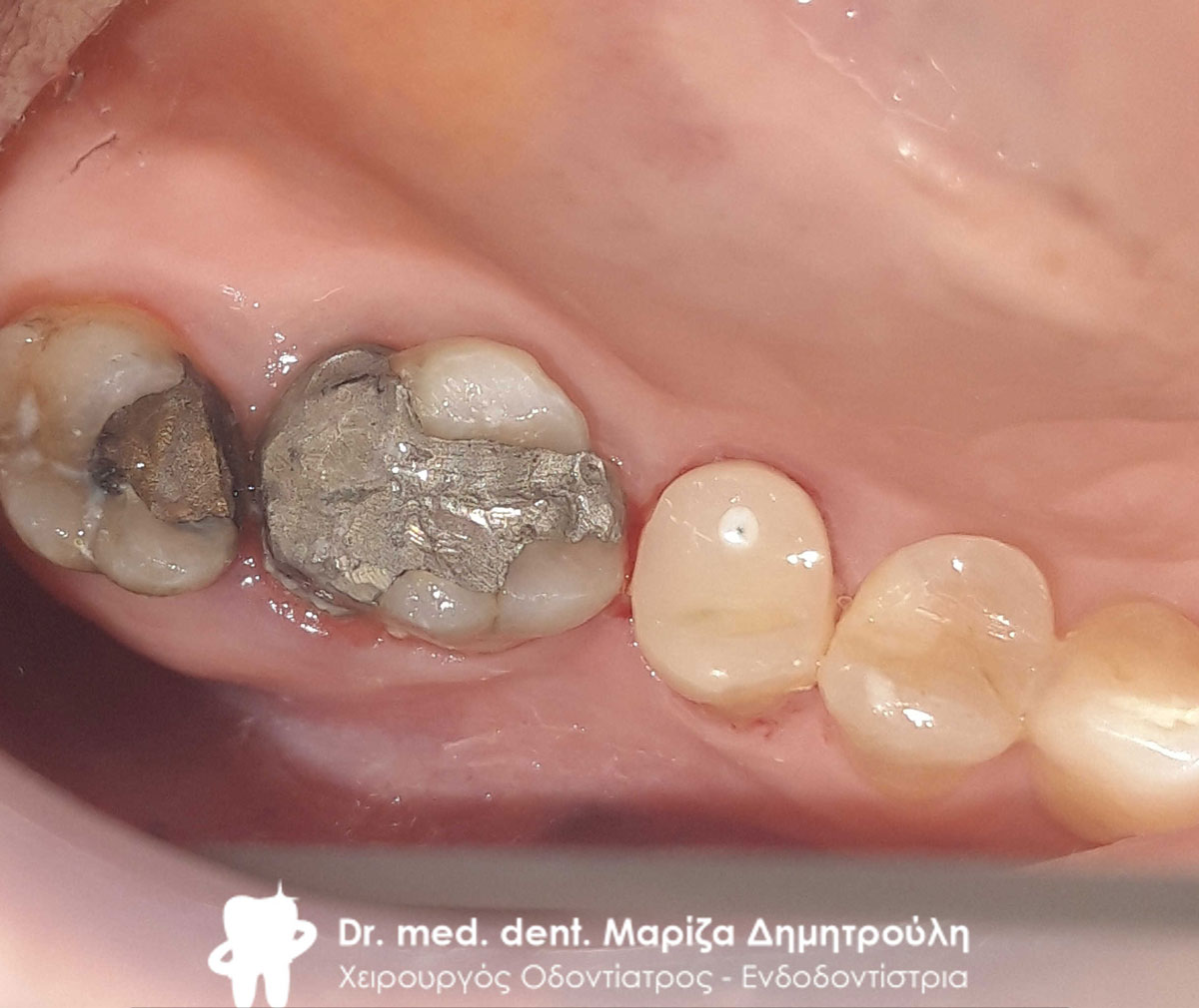
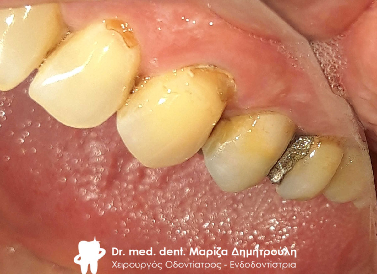
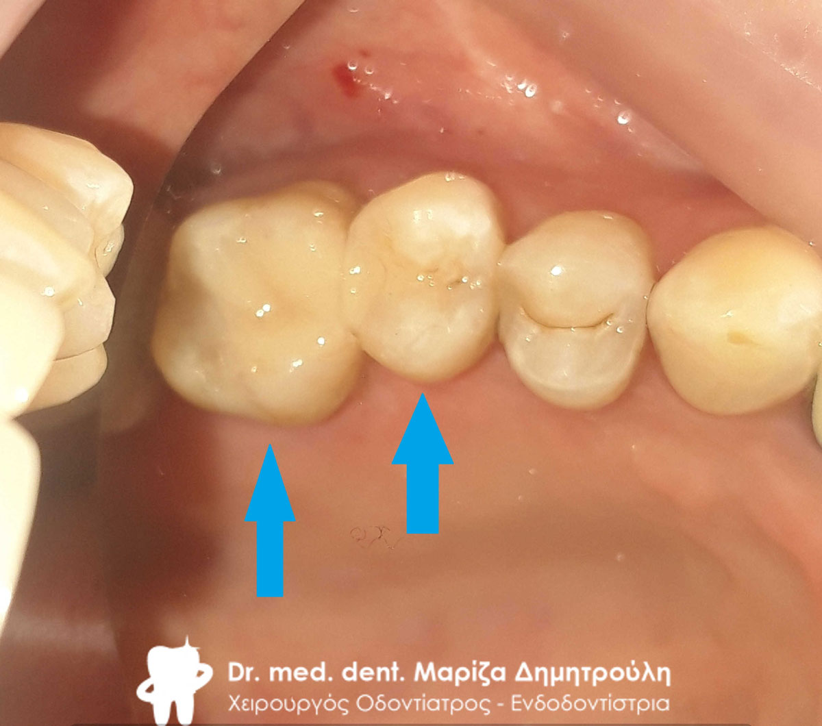
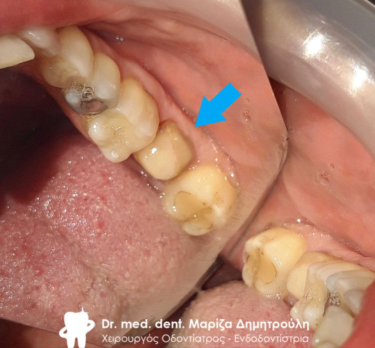
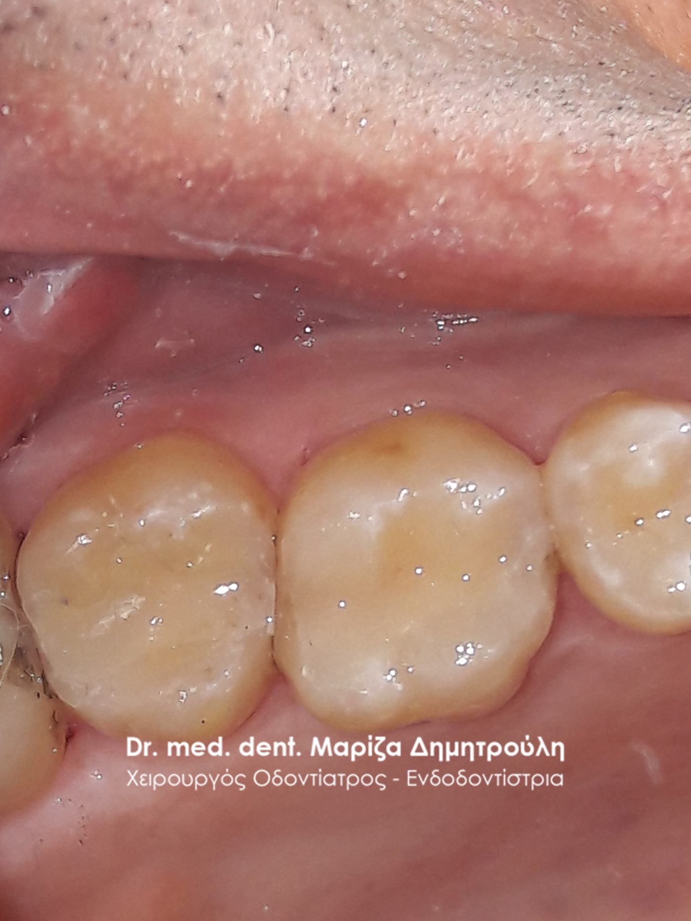
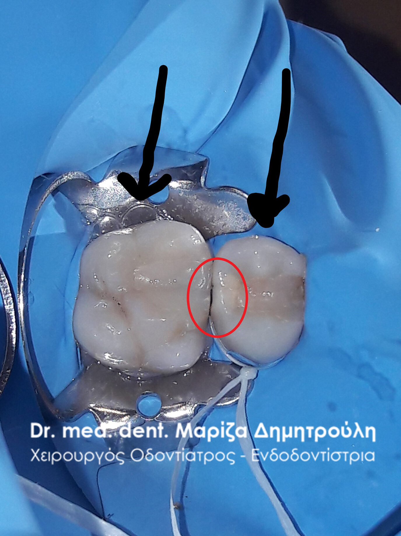
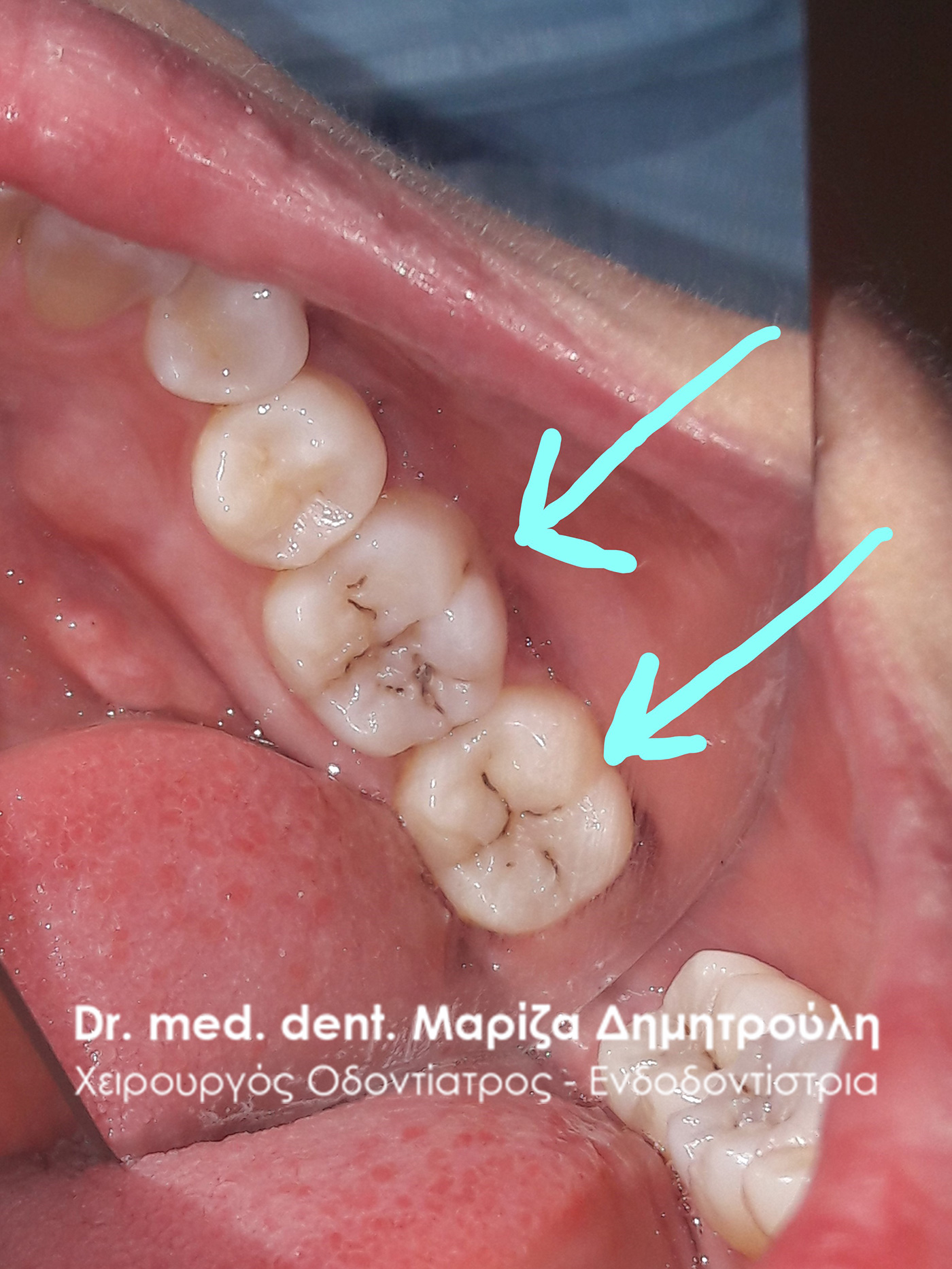
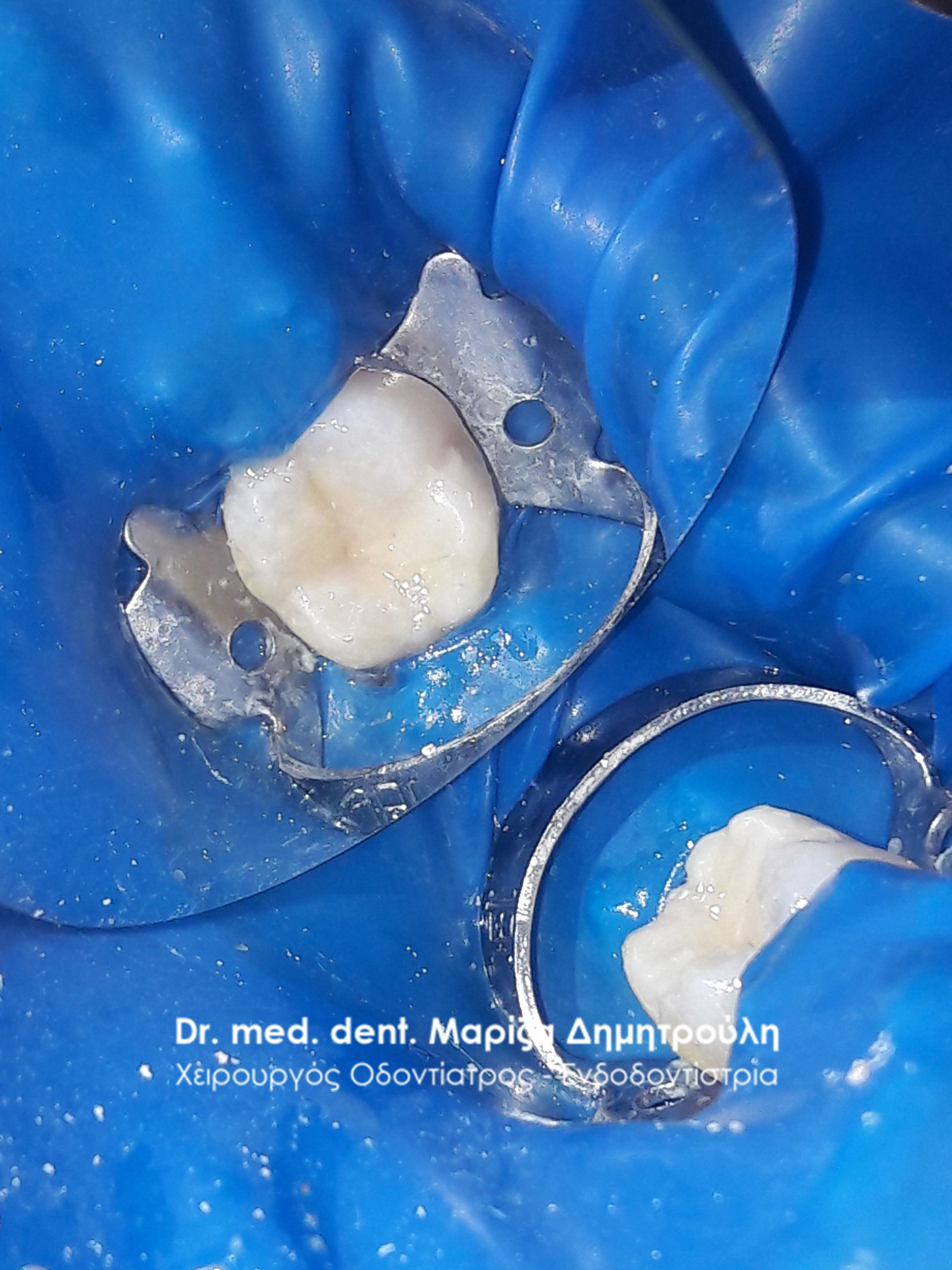
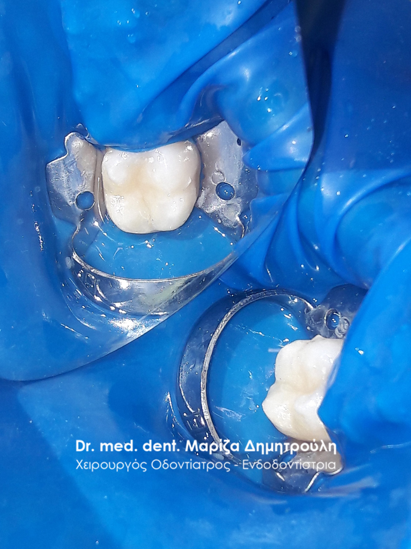
Original image of decayed teeth on the right side of the lower jaw
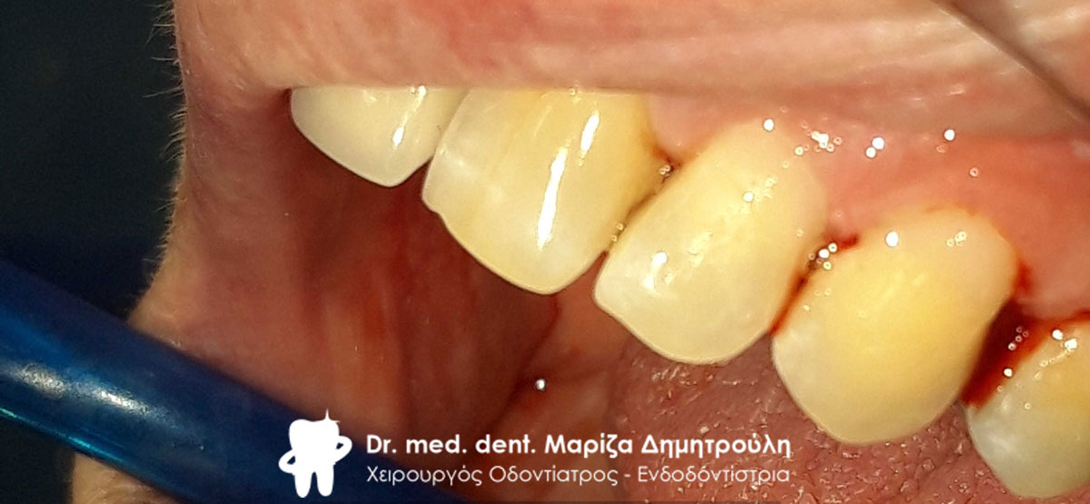
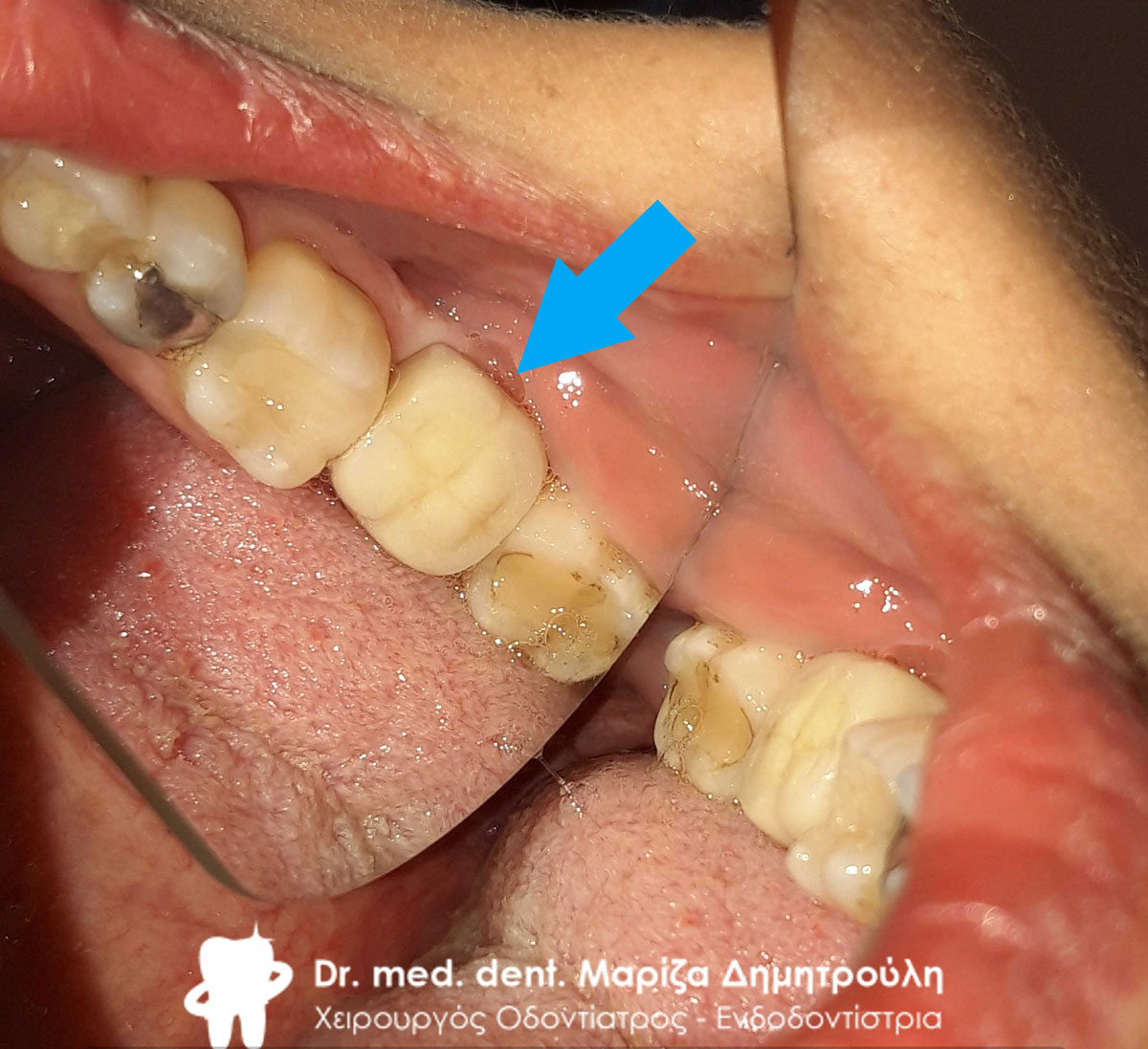
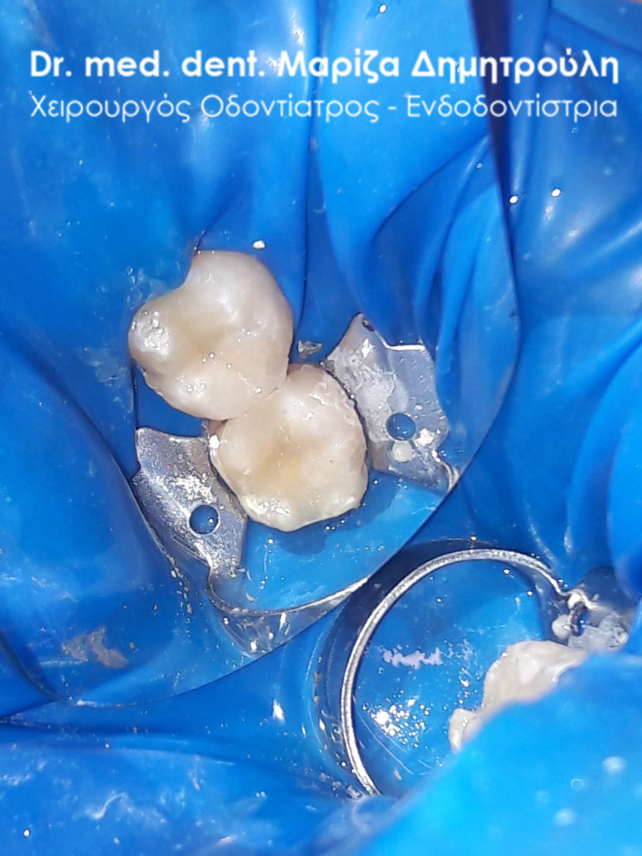
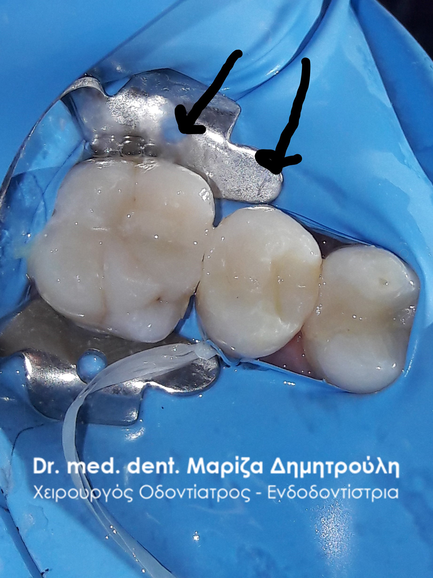
Final image of the new white fillings on the 2 premolars on the right side of the lower jaw
Case – Full Restoration of Dental Fillings
The patient had old fillings and his desire was the total restoration of his mouth and the replacement of his old fillings with new white composite resin fillings.
BEFORE
META
Cervical seal – original image
Nervical sealing – final image
BEFORE
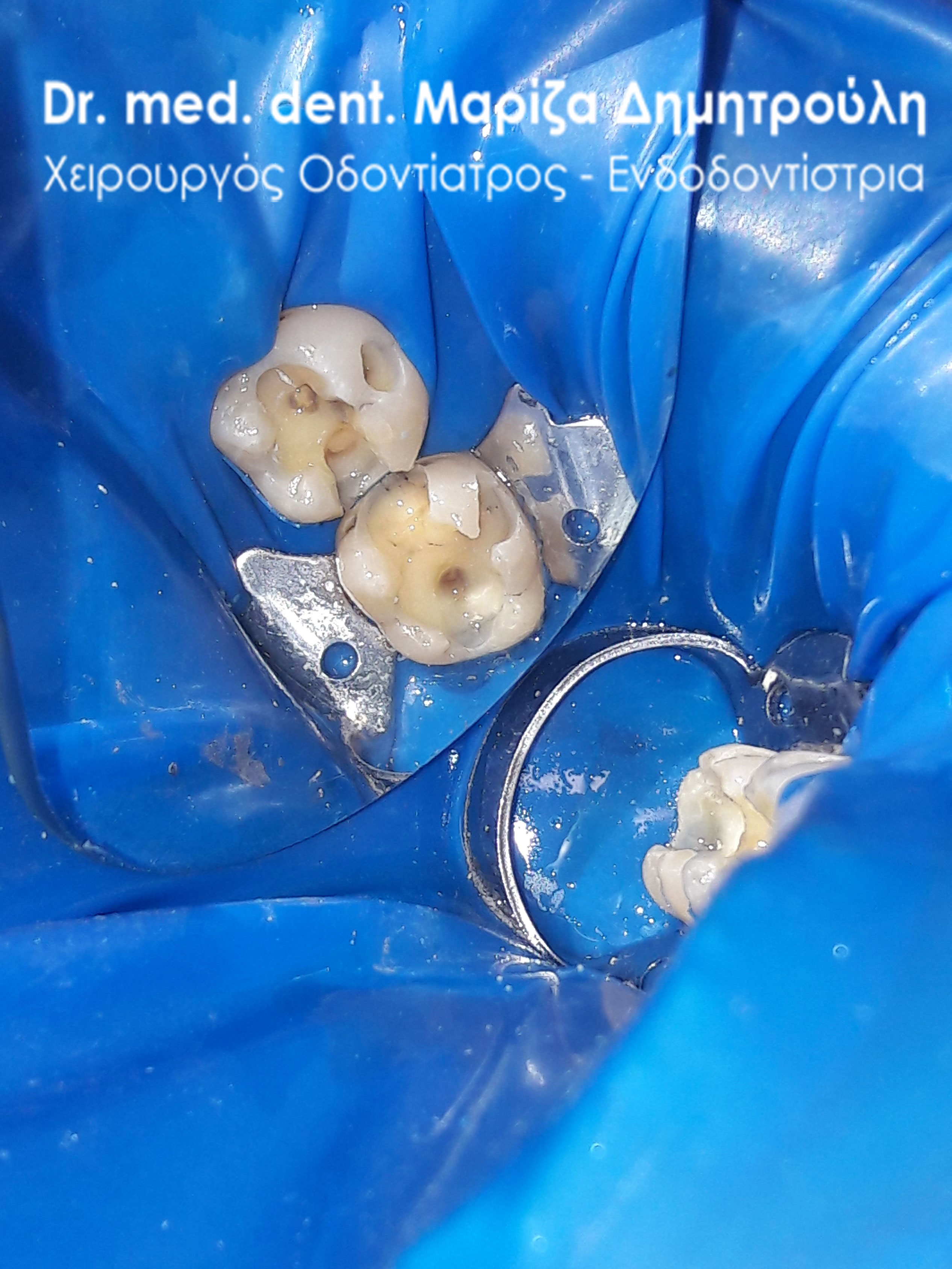
Image of teeth after removal of black fillings
META
META
Case – Full Restoration of Dental Fillings
The patient came to the clinic with the intention of replacing some old fillings, which occasionally gave her symptoms of pain. as the photos show the teeth were restored with new white fillings.
BEFORE
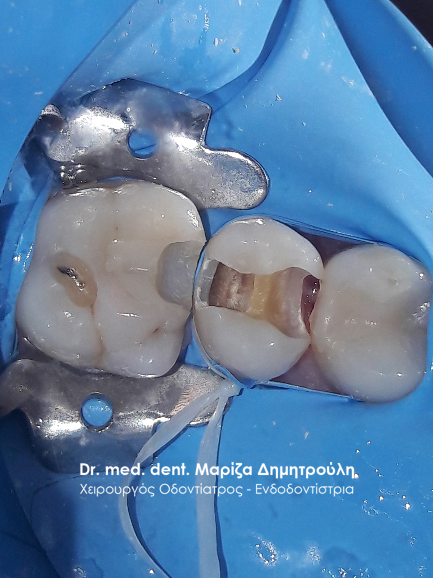
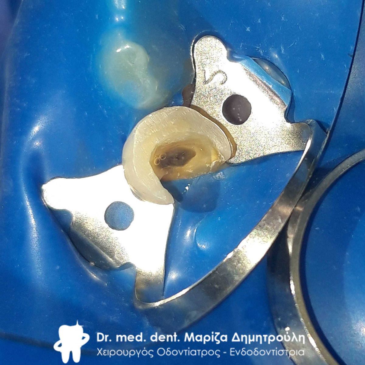
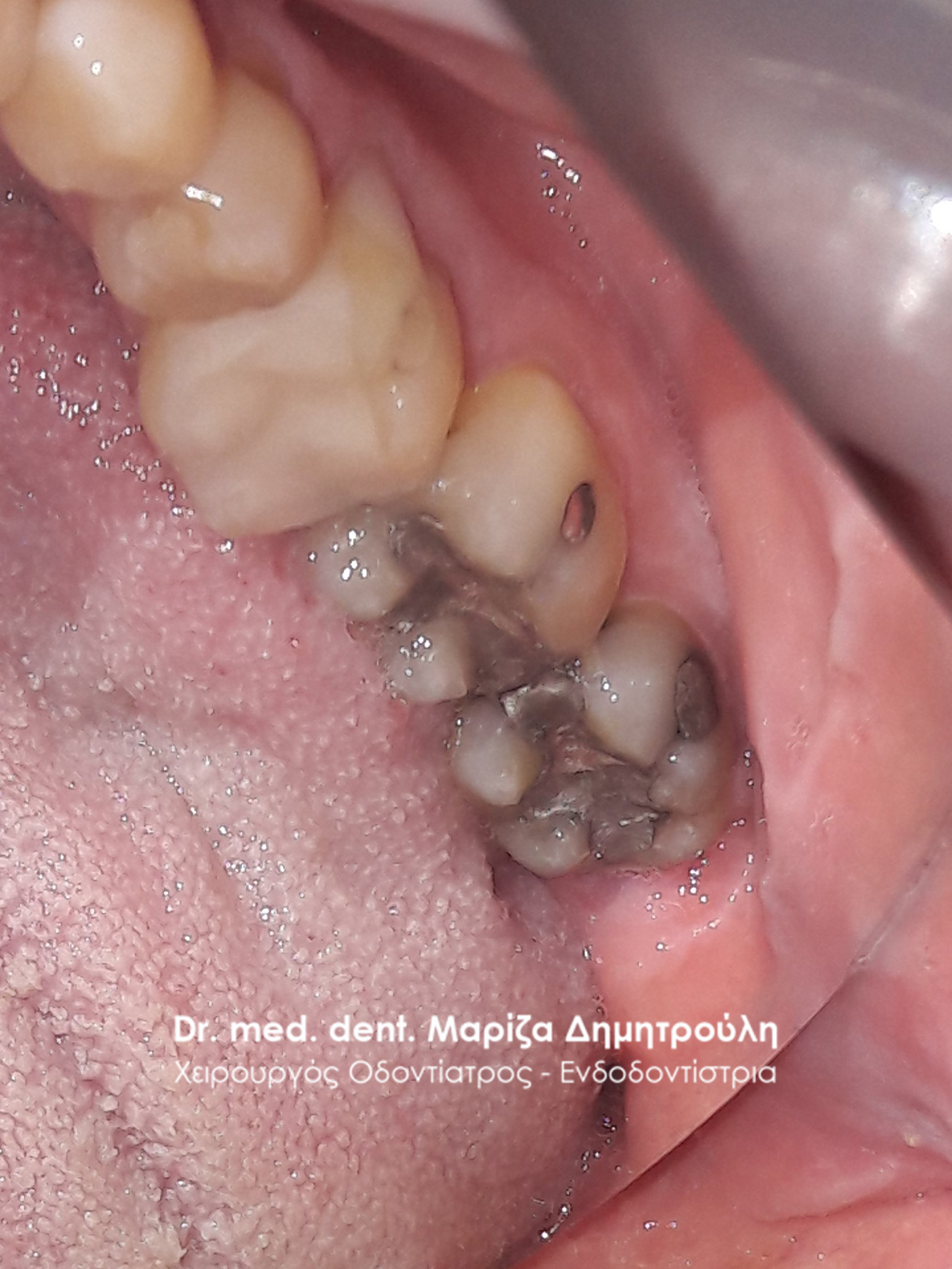
Image of caries cavity after tooth extraction
META
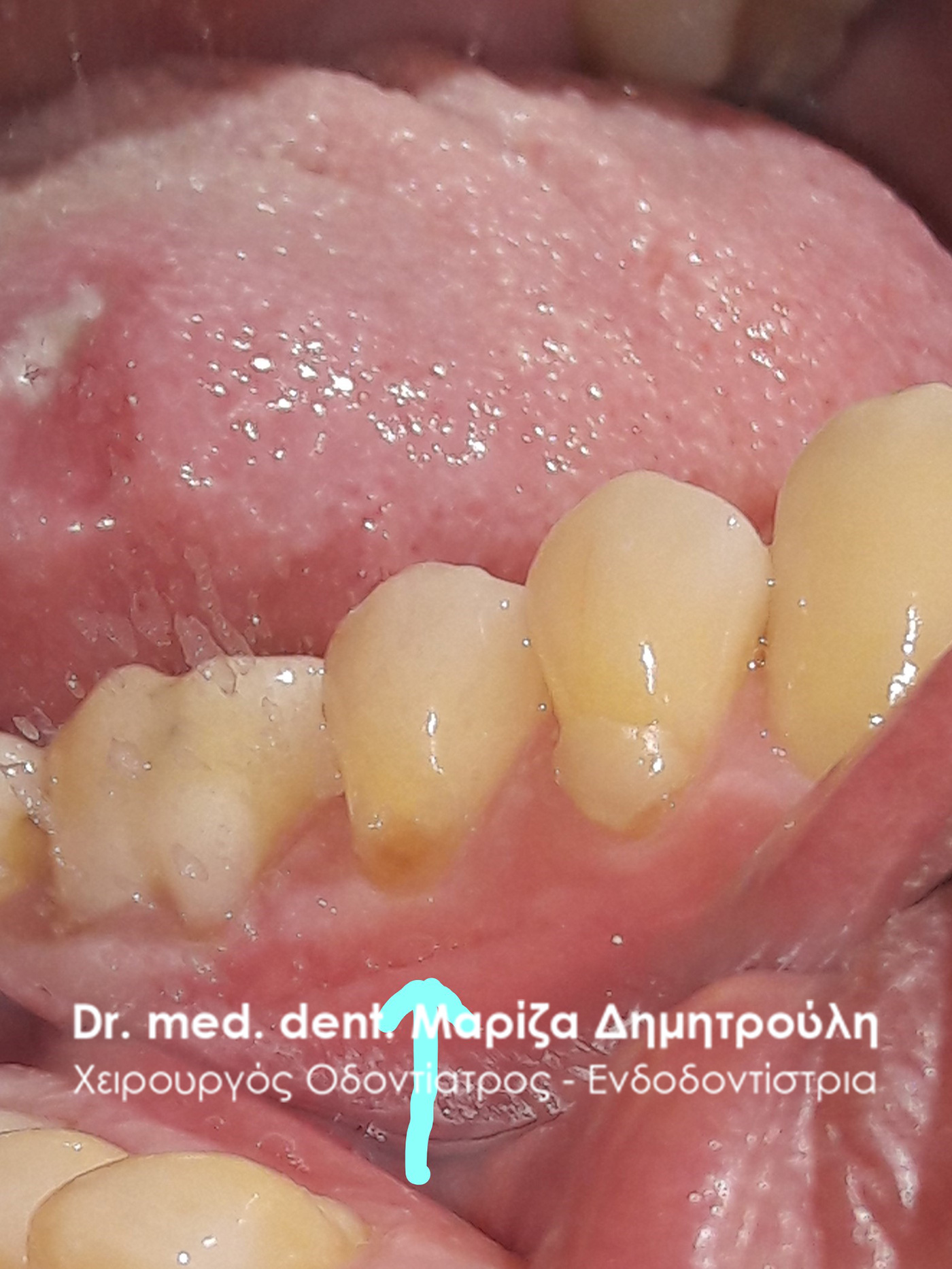
Original image of lower left molars
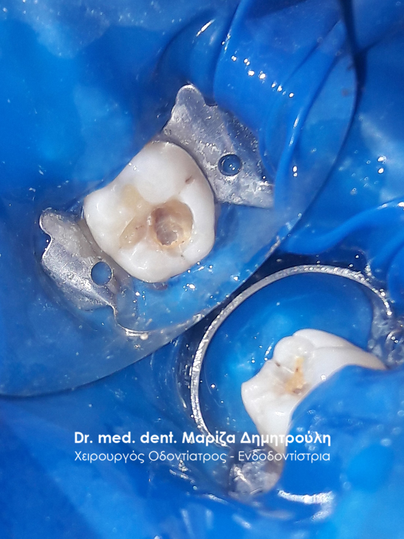
Image of 1st lower left molar after its eruption
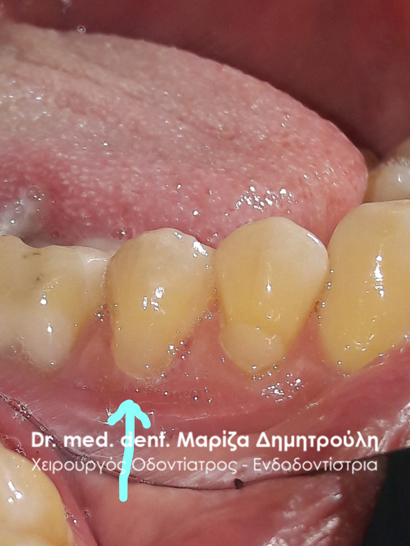
Final image of 1st lower left molar
Final image of 2nd lower left molar
Widget nach Inhalt

Dr. med. dent. Mariza Dimitrouli
Dr. med. dent. Mariza Dimitrouli hat sich in der Endodontologie (Wurzelkanalbehandlung) an der Medizinische Hochschule Hannover (MHH) spezialisiert und promoviert. Außerdem hat sie in einer Zahnklinik in Berlin gearbeitet. Sie unterhält ihre eigene Zahnarztpraxis in Thessaloniki, Thermi.