Incident – Cleaning Black Spots
The little patient had the desire to clean his little teeth from black spots. A thorough teeth cleaning was done and the little boy had a beautiful and bright smile again. The one and only tooth that was not cleaned (lower right front tooth) was removed the same day in the office because it was shaking and ready to fall out.
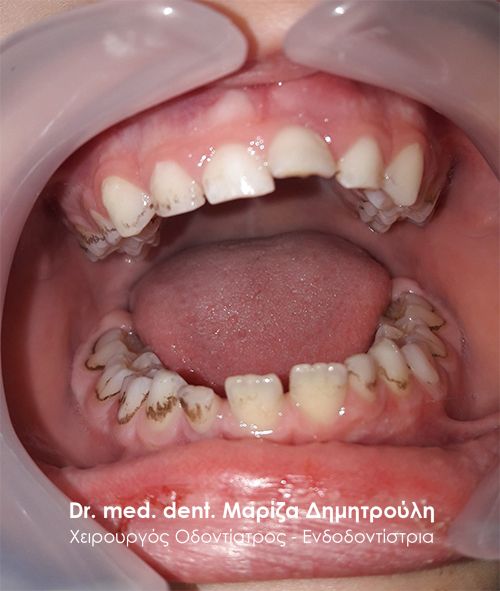
BEFORE
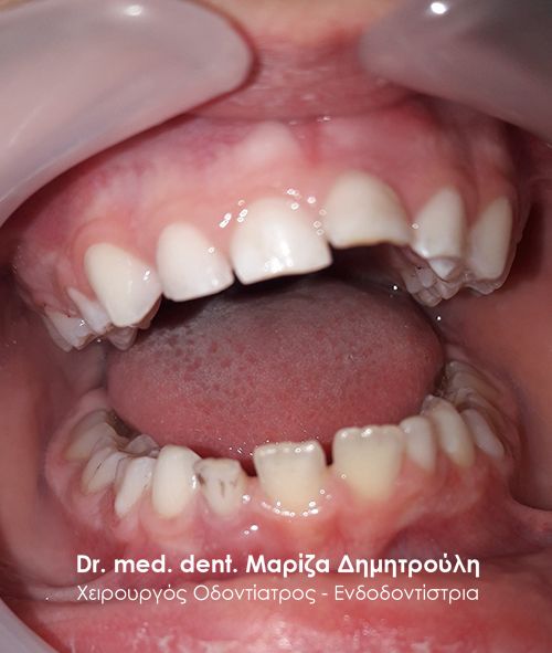
AFTER
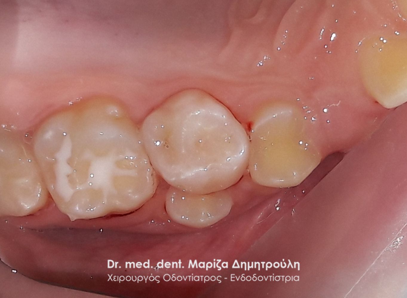
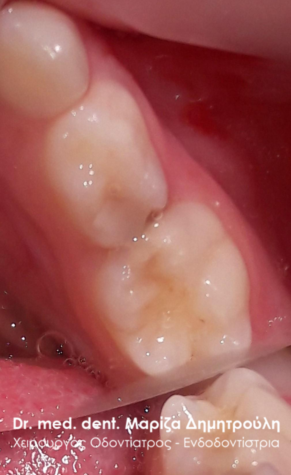
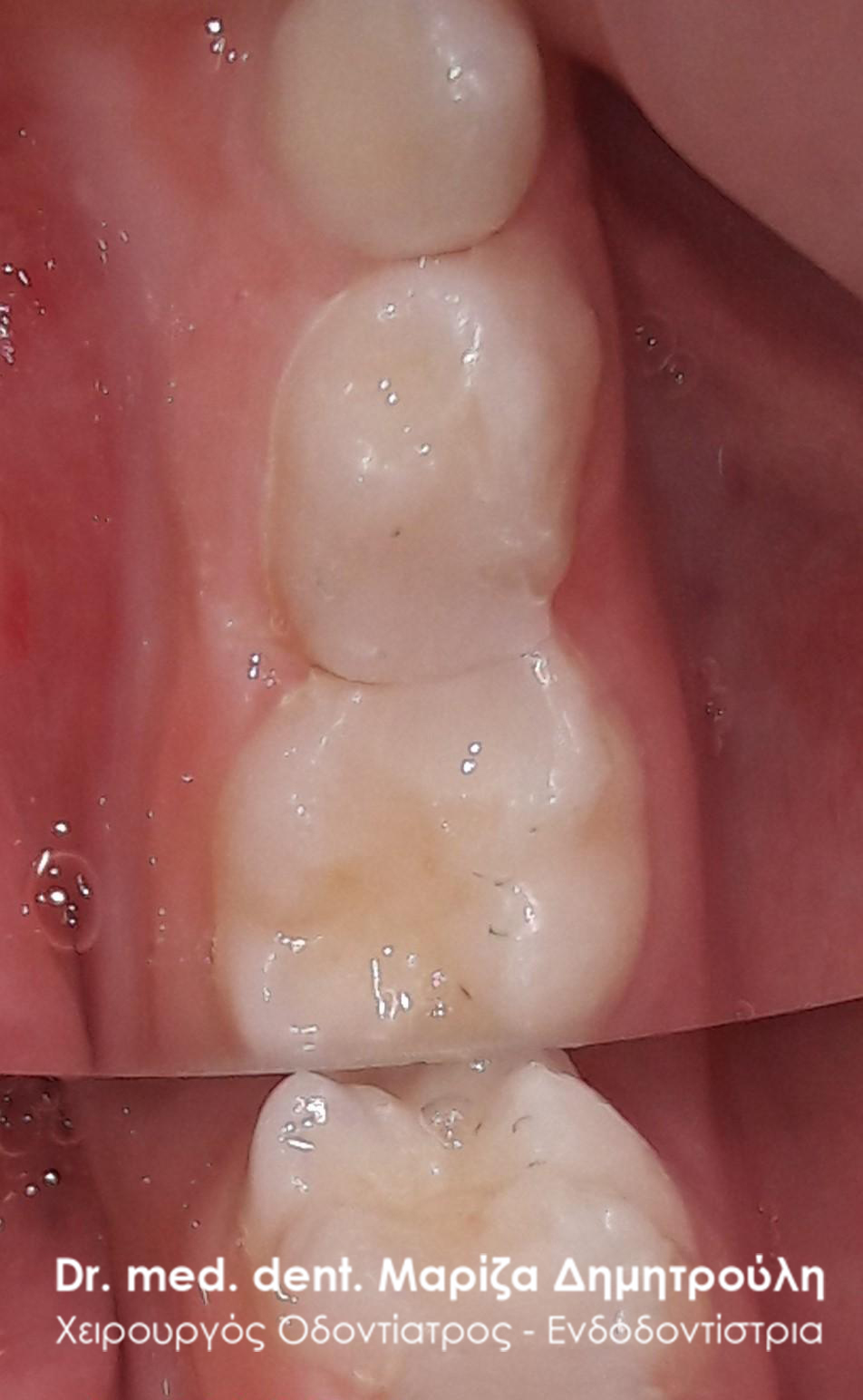
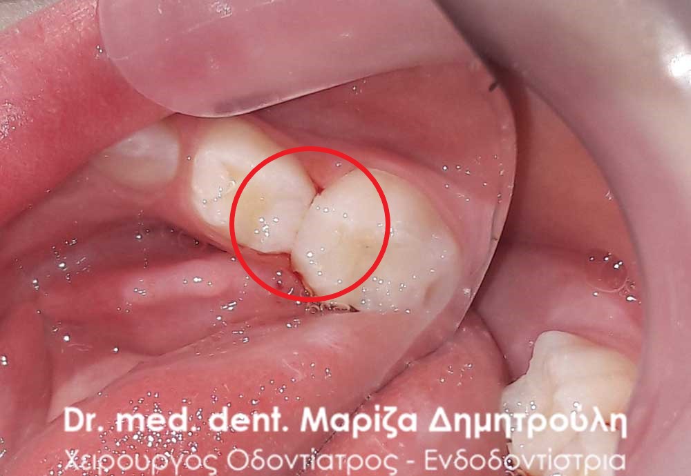
Incident – Extraction of a child tooth (double tooth / remaining baby tooth)
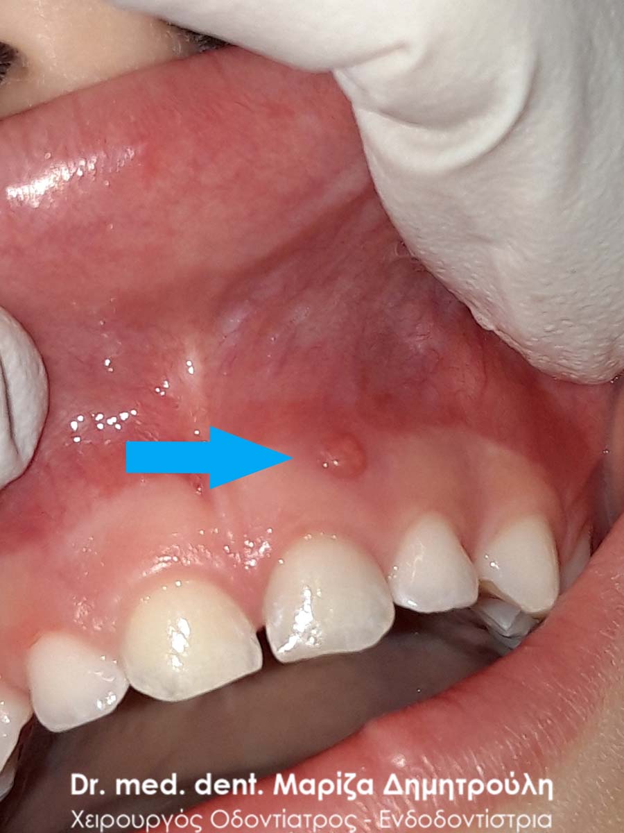
The young patient complains that a tooth in her upper jaw is bothering her. After the clinical examination, it was found that a new tooth remained, which, although it was mobile, did not fall out. Above the baby tooth there is already the permanent one, which cannot take its final position in the mouth, because the baby tooth prevents it.
After the administration of local anesthesia, the tooth is extracted, without the patient feeling pain or discomfort.
Therefore, in cases where the baby teeth remain in the dental barrier while the corresponding permanent teeth have already appeared, the removal of the baby teeth is necessary. Otherwise, the permanent tooth cannot fully emerge in the child’s mouth and it is possible in advanced cases to create orthodontic problems.
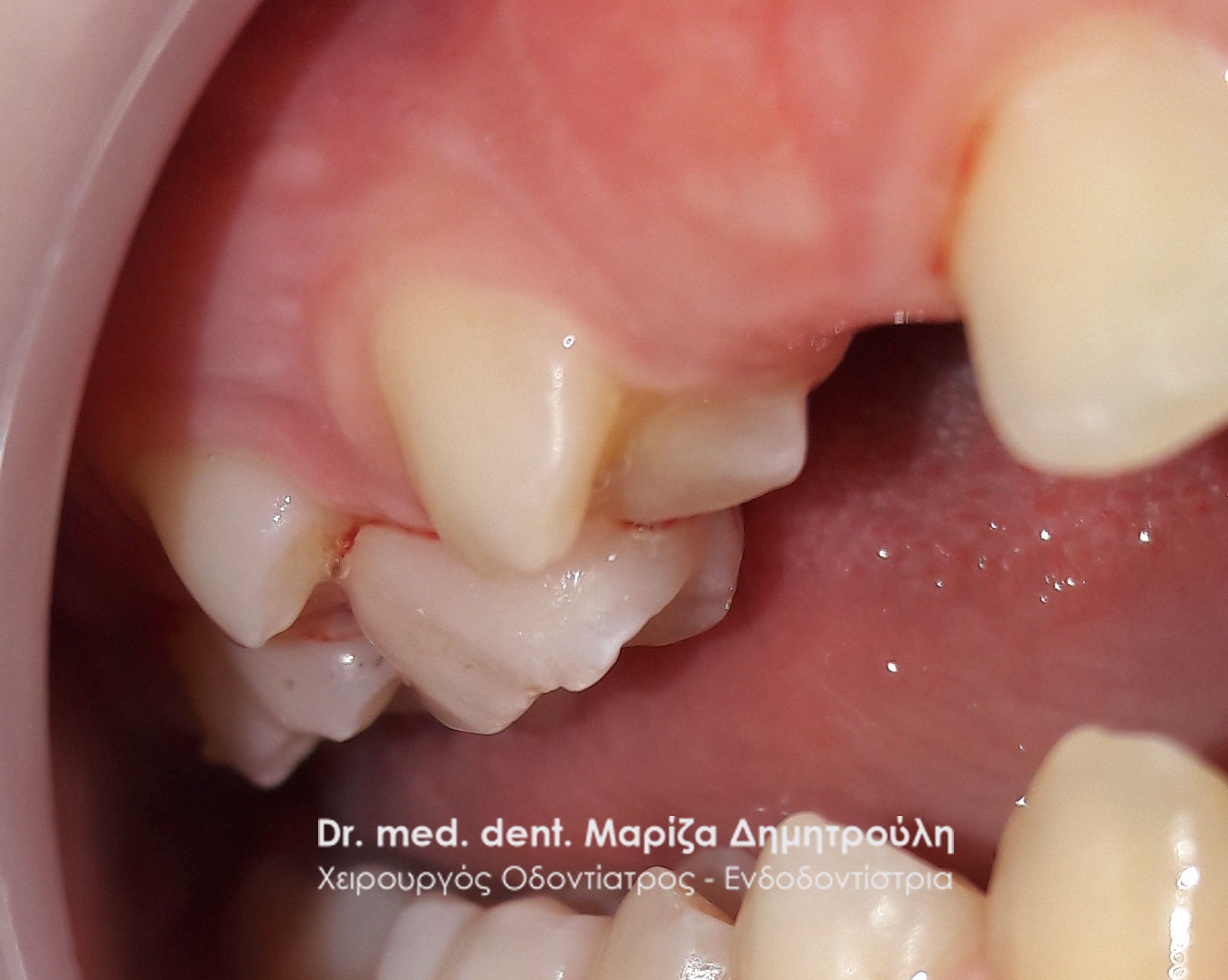
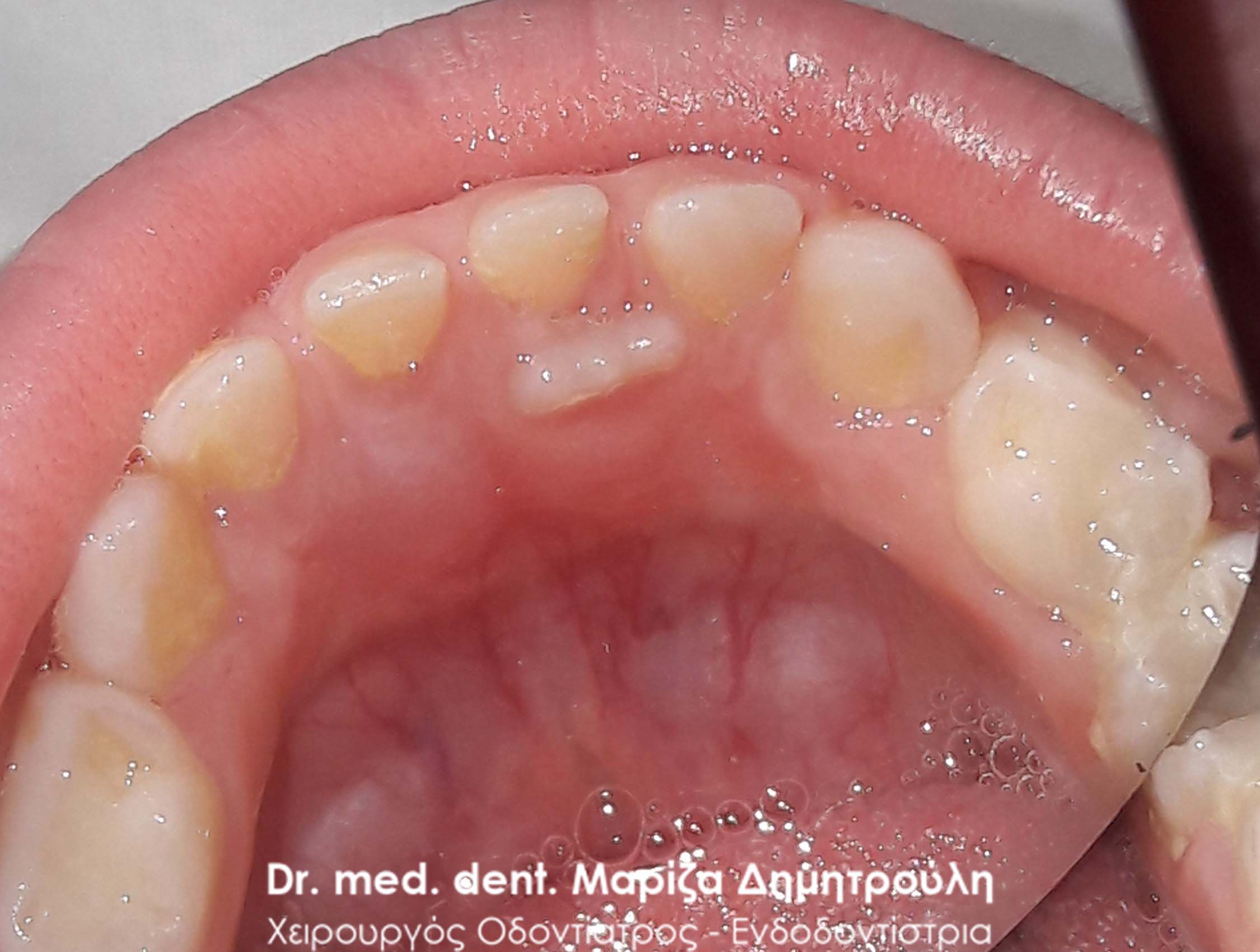
Incident – Extraction of a child tooth (double tooth / remaining baby tooth)
In this small patient, the child sector remains in place, while at the same time the corresponding permanent one has already made its appearance in the child’s mouth. In cases like these, it is necessary to remove the children’s teeth by the dentist, so that the permanent tooth can fully emerge in the child’s mouth and “take” its final position in the dental barrier.
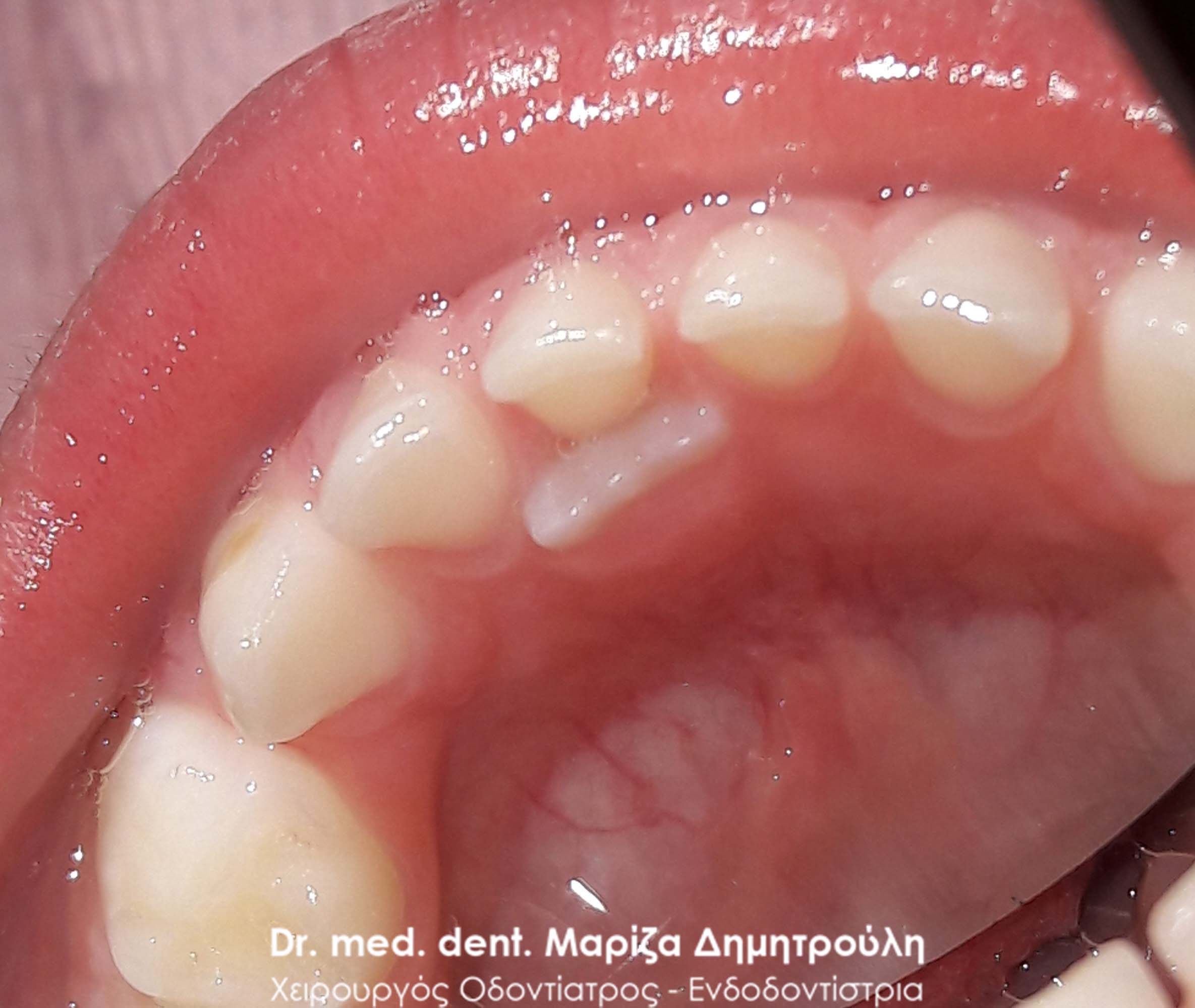
As the photos show, just one week after the extraction of the baby sector, the corresponding permanent tooth has changed position and has begun to take its final position slowly inside the child’s mouth. One month after extraction, both permanent central sectors rose normally in the mouth without any problems.
BEFORE
One week after the extraction of the baby tooth
A month or so after the extraction of the baby tooth
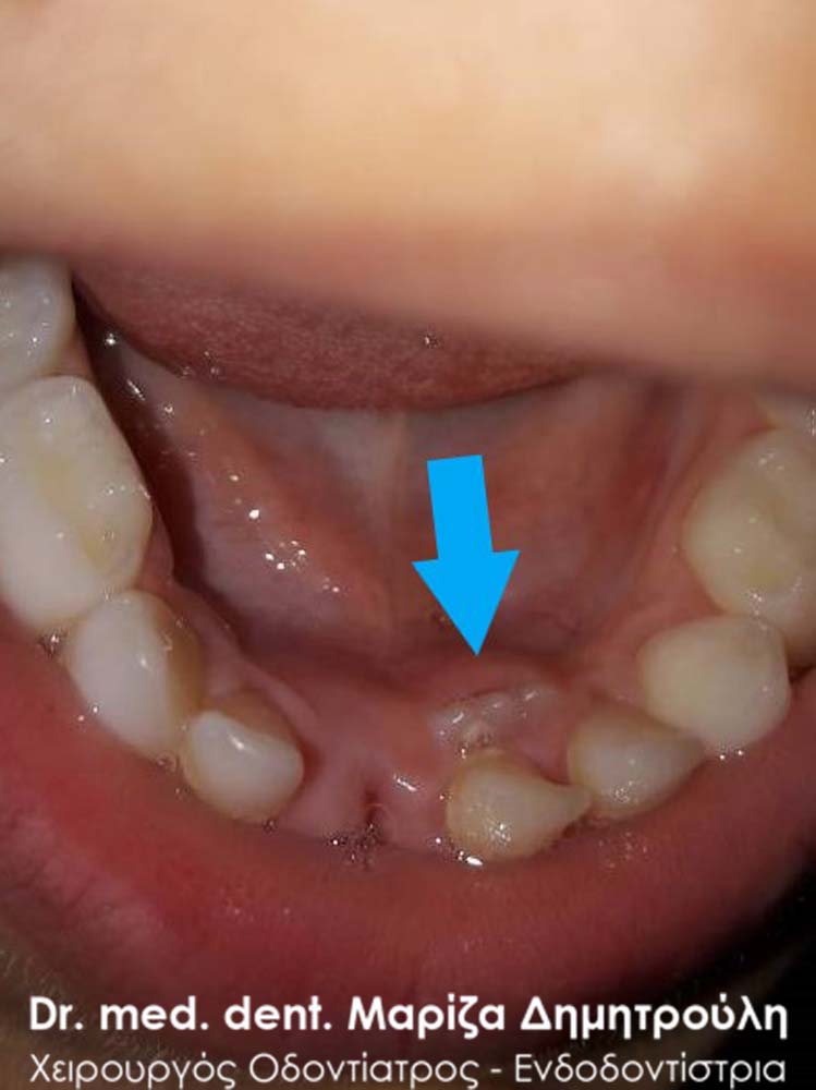
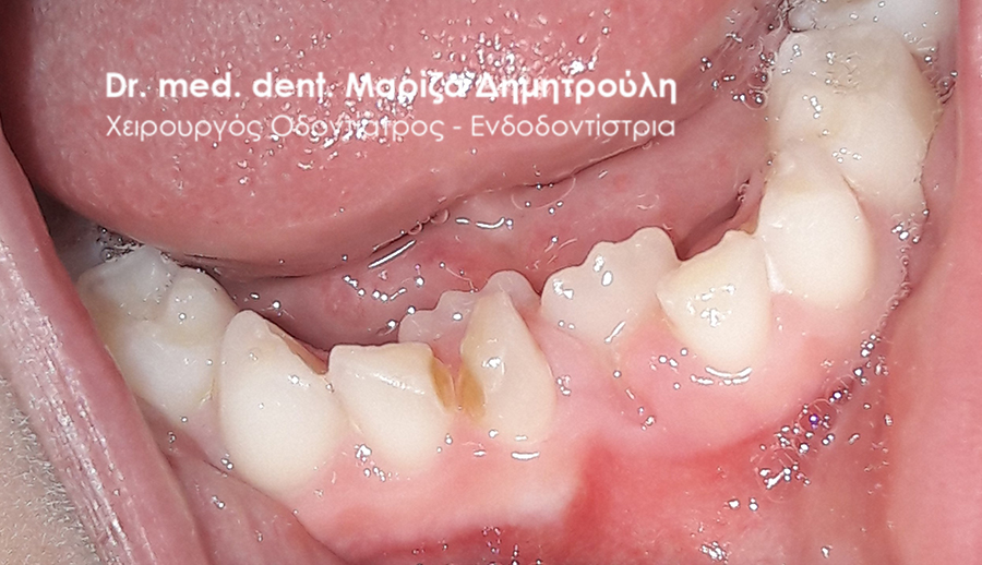
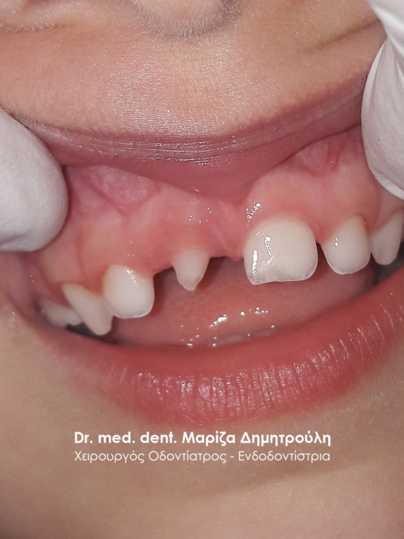
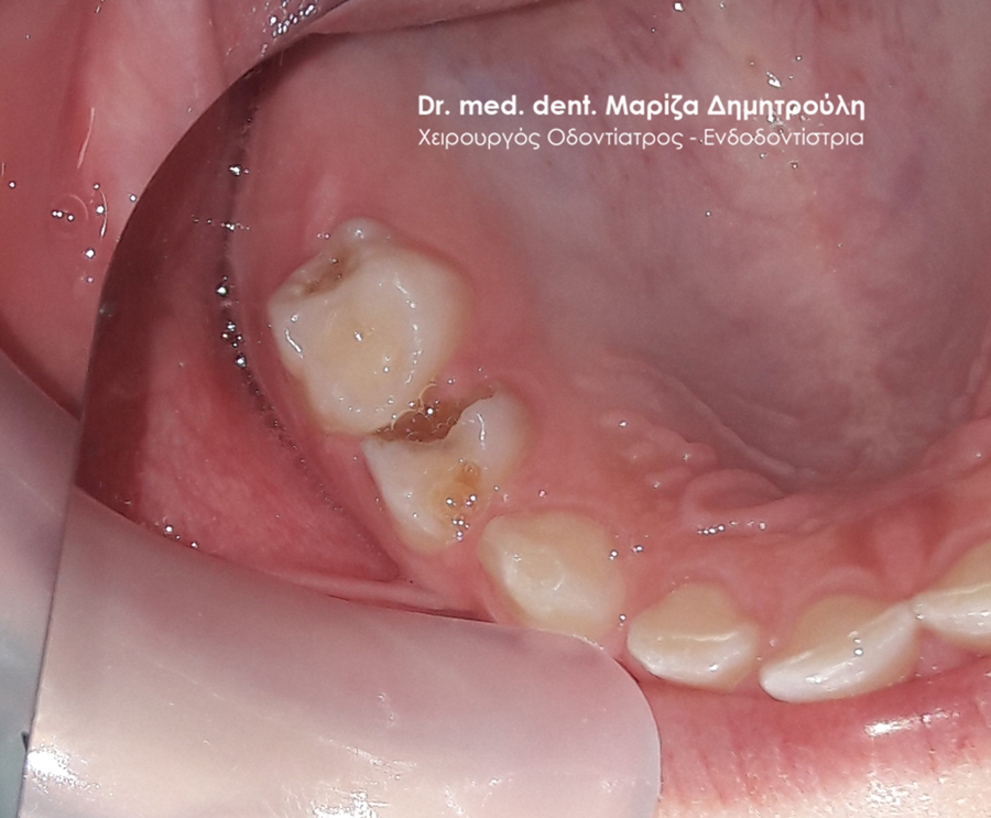
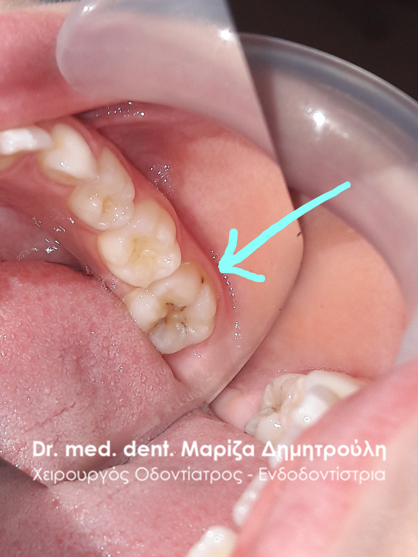
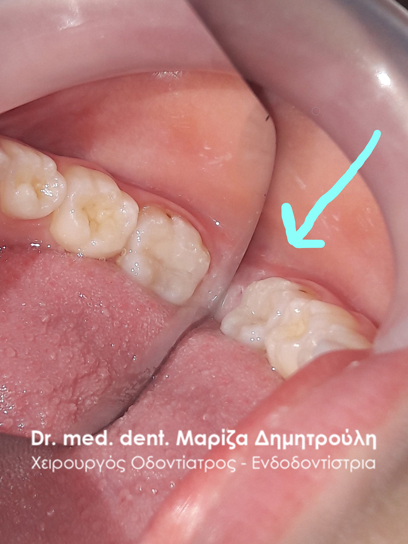
Incident – Extraction of a child tooth (double tooth / remaining baby tooth)
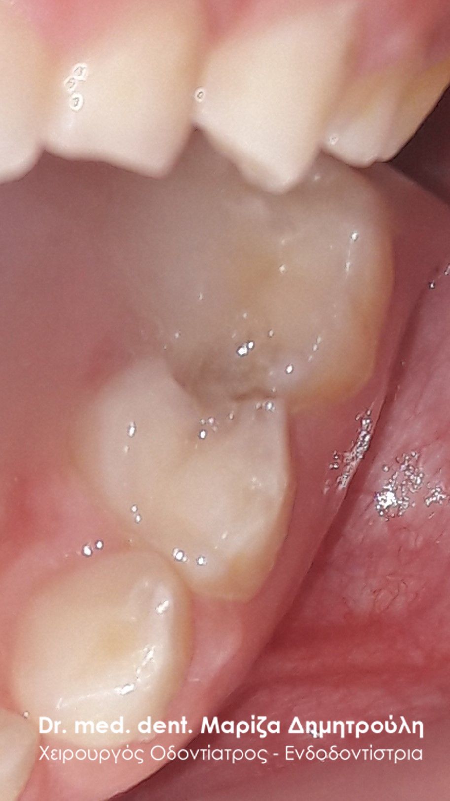
The little girl complains that it sometimes hurts in the front area of the lower jaw when biting food. The parents, after the child’s complaints, noticed the presence of the new lower incisor, even though the corresponding permanent tooth has erupted in the mouth. In this case, the immediate extraction of the baby tooth by the dentist is required.
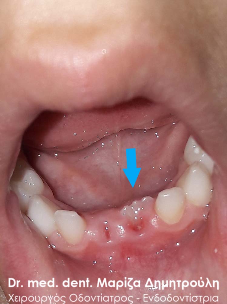
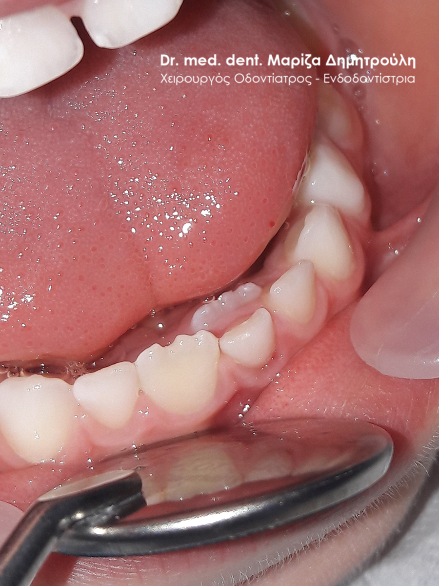
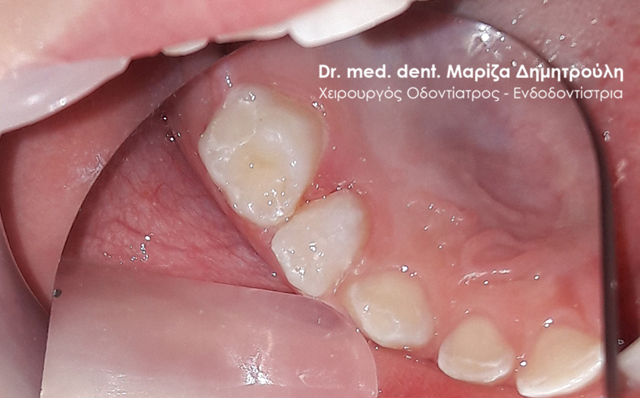
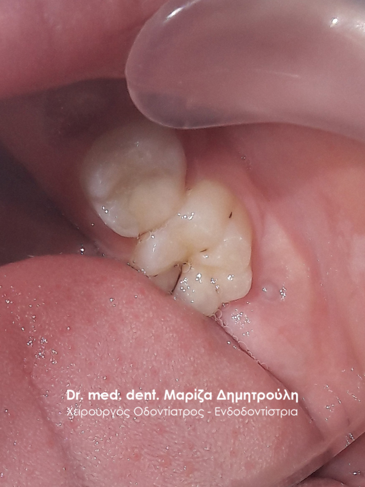
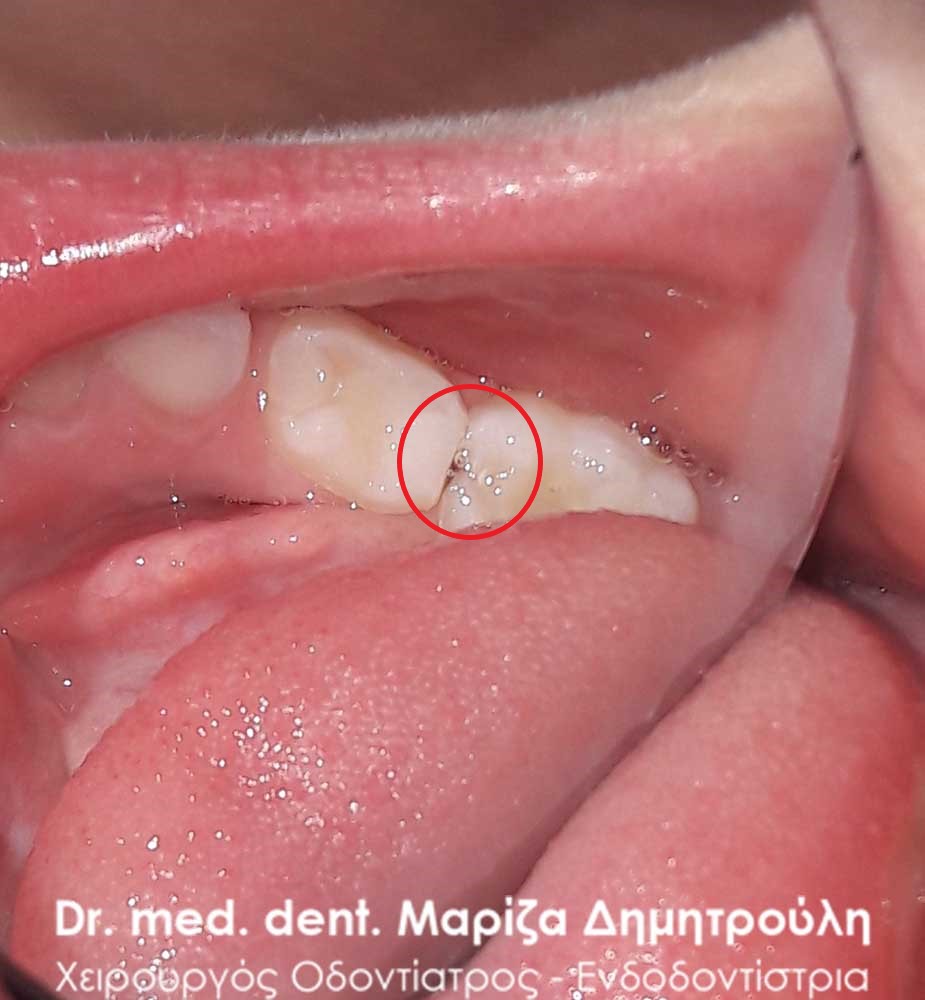
Incident – Extraction of a child tooth (double tooth / remaining baby tooth)
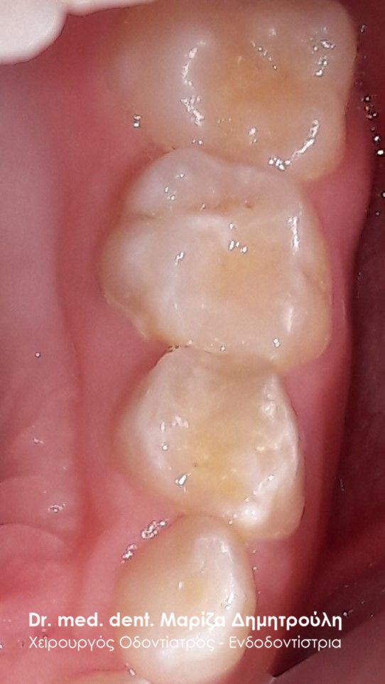
The parents noticed that the left baby sector did not “move” even though the two permanent lower front teeth had made their appearance. After the clinical examination, it was confirmed that the child’s tooth had no mobility, so it was decided to remove it in the doctor’s office, after administration of local anesthesia.
There are many times when the baby teeth remain in the dental barrier even though the corresponding permanent teeth have already erupted in the child’s mouth. In this case, it is necessary to remove the children’s teeth by the dentist, so that the permanent tooth can fully emerge in the child’s mouth and “take” its final position in the dental barrier.
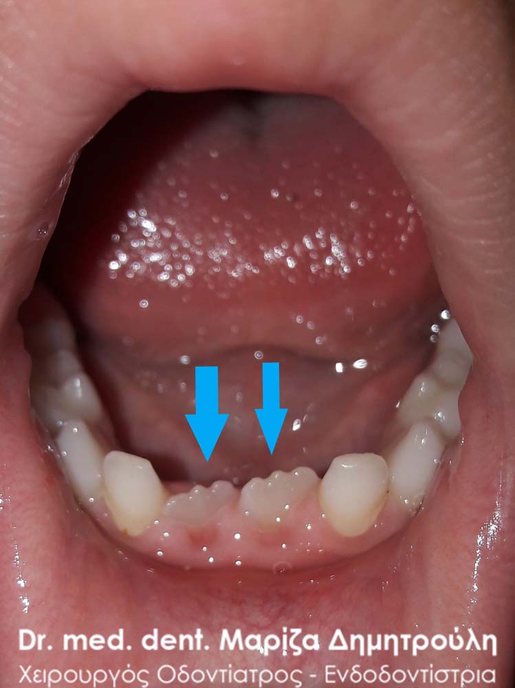
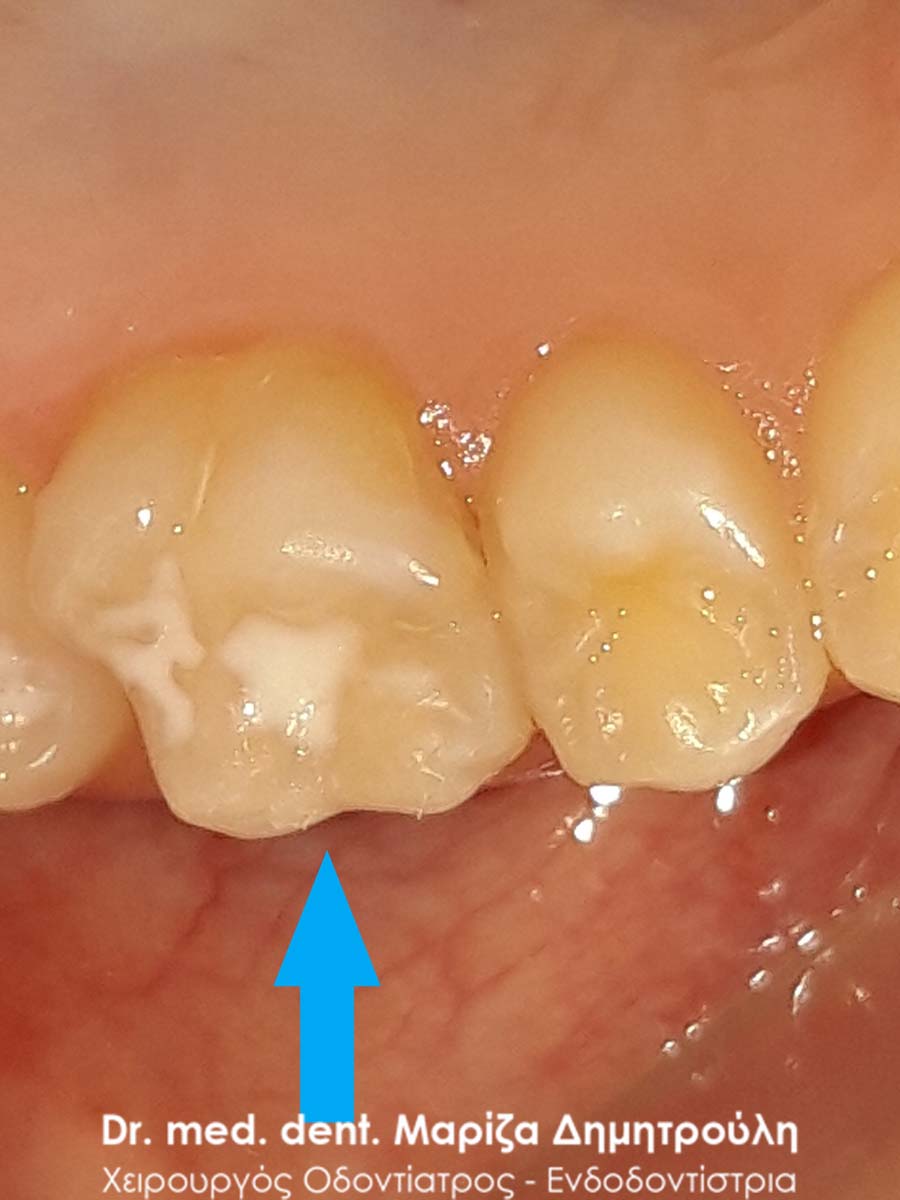
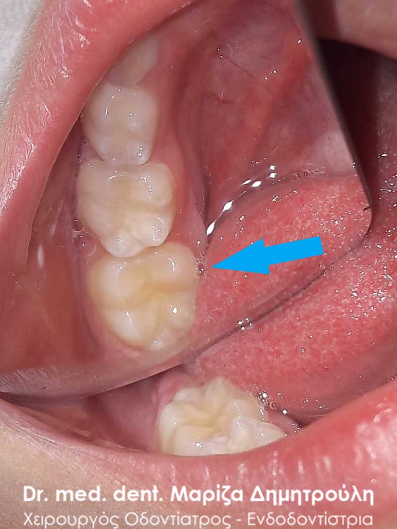
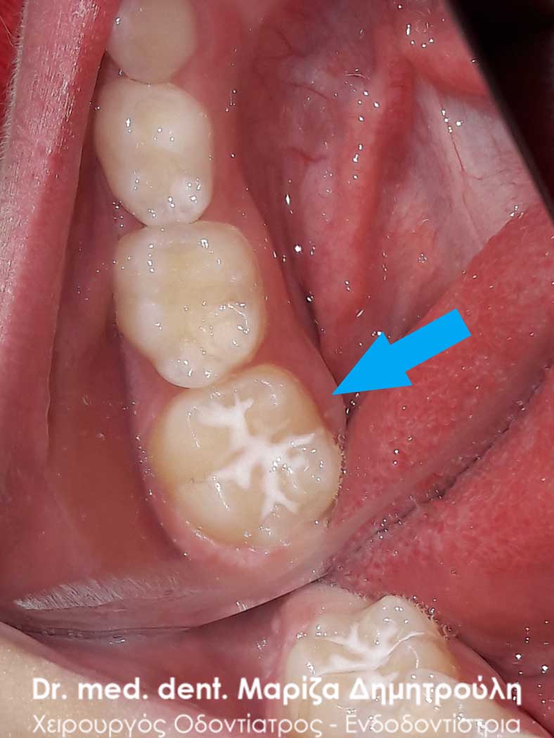
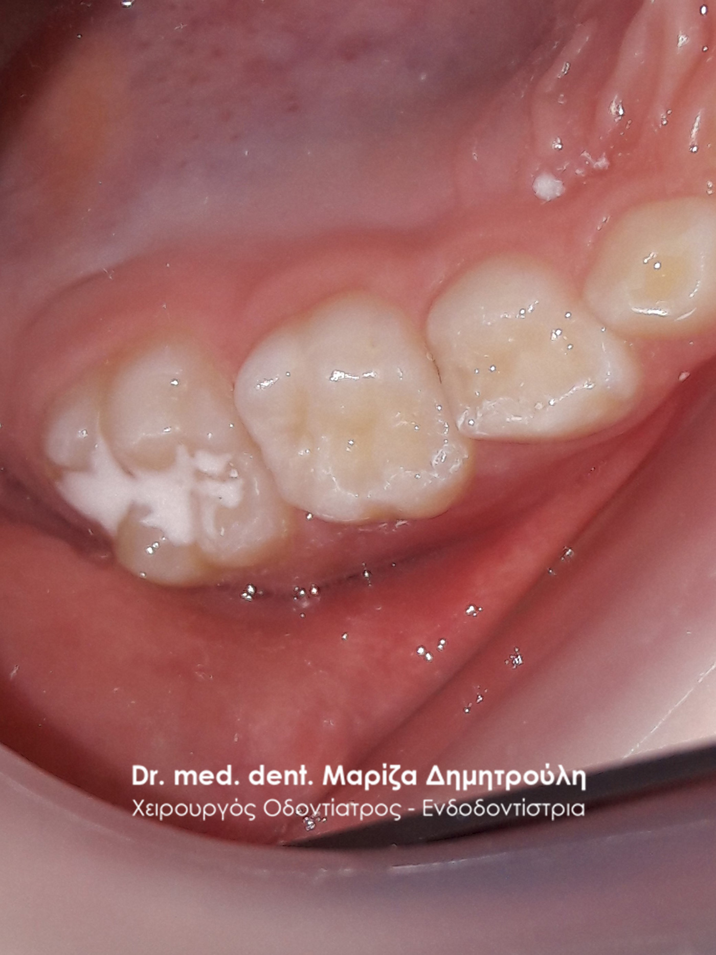
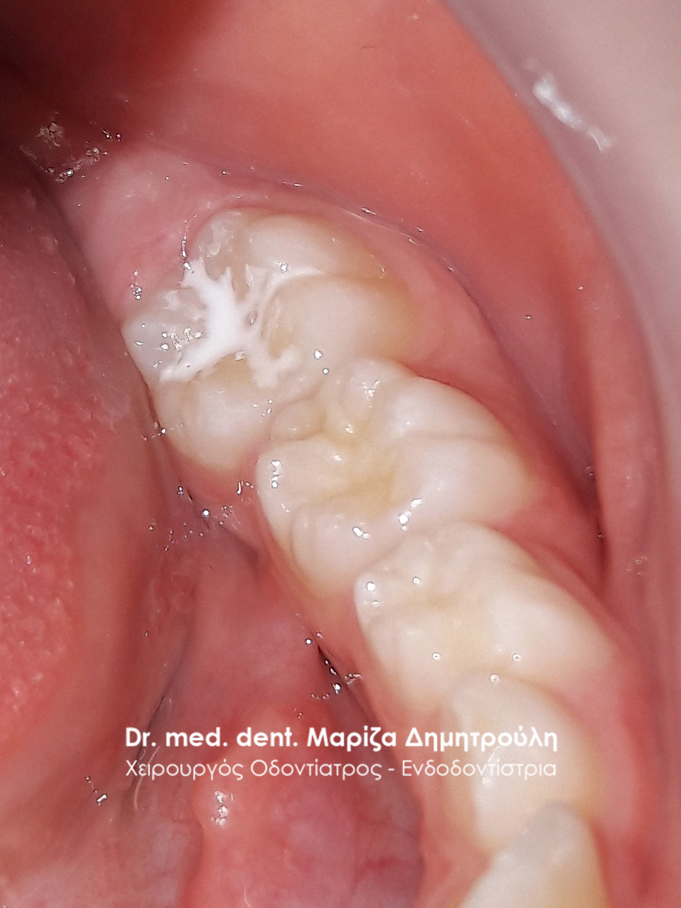
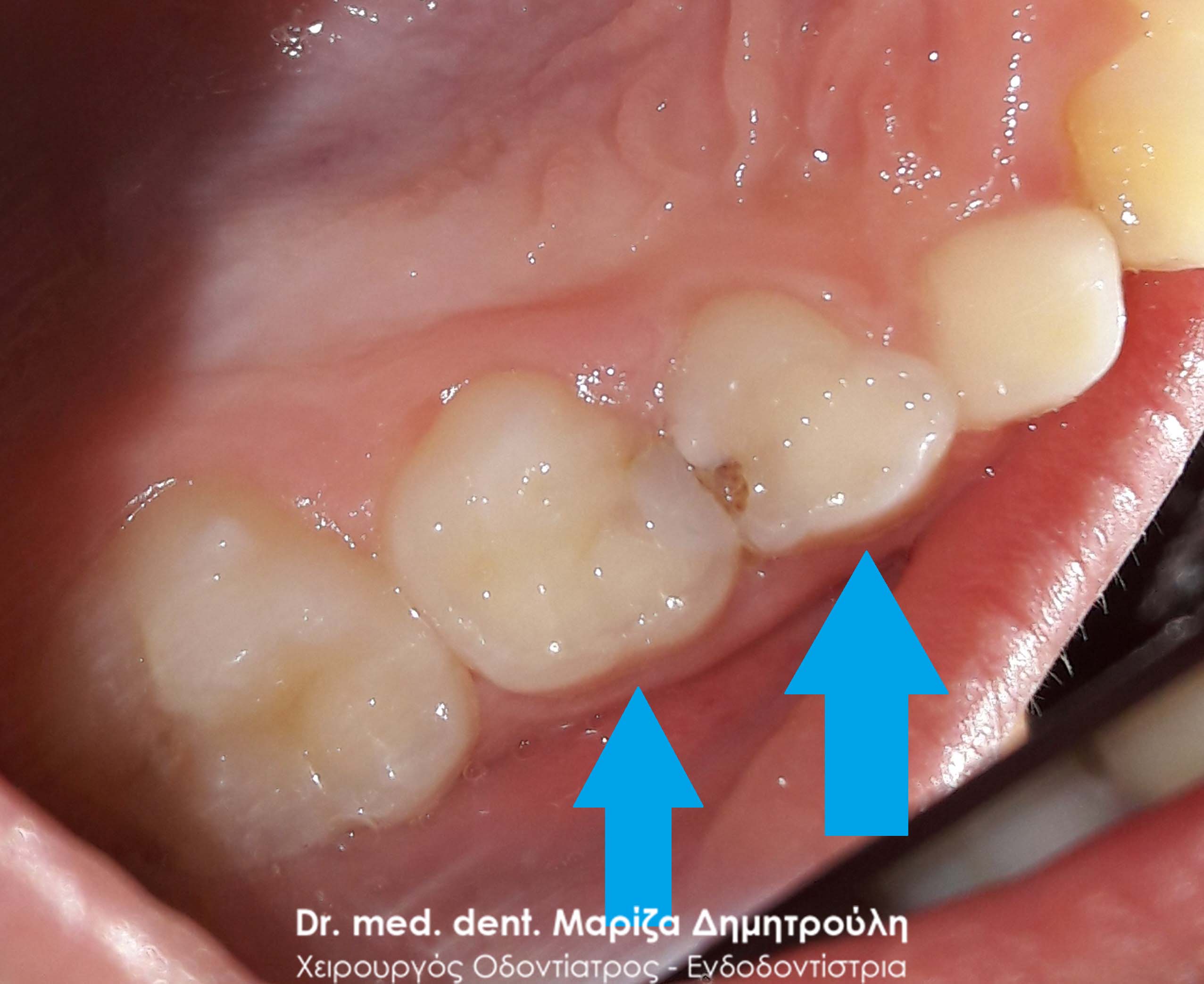
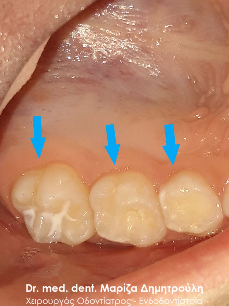
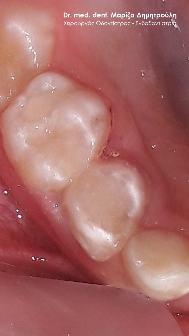
Incident – Extraction of a child tooth (double tooth / remaining baby tooth)
In the little patient’s mouth, the lower, young central sector remained in place, even though the corresponding permanent tooth had erupted. For this reason, the baby tooth was removed so that the permanent central sector could take its final position in the child’s mouth.
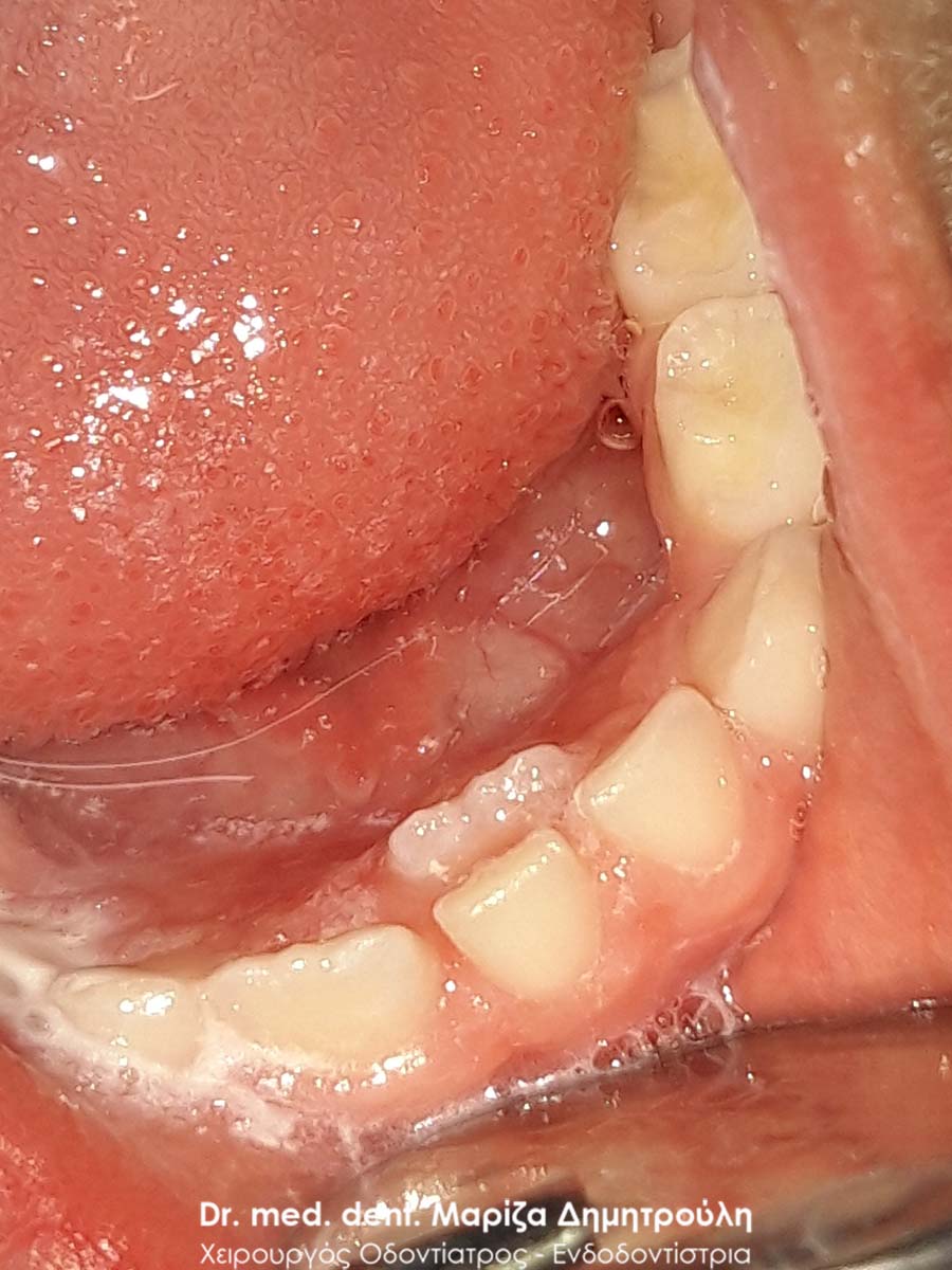
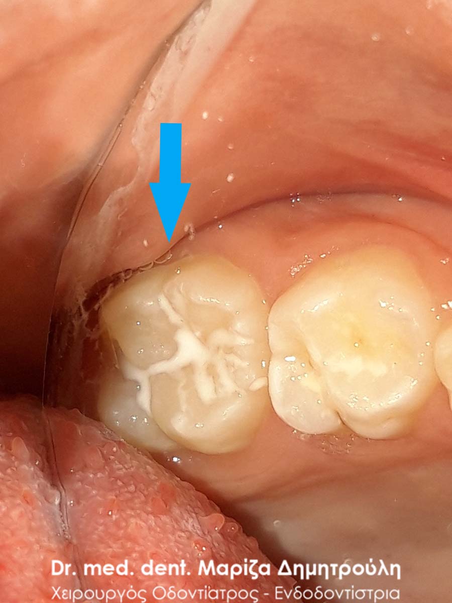
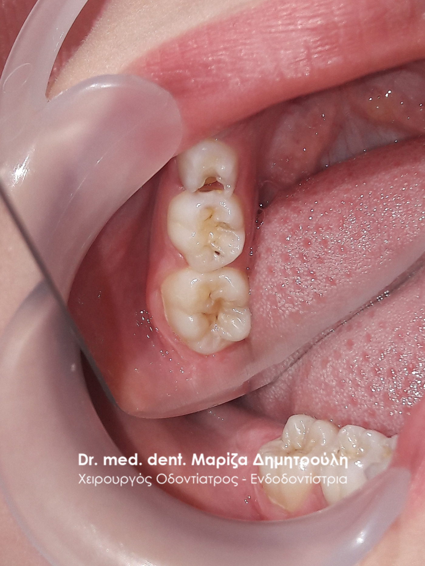
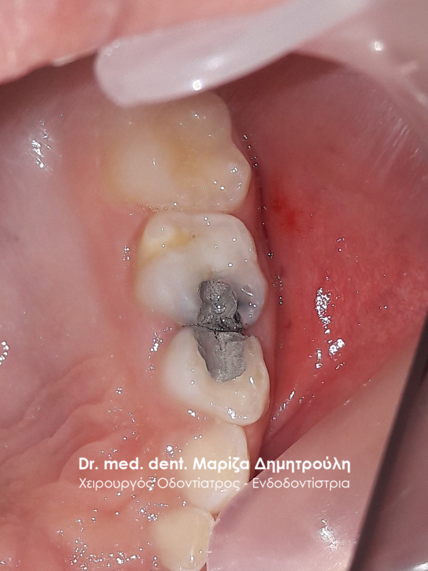
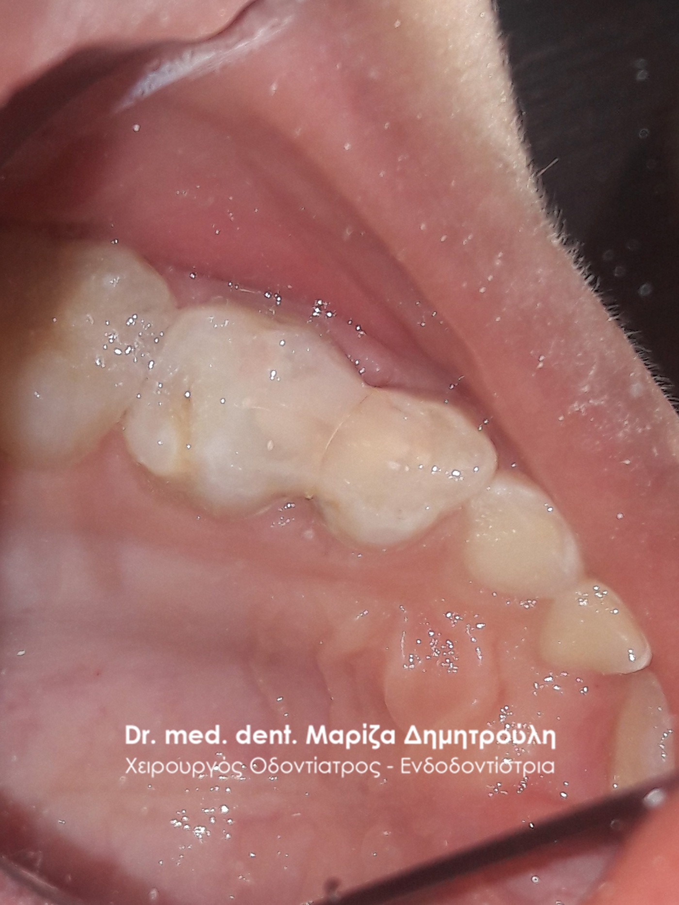
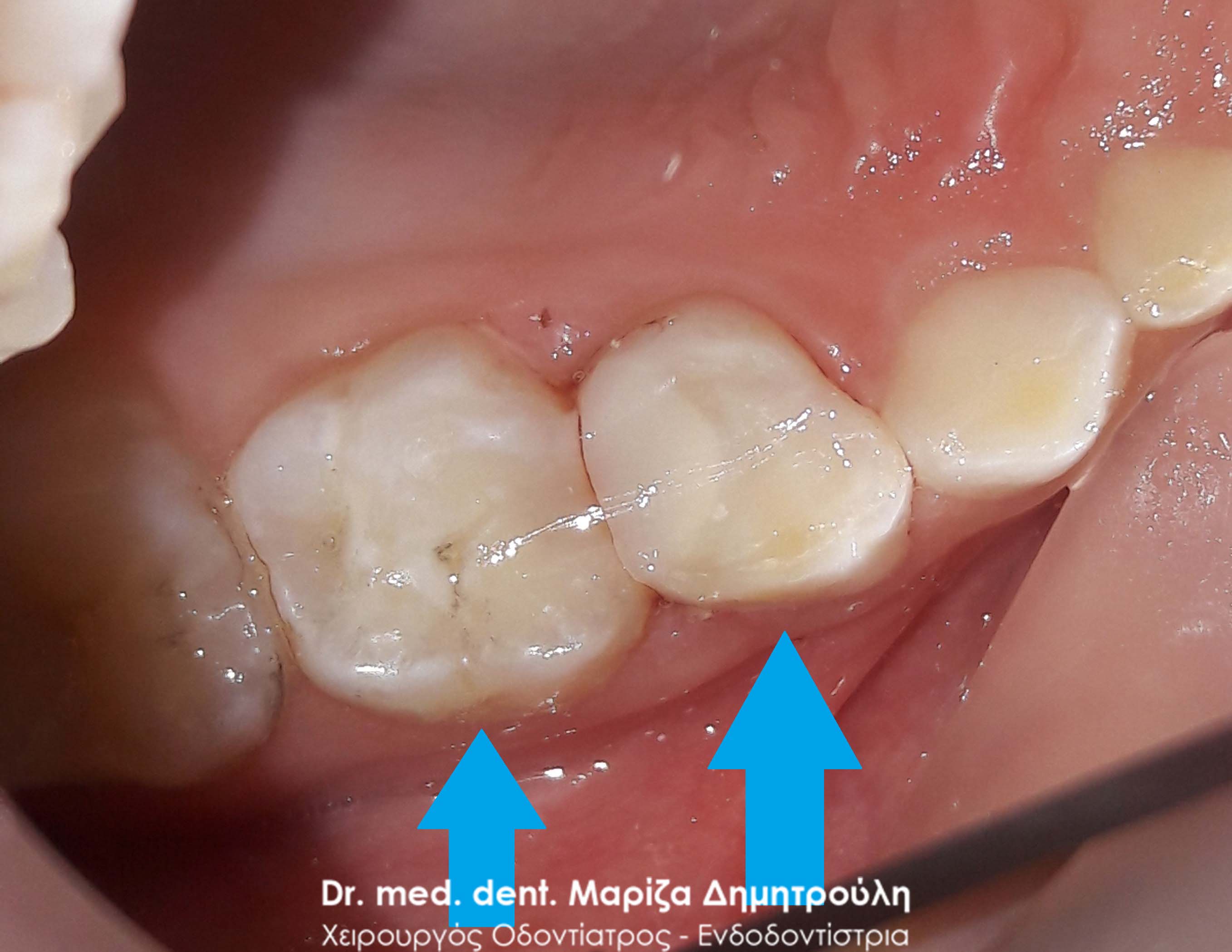
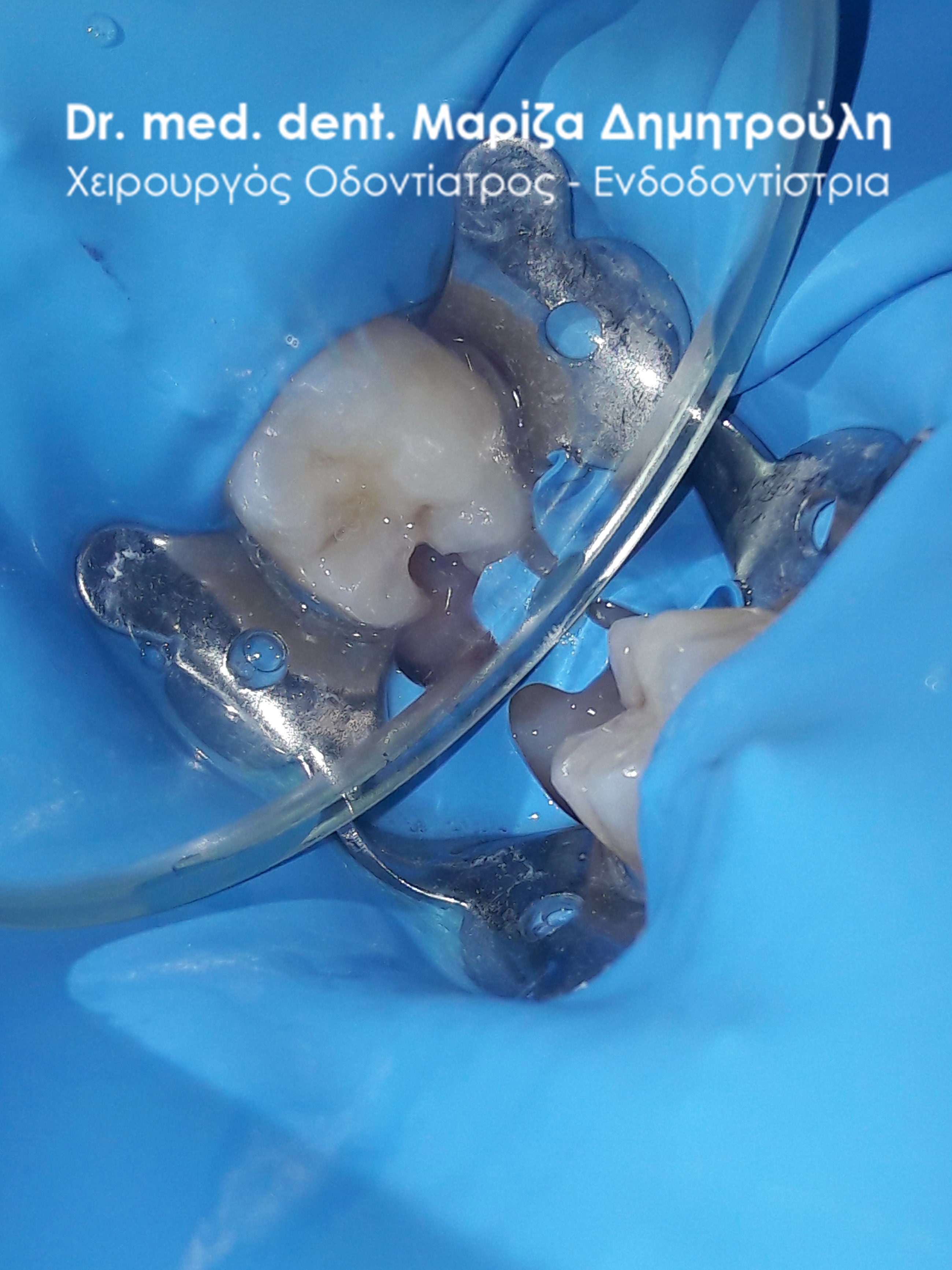
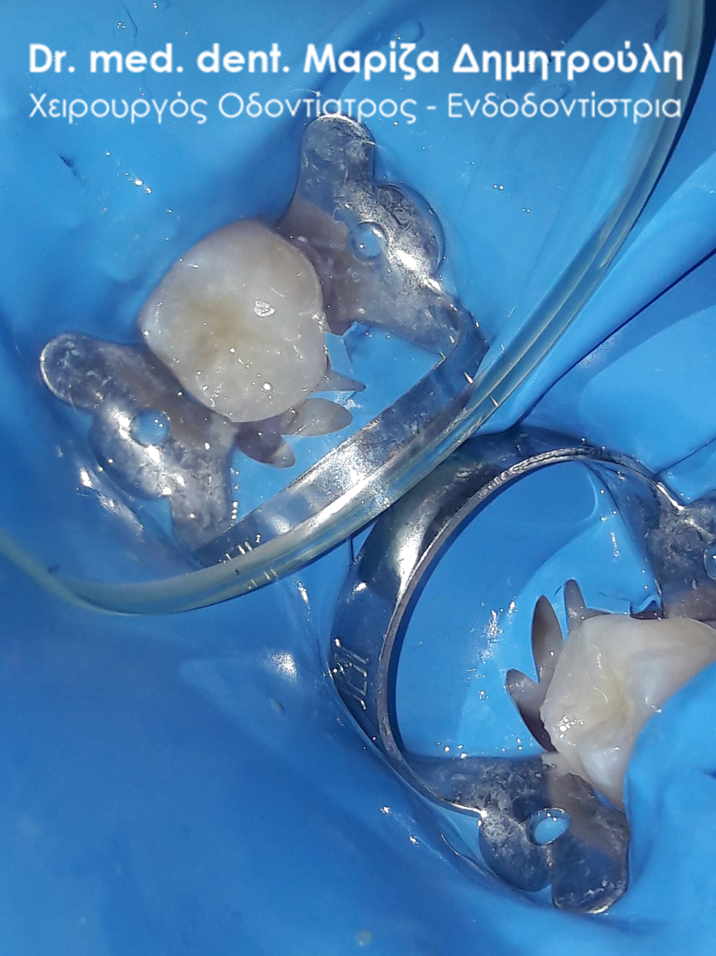
Incident – Extraction of a child tooth (double tooth / remaining baby tooth) and filling of a permanent tooth
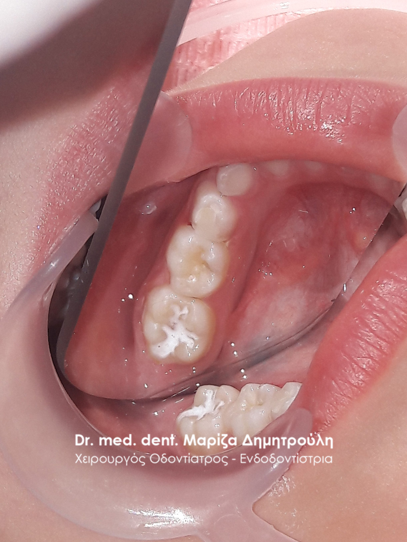
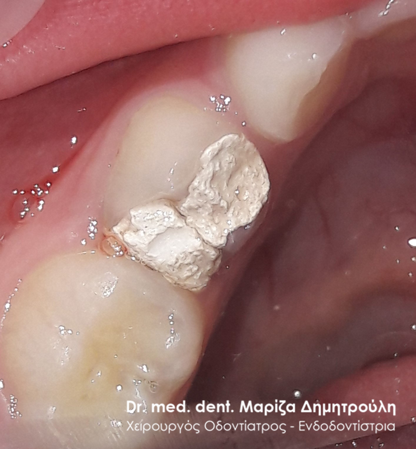
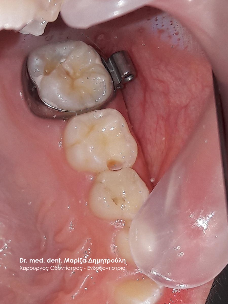
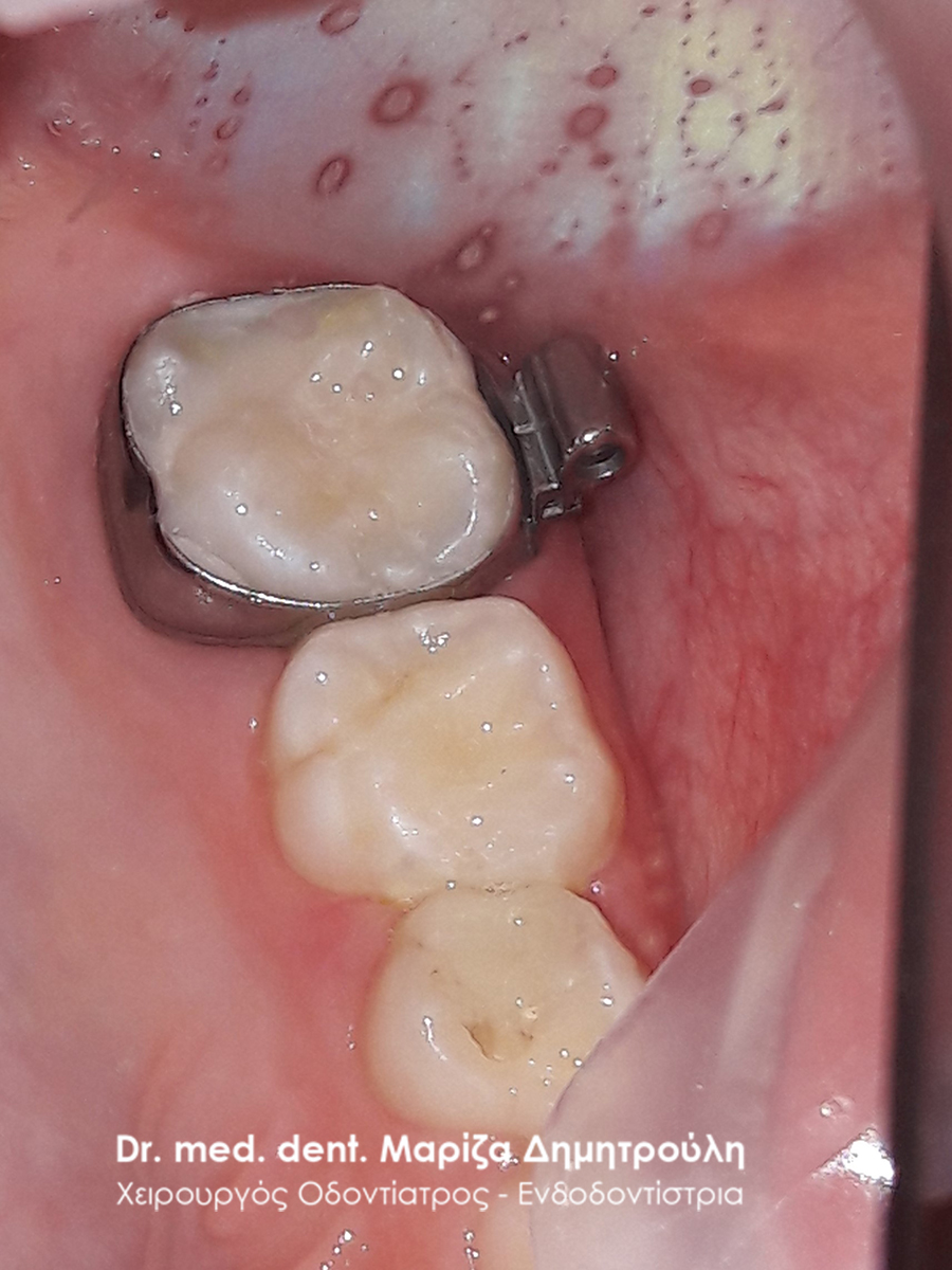
The little patient visited the clinic for the purpose of extracting the upper left first molar, which remained in the mouth even though the corresponding permanent tooth had erupted. In addition to the aforementioned, during the clinical examination, caries of the upper first permanent molar was found. After the administration of the local anesthesia, a white seal was performed on the permanent molar, followed by the extraction of the baby tooth.
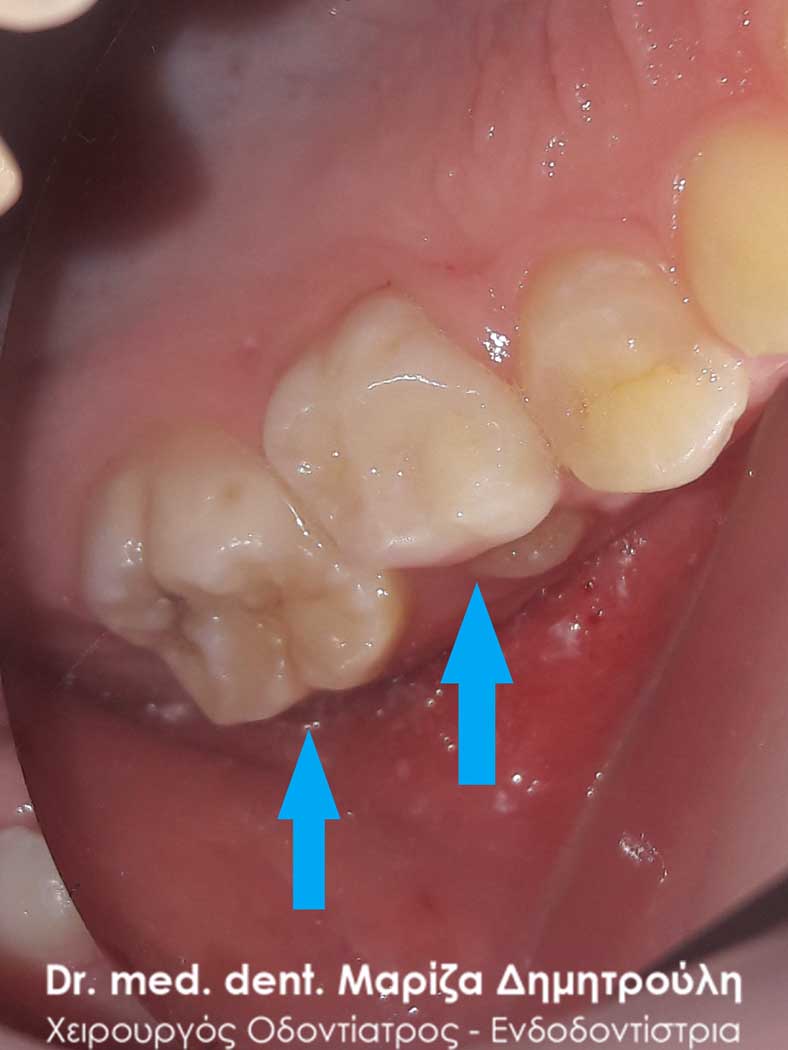
BEFORE
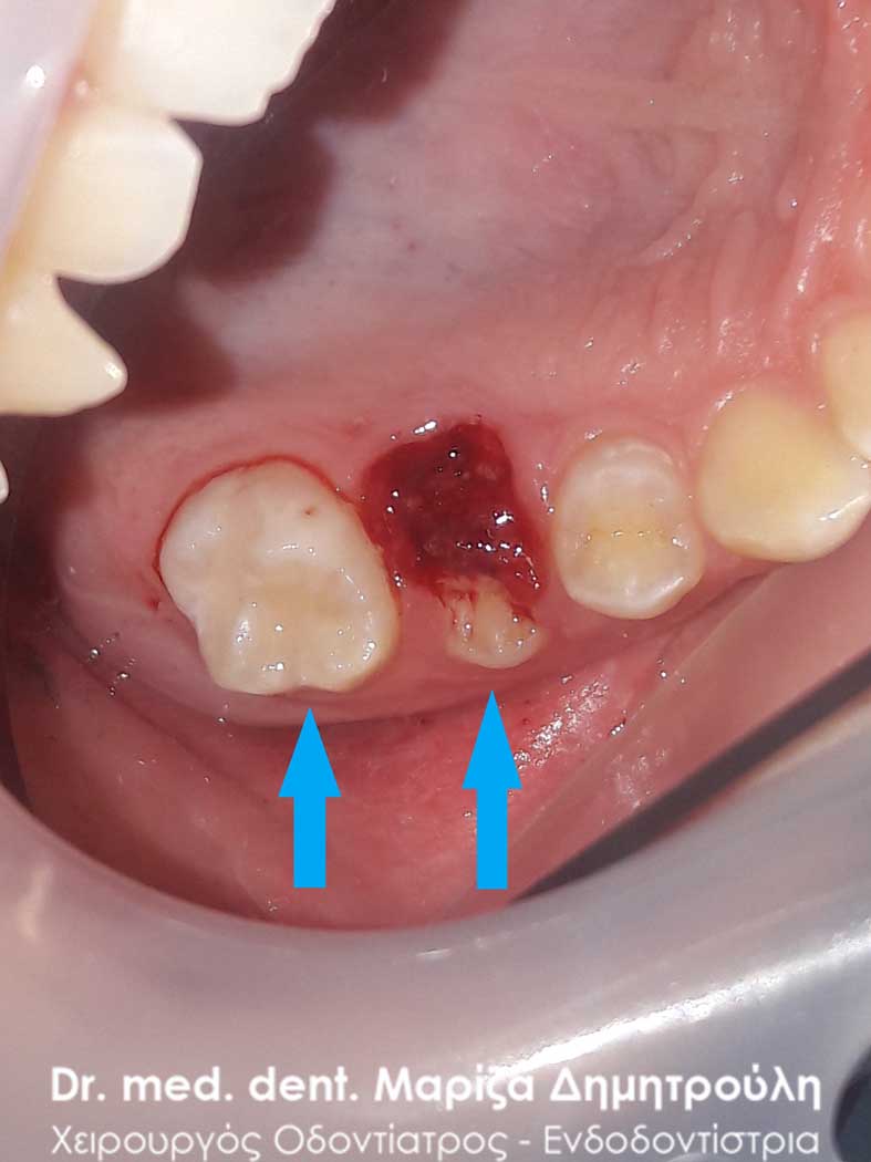
AFTER
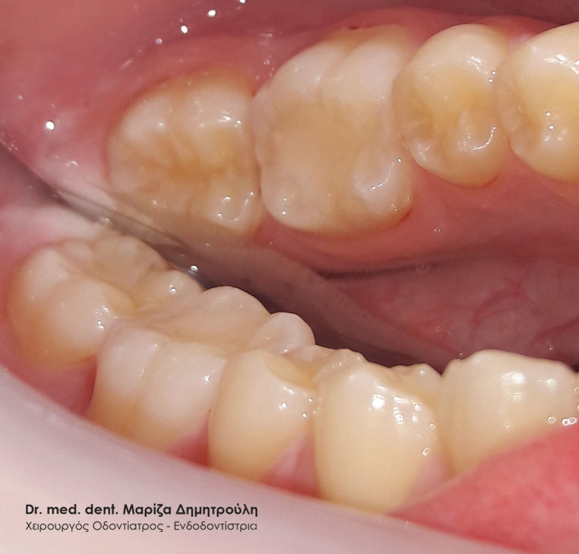
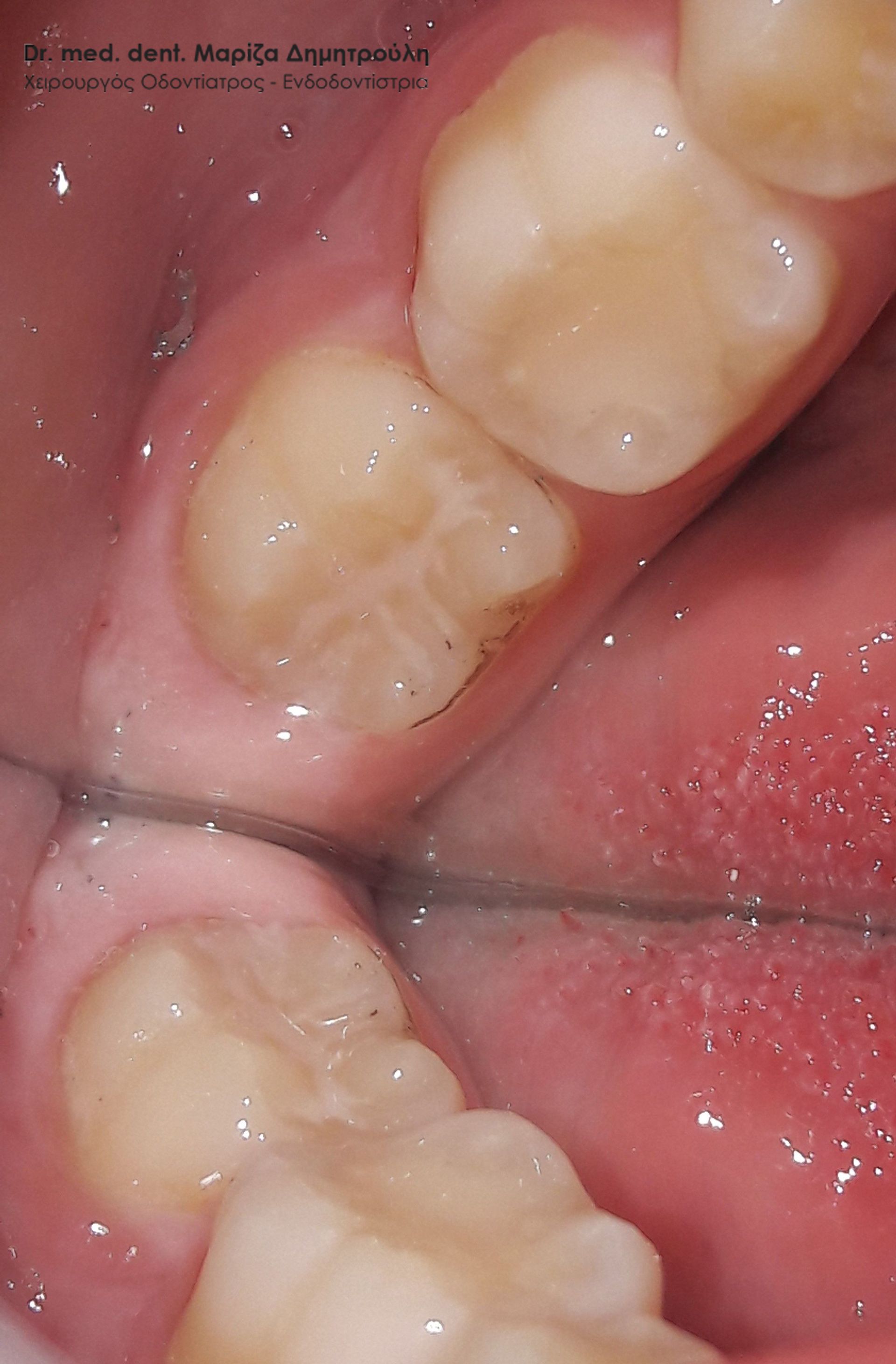
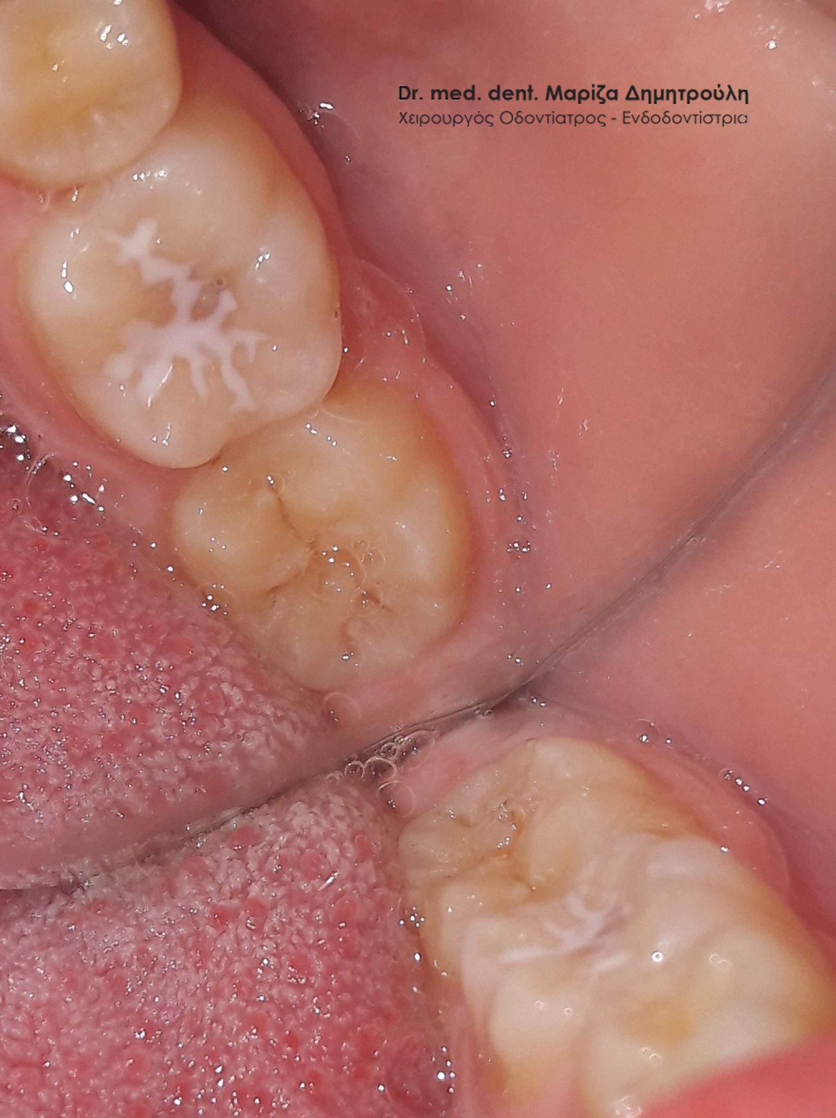
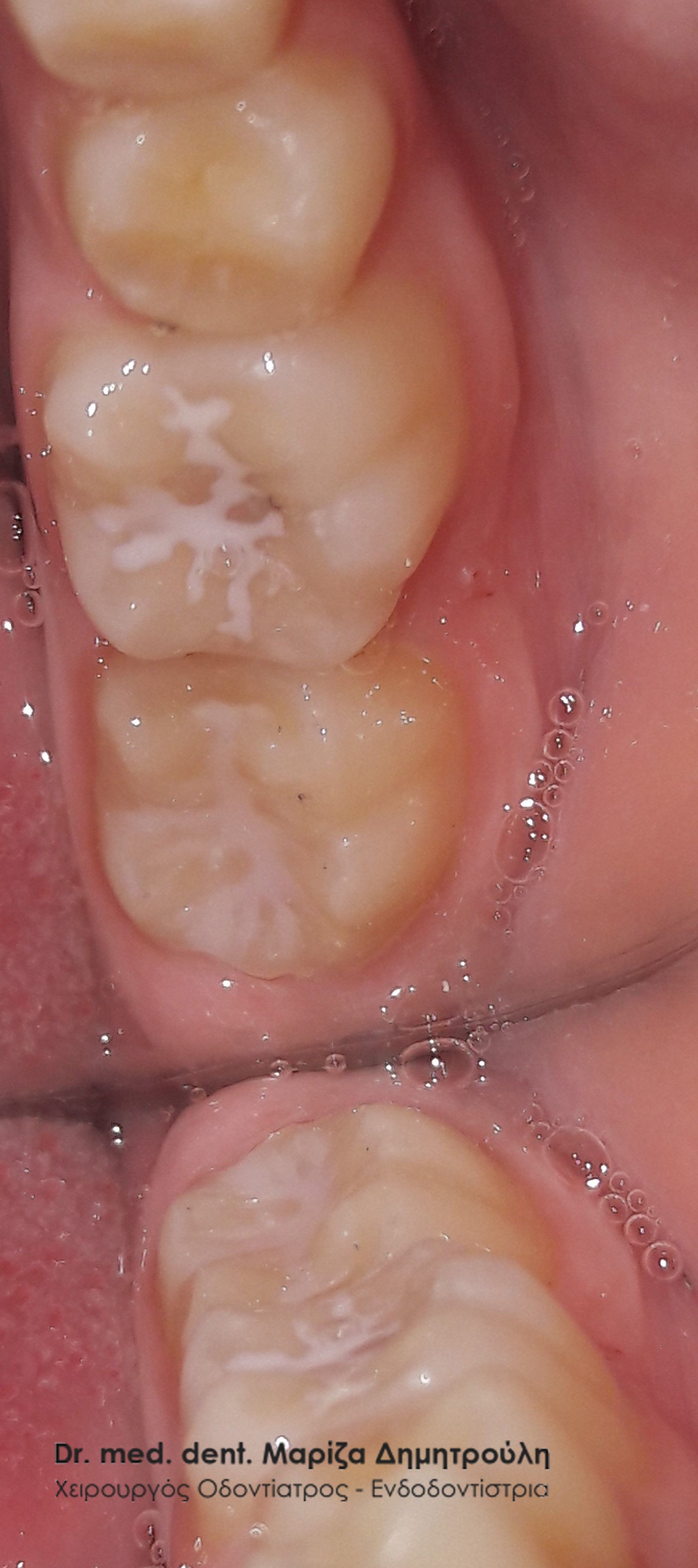
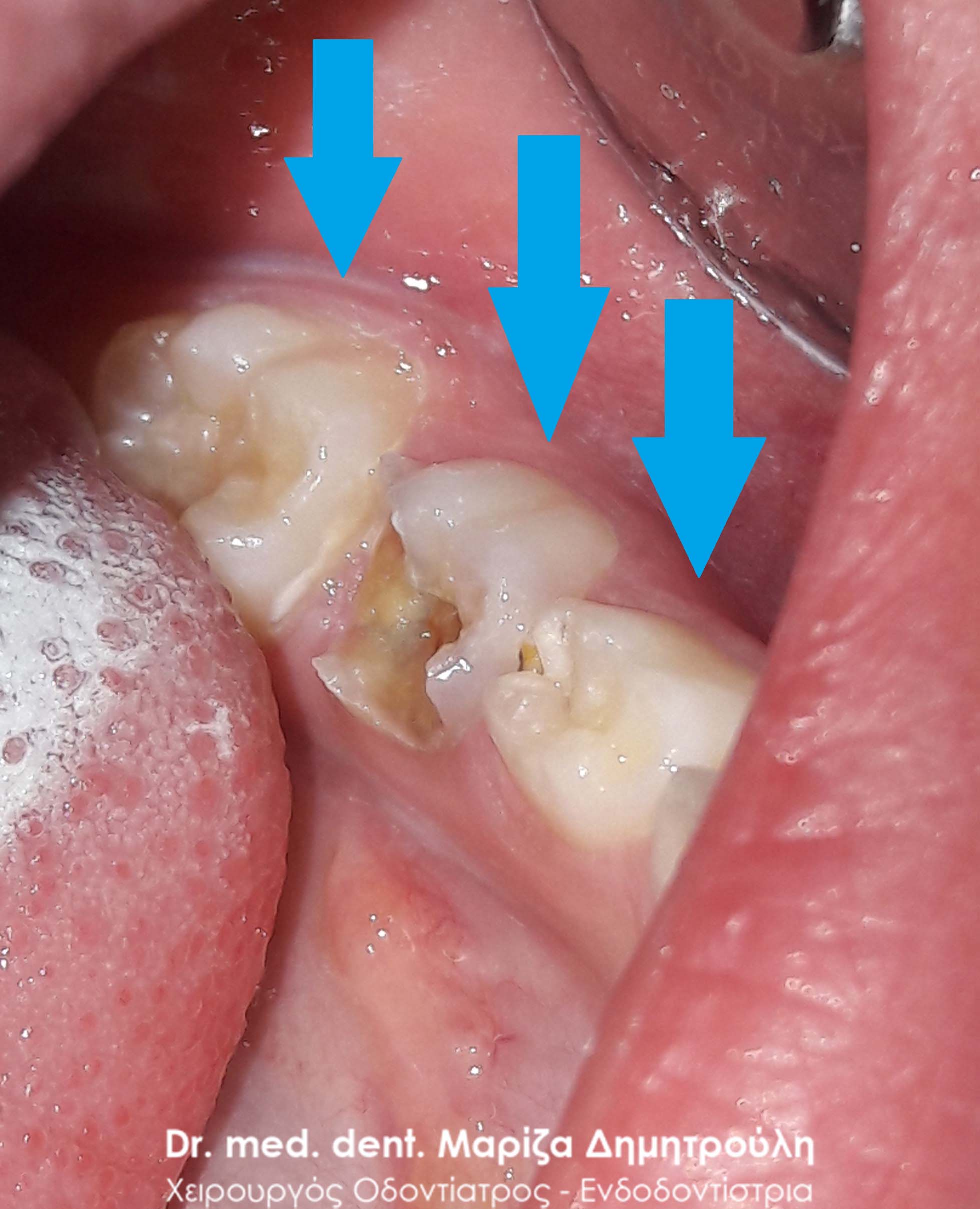
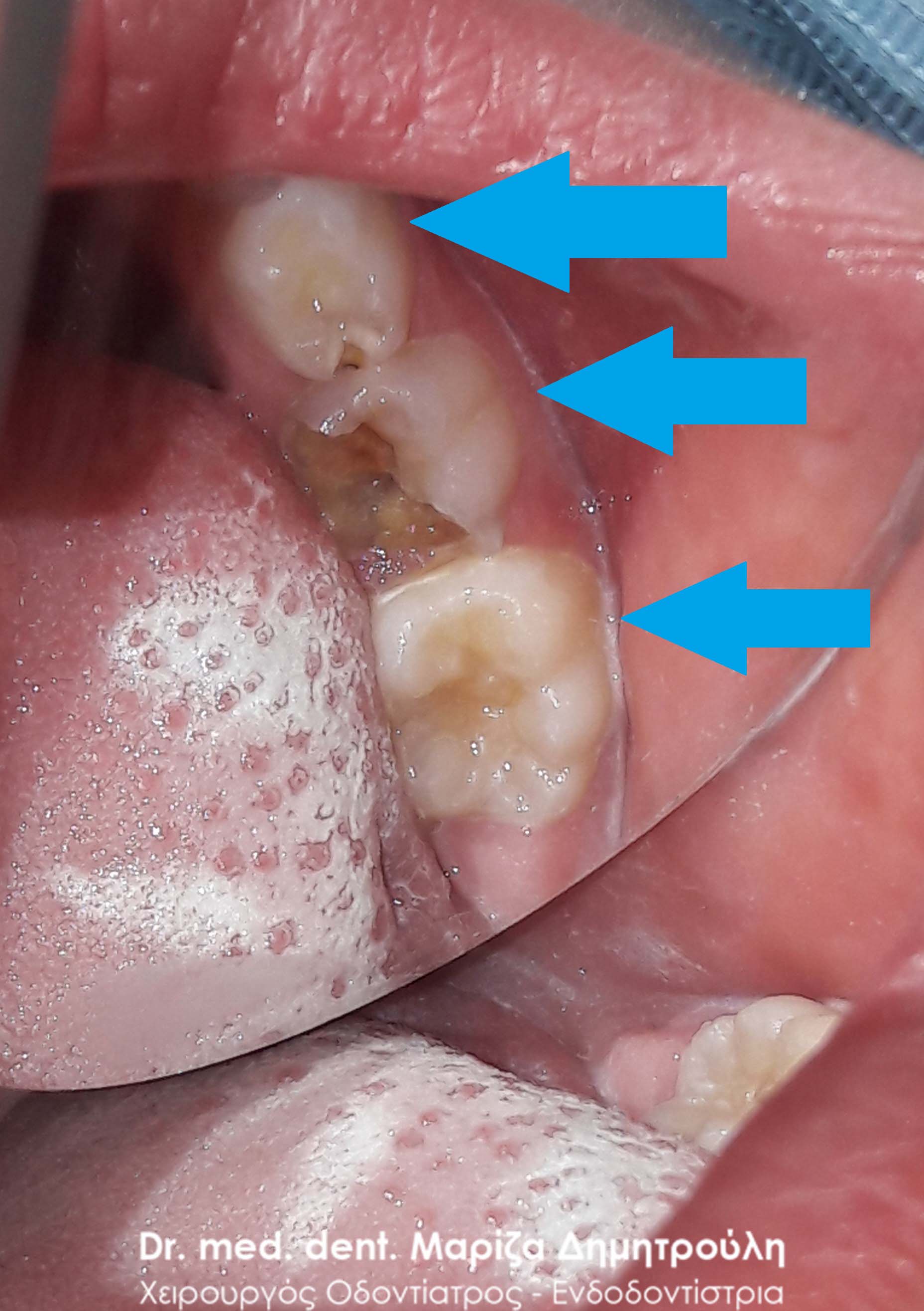
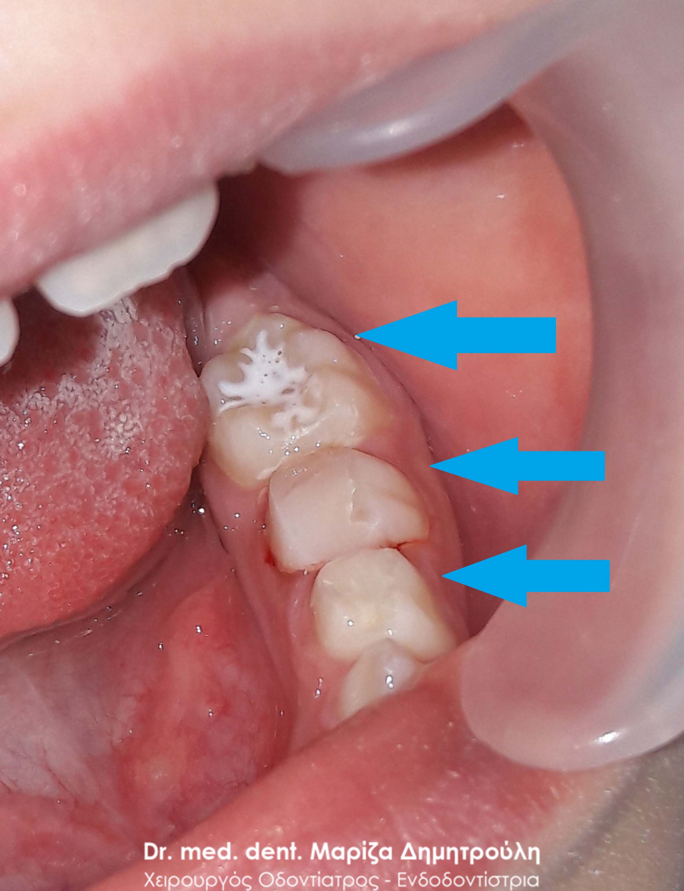
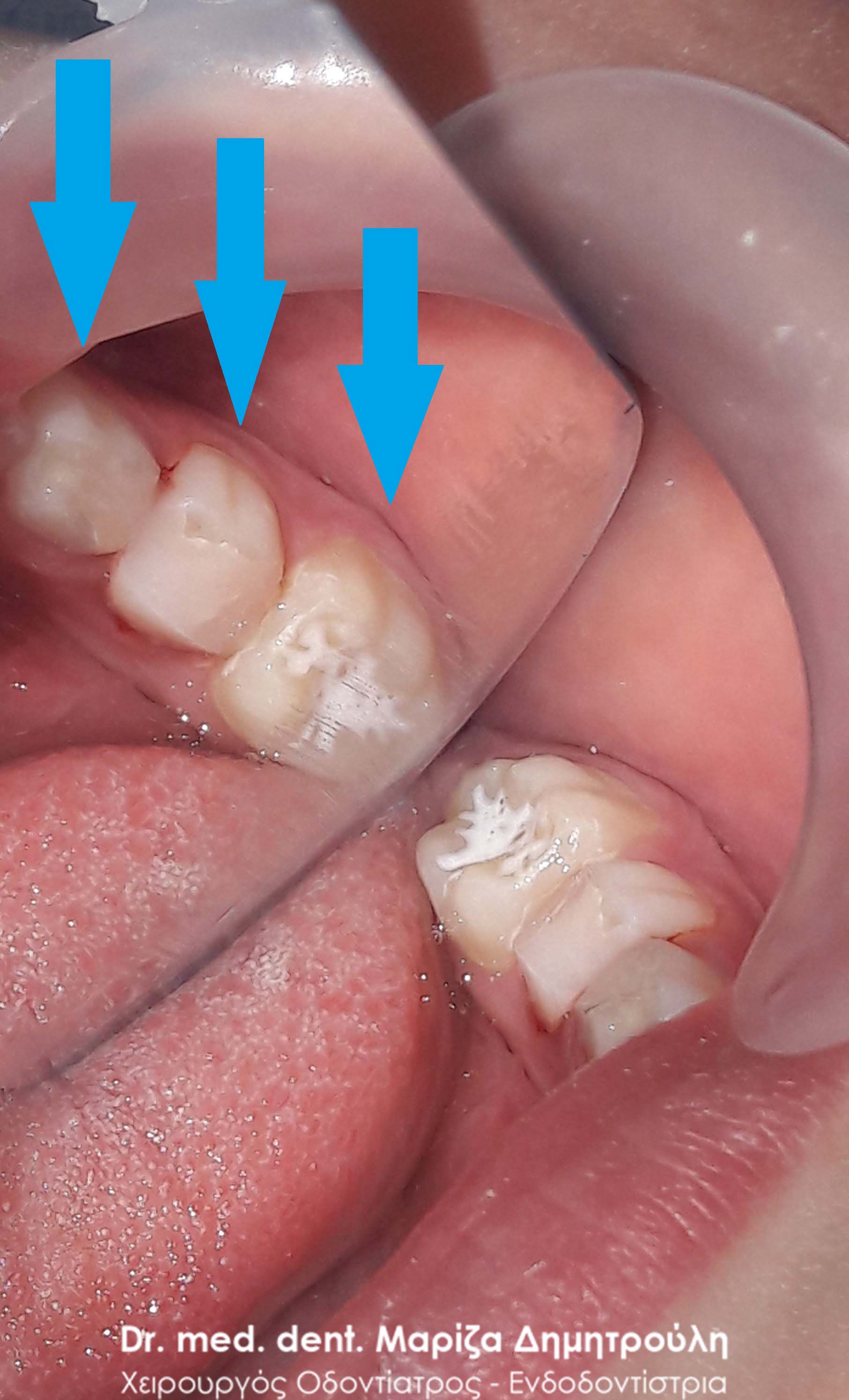
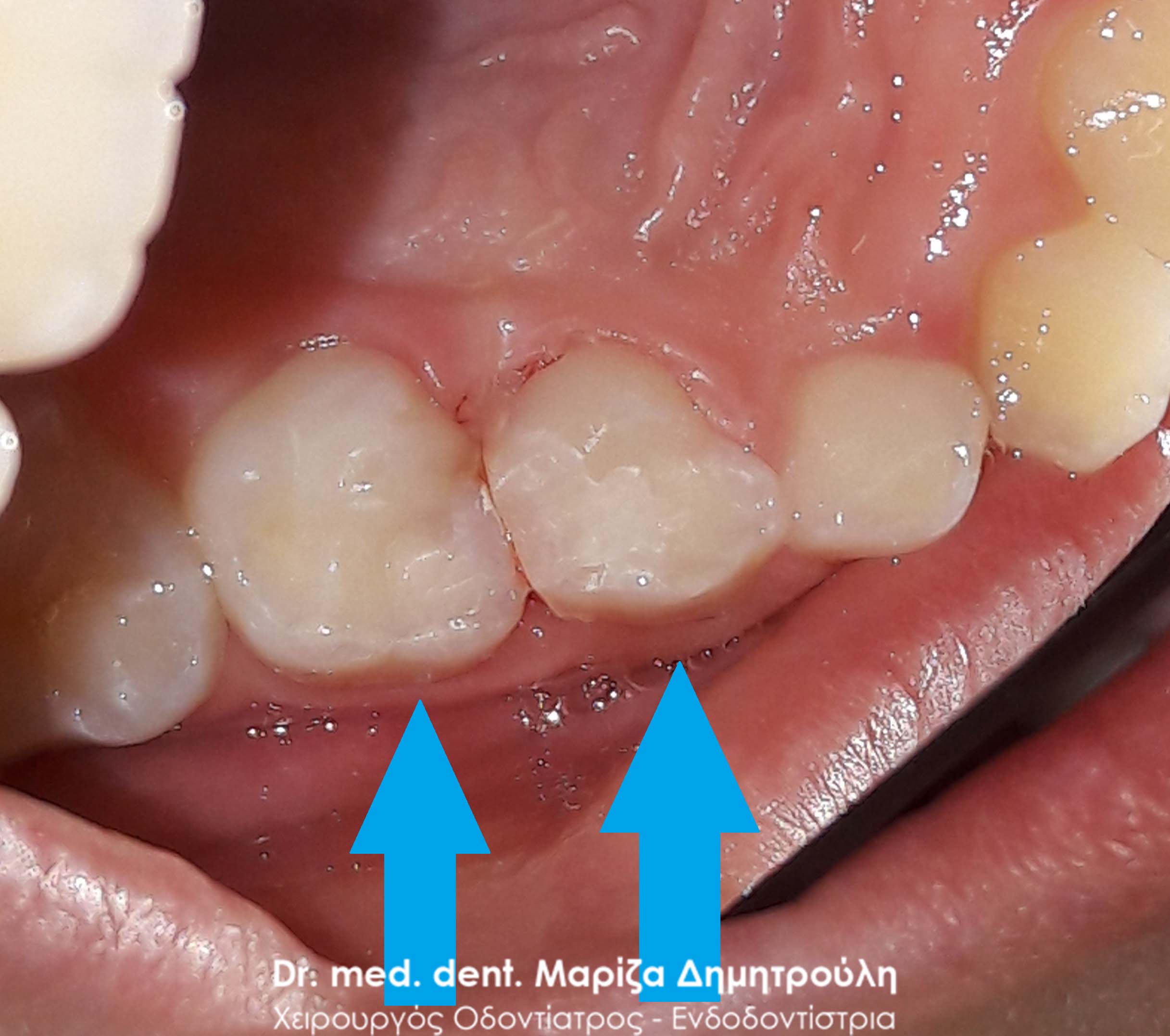
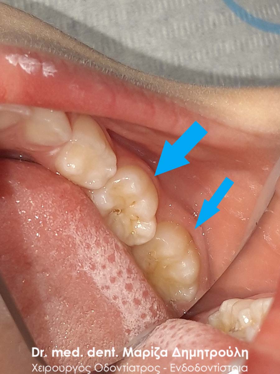
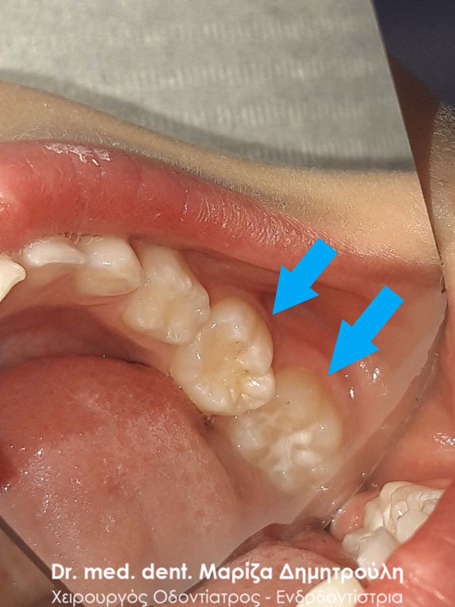
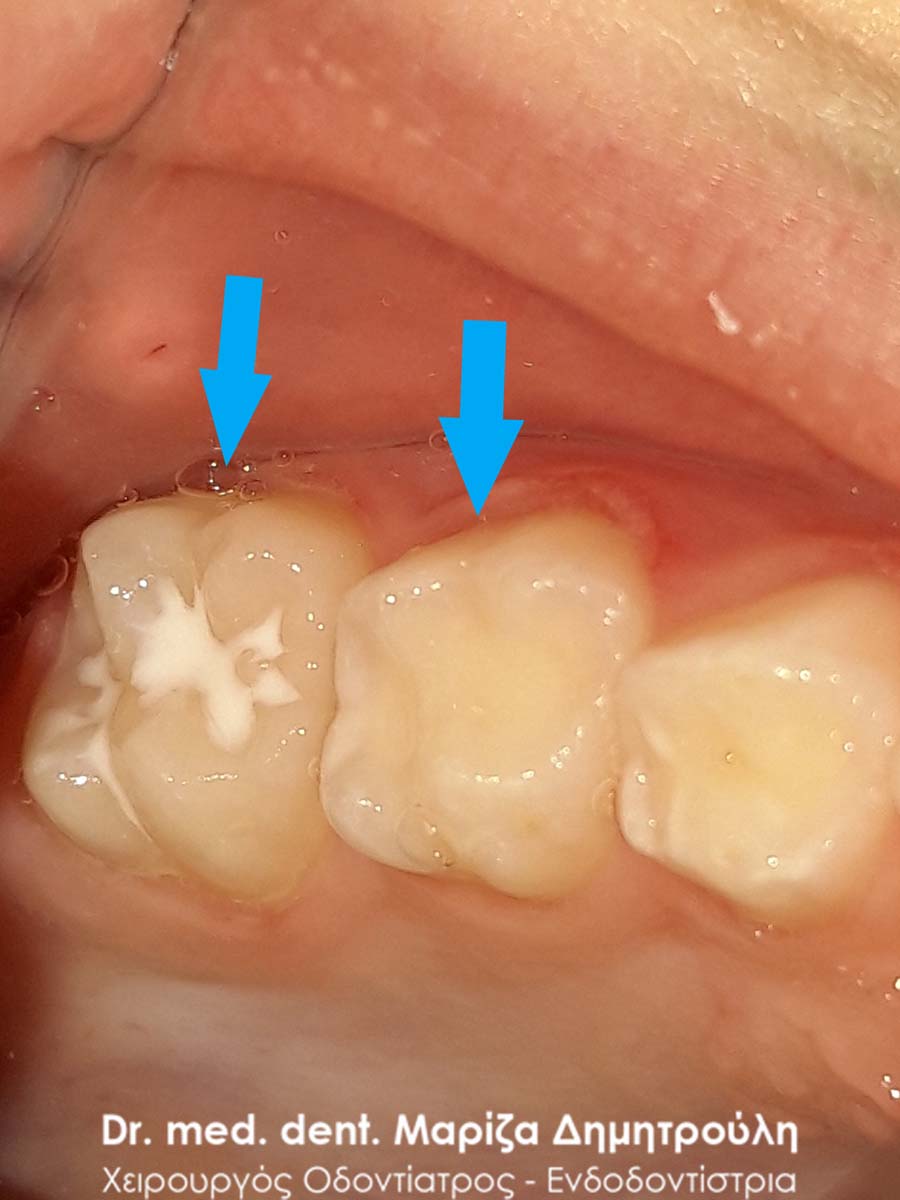
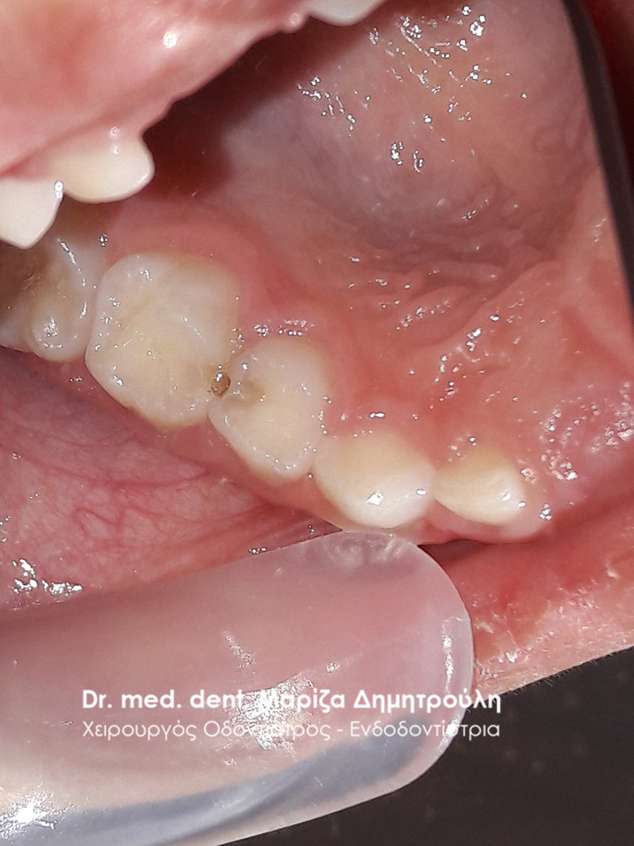
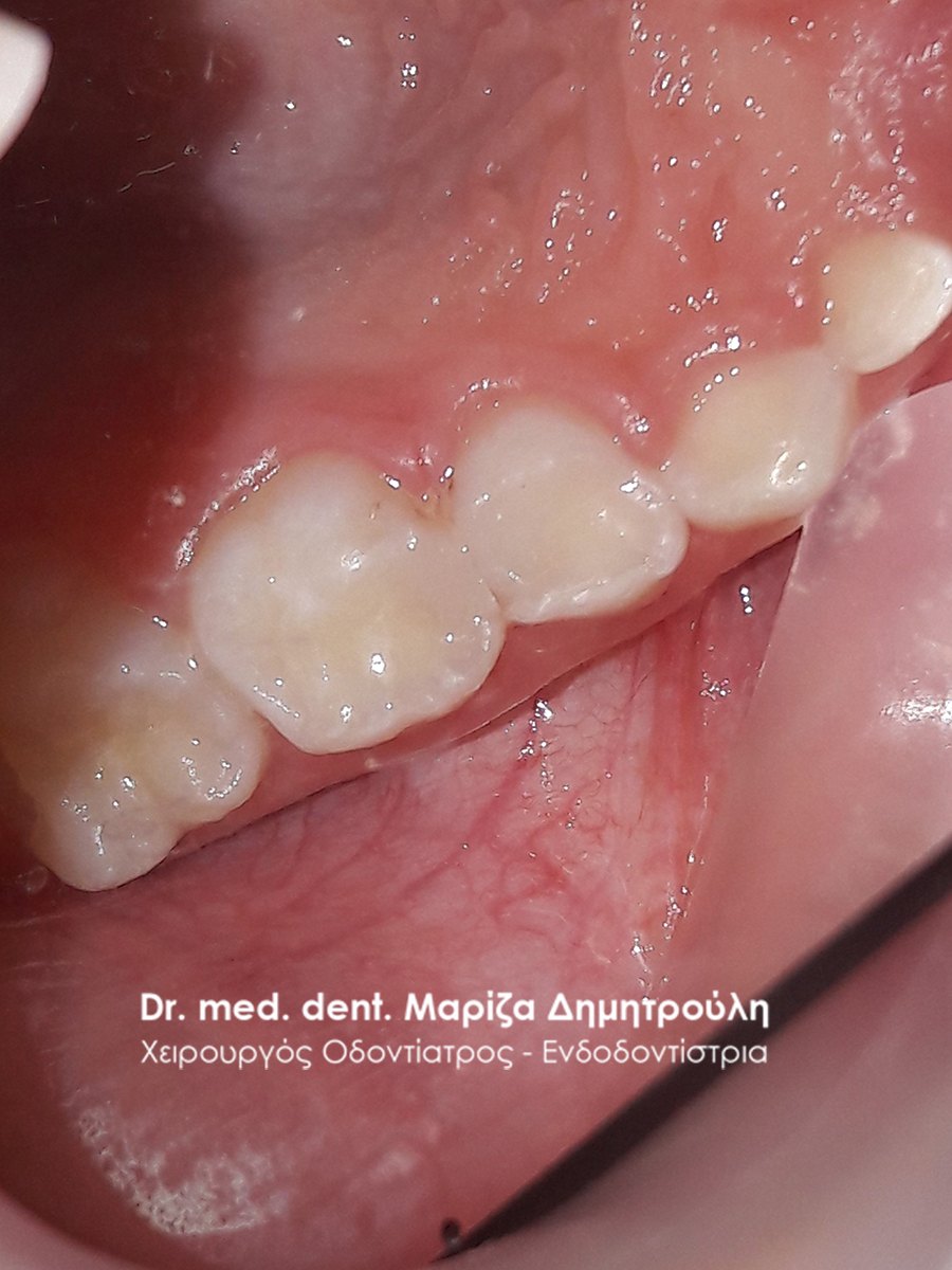
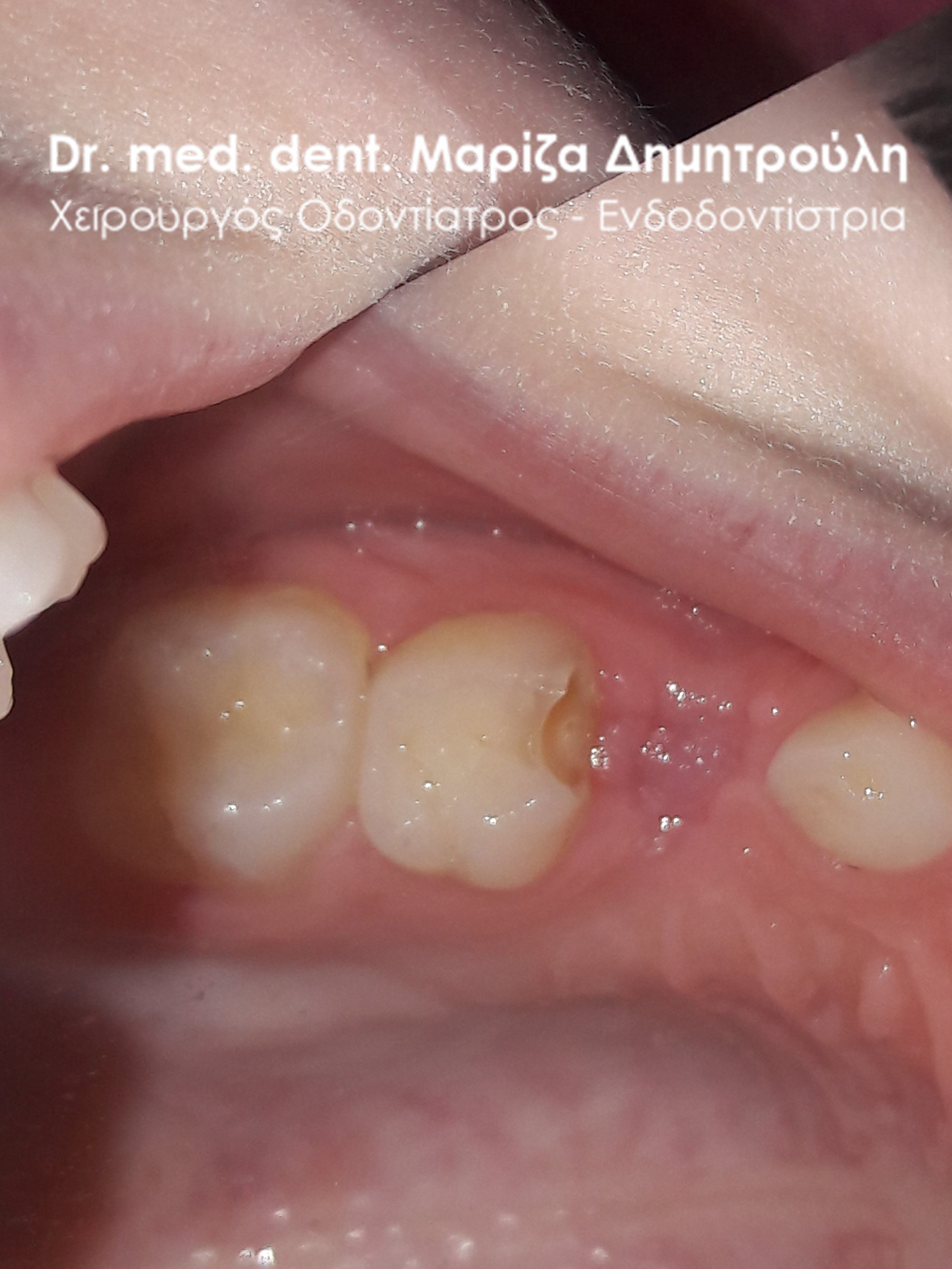
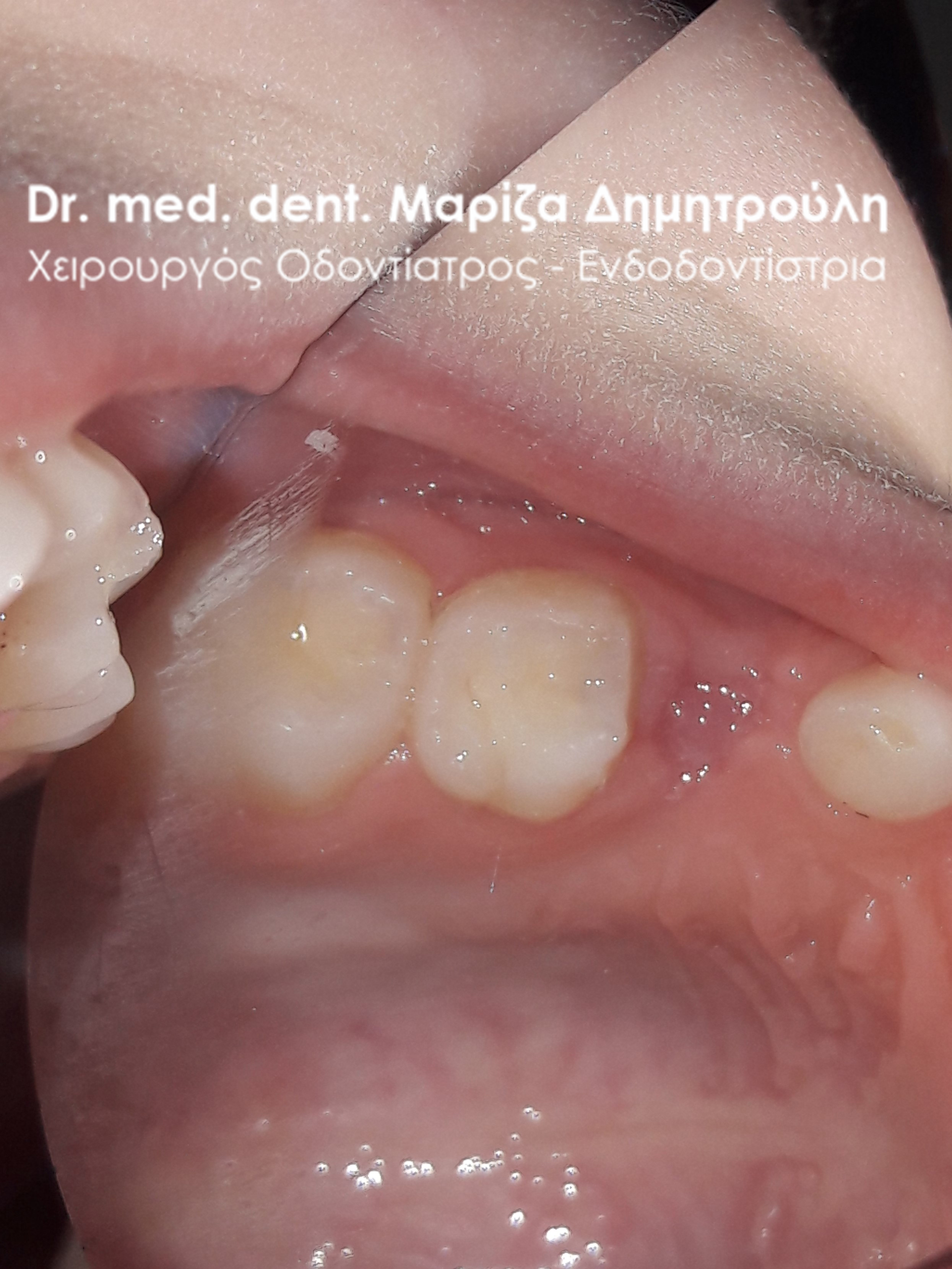
Incident – Extraction of a child tooth (double tooth / remaining baby tooth)
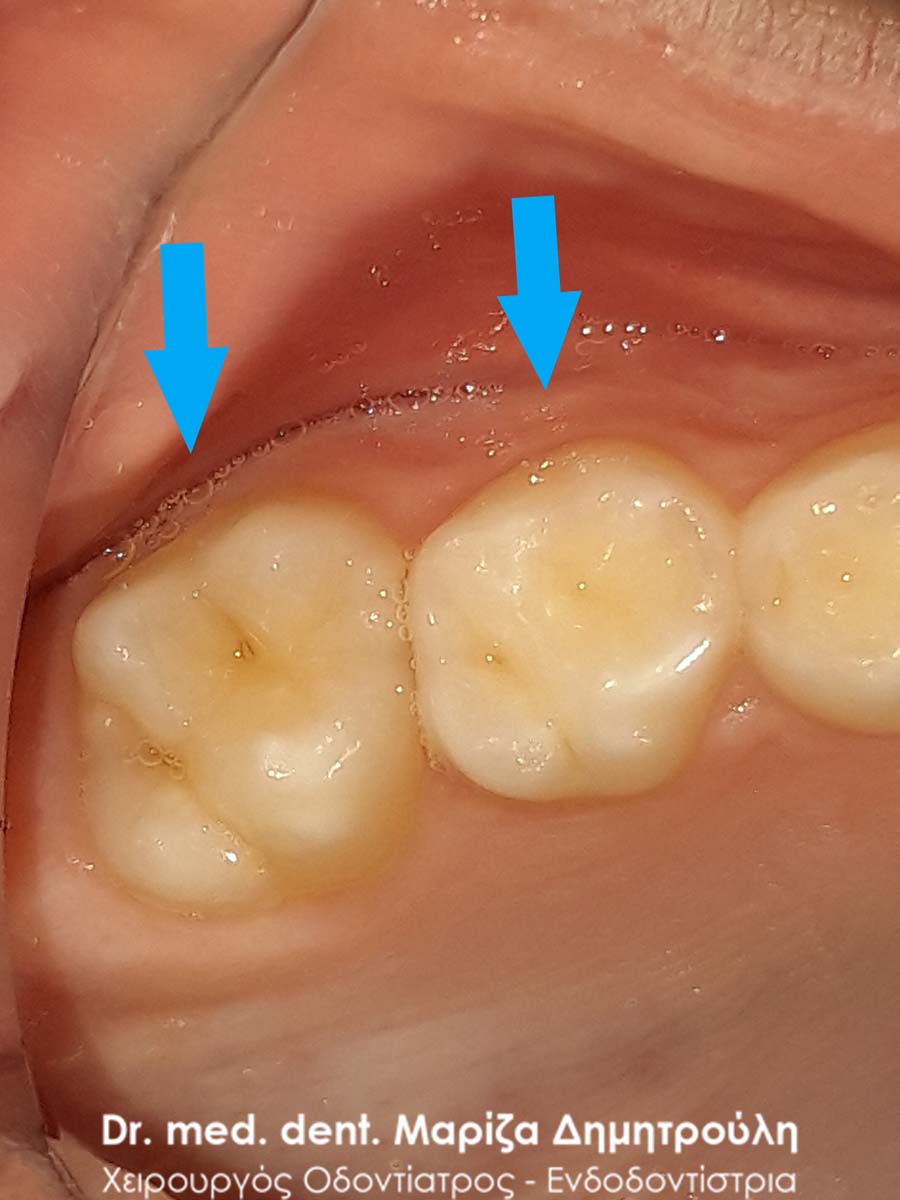
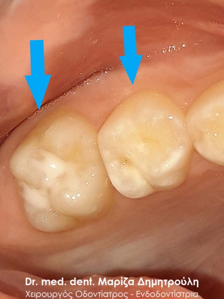
In the following two different cases, photographs are shown, showing the presence of the young lower central incisor even though the corresponding permanent tooth had already erupted in the mouth of the young patients. In this case, the child’s tooth is removed by the dentist, so that the permanent tooth, with the help of the tongue, takes its final position in the child’s mouth.
Incident – Supernumerary baby tooth
Many times, extra teeth appear in children’s mouths than the normal number of teeth (child or permanent). These supernumerary teeth are called incisors and appear more frequently in the upper jaw, usually between the upper central sectors. Morphologically, they are smaller than normal teeth and their presence can cause aesthetic as well as functional problems.
The appearance of supernumerary teeth has a genetic cause.
Regarding their treatment, it is recommended to remove them, so that the eruption of the adjacent teeth is not hindered.
Incident – Sealant / preventive covering of holes and cracks
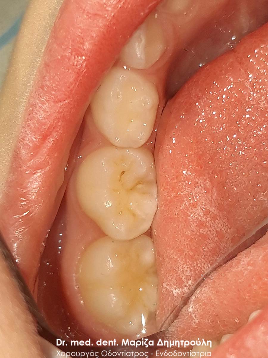
This young lady had a preventive masking of the occlusal surface of the permanent upper first molar, to protect the tooth from caries. Since the chewing surface of a tooth is covered with a sealant material, the risk of attack by caries-causing microbes is reduced, as fewer microbes and food residues accumulate on the chewing surface, while at the same time the child can brush more efficiently and easily. The lifespan of a sealant ranges from 2 – 5 years.
BEFORE
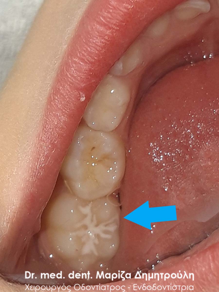
AFTER
Incident – Sealant / preventive covering of holes and cracks
In this particular little girl, as part of the annual teeth cleaning, the existence of a small incipient caries was found on the first left molar of the upper jaw. It was decided to cover the occlusal surface of the particular tooth as a preventive measure, since previously the caries, which was in its initial stage, was properly cleaned on the distal surface of the occlusal surface.

BEFORE
AFTER
Incident – Sealant / preventive covering of holes and cracks
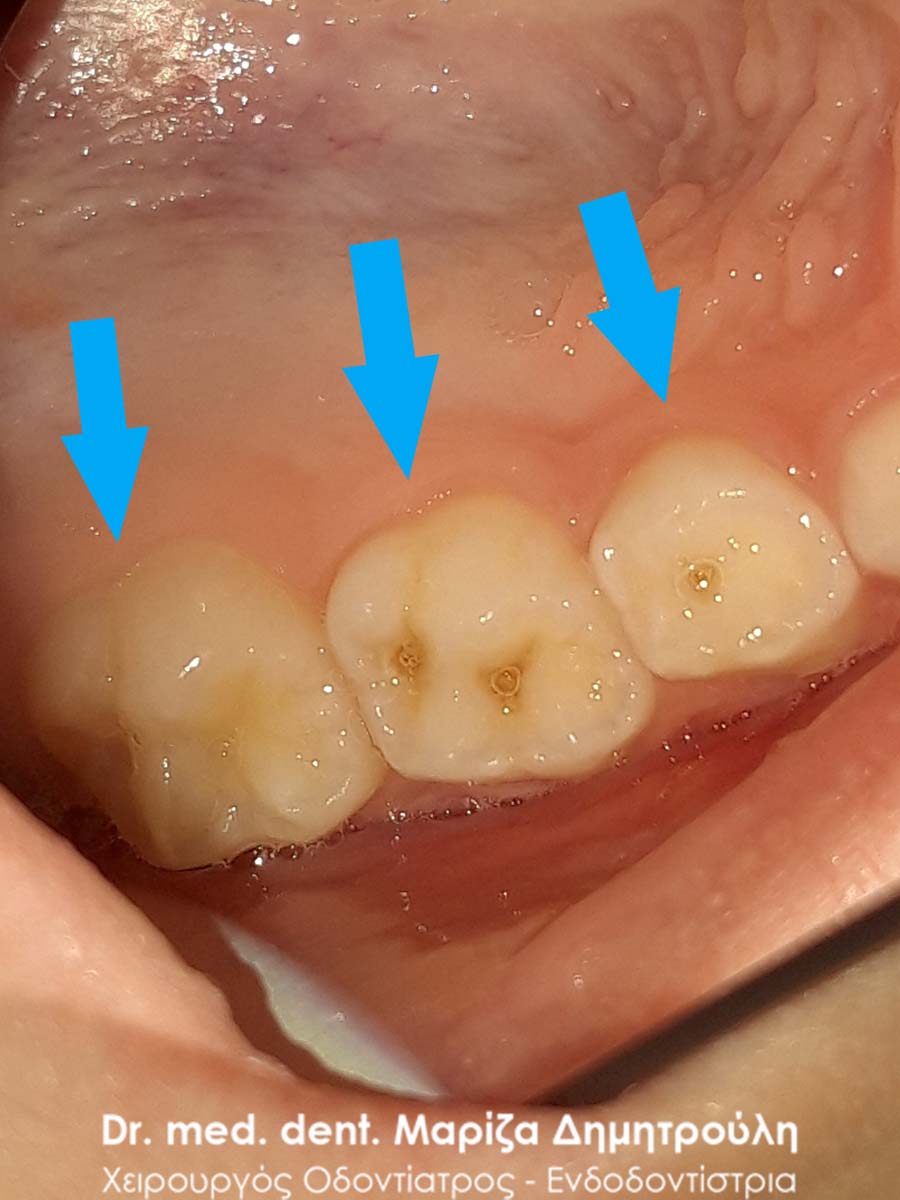
The little patient came in for her standard annual check-up. The preventive covering of the second permanent molars (sealant) was recommended, since the girl already had 4 sealed teeth and therefore there is caries activity in her mouth. With the preventive covering of the teeth, their chewing surfaces are protected from possible caries. Sealants are good to be applied to all the permanent molars of the teeth. Prevention is the best cure.
BEFORE
AFTER
BEFORE
AFTER
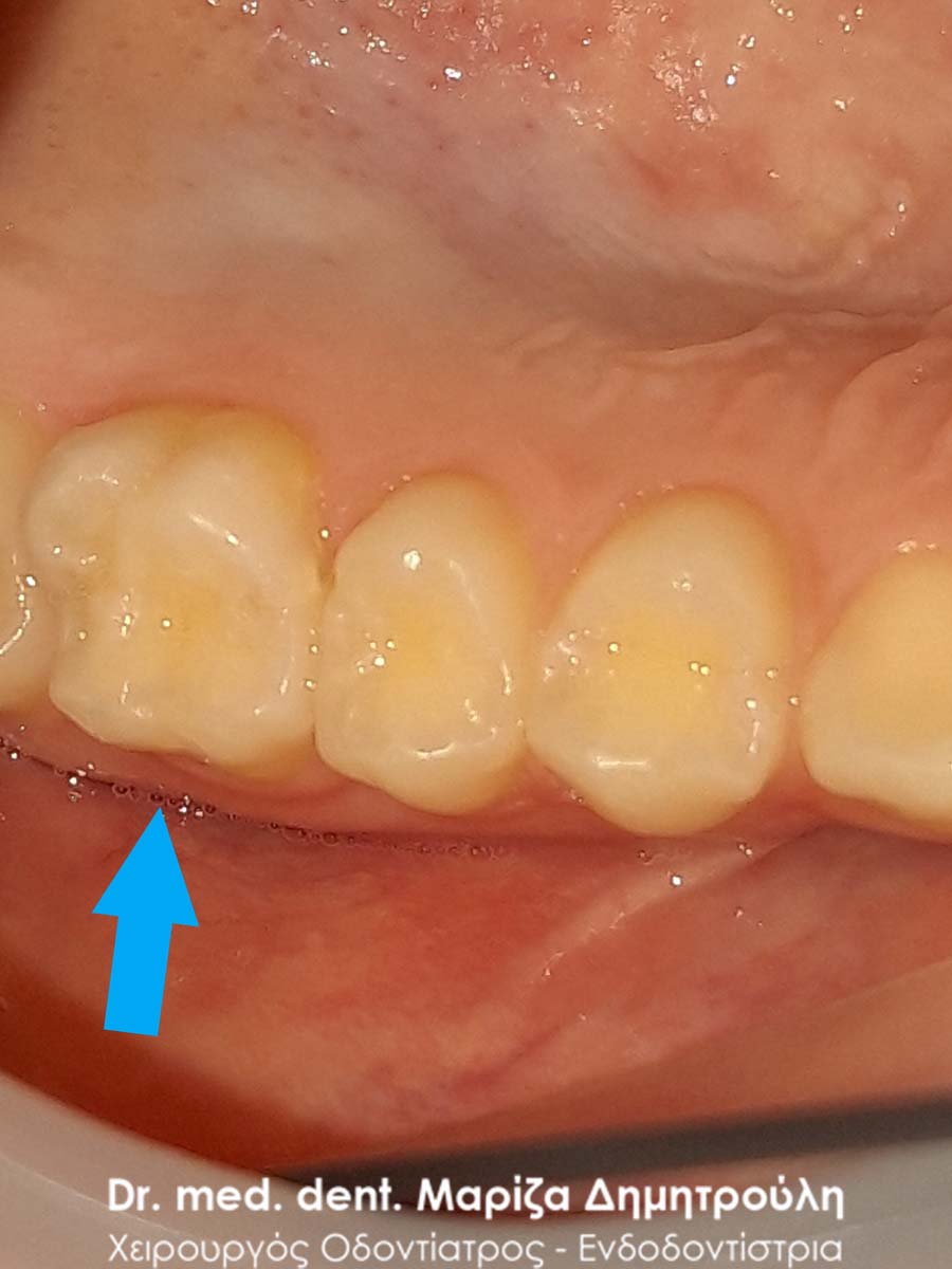
Incidents – Sealant / preventive covering of holes and cracks
As part of the annual preventive dental check-up, the little patient had preventive covering of holes and fissures (sealant) on the first left permanent molar of the lower jaw.
BEFORE
AFTER
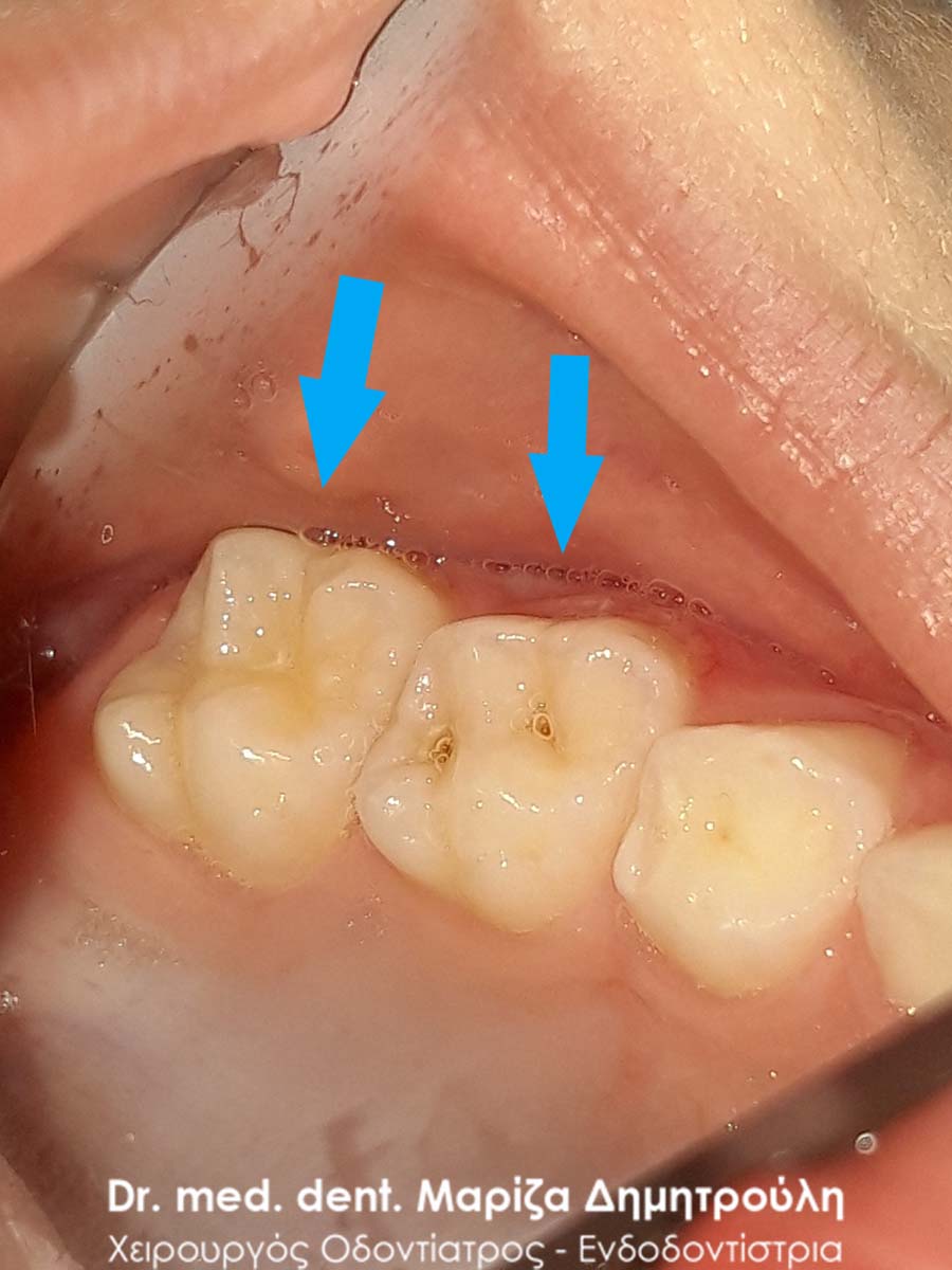
Incidents – Sealant / preventive covering of holes and cracks
Various cases of incidents are presented, in which preventive covering of the holes and cracks (sealant) was performed on the chewing surface of the permanent molars.
Incidents – Sealant / preventive covering of holes and cracks
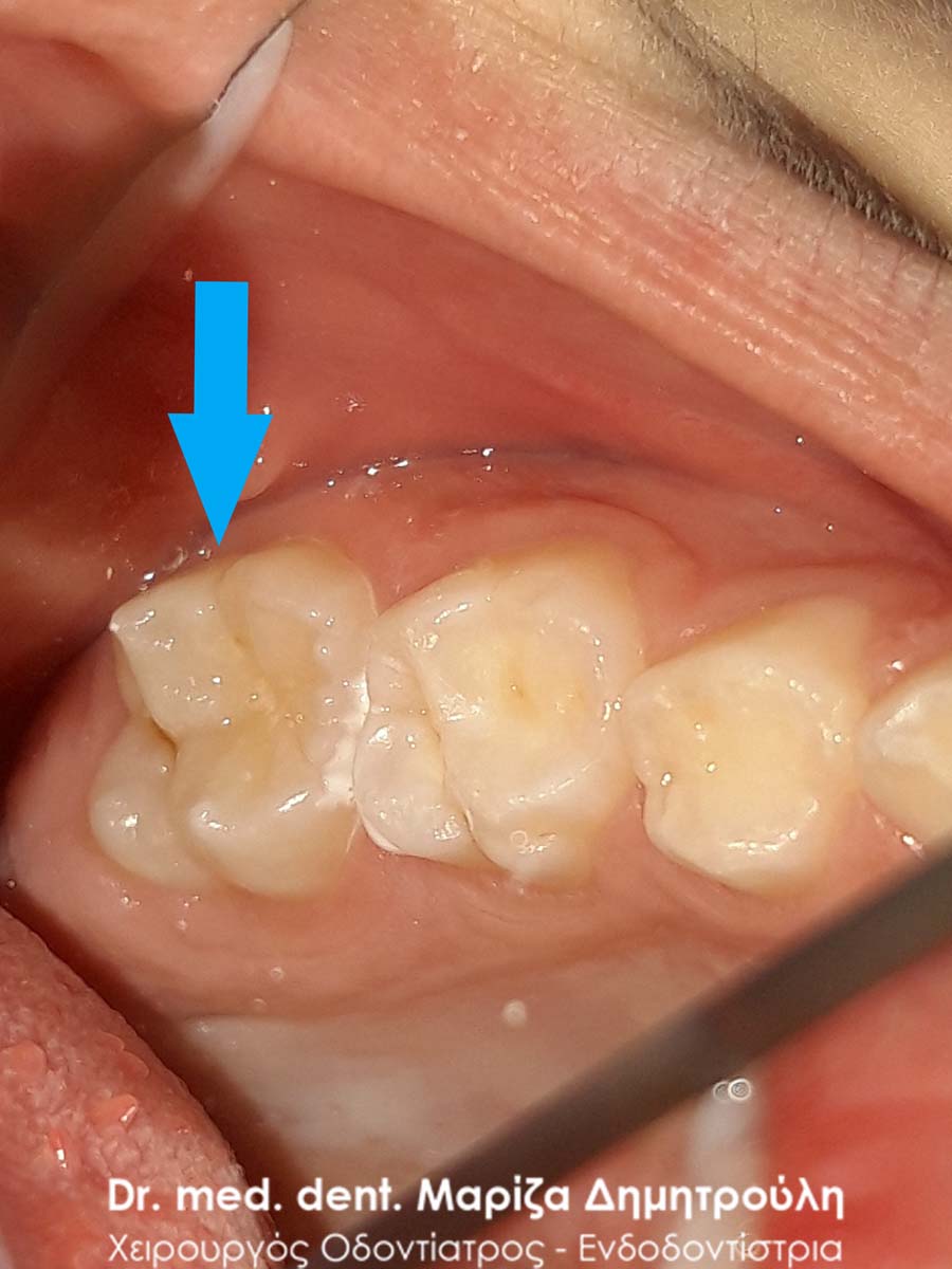
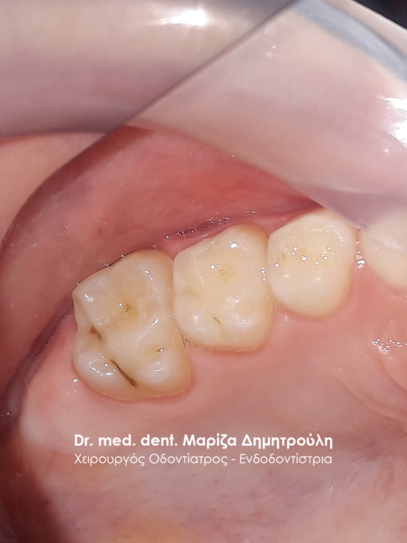
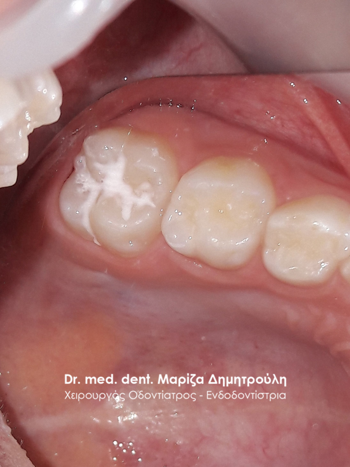
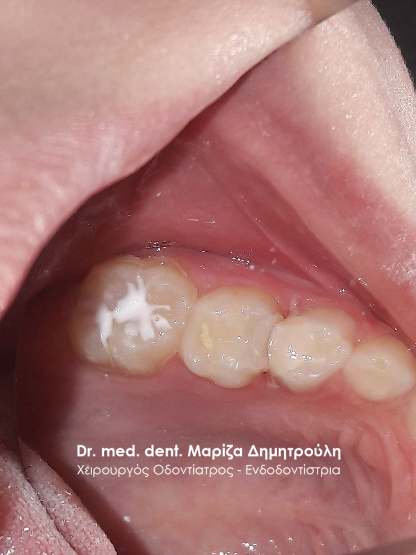
In the context of the standard dental examination, the existence of a small carious cavity in the first left permanent molar was established. It was decided to remove the surface incipient caries and preventive covering of holes and fissures (sealant) on the occlusal surface of the permanent upper left first molar.

Incidents – Sealant / preventive covering of holes and cracks
In this particular patient, removal of the surface incipient caries andpreventive covering of holes and fissures (sealant) on the masticatory surface of the upper left first molar was performed at the same time.
Incident – Sealing of a baby tooth and sealant / preventive covering of holes and fissures
During the clinical examination of this little boy, a small incipient caries was found in the first left permanent molar of the upper jaw, while in the second molar there was a small cavity that is not clearly visible in this particular photo. The incident was treated with preventive sealing of holes and fissures (sealant) on the occlusal surface of the first permanent molar and the creation of a white seal on the second molar.
Incident – Seals of new teeth and sealant / preventive covering of holes and fissures
In this specific case, 2 white fillings were performed on the last 2 posterior molars and at the same time preventive covering of holes and fissures (sealant) on the occlusal surface of the first permanent molar. The photo of the original state of the teeth is missing.
Incident – Seals of new teeth and sealant / preventive covering of holes and fissures
The patient had pain when chewing on the right side of the lower jaw. After the clinical examination it was established the existence of 2 carious cavities in the 2 rear baby teeth. 2 white fillings were performed on the carious teeth. For preventive reasons, the preventive covering of holes and fissures (sealant) was placed on the first lower permanent molar.
BEFORE
AFTER
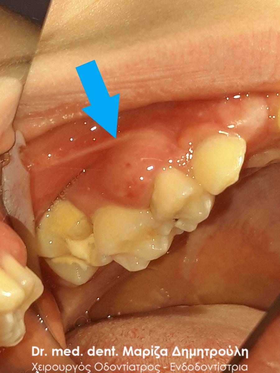
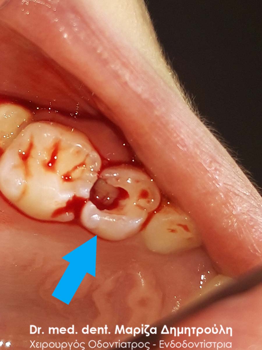
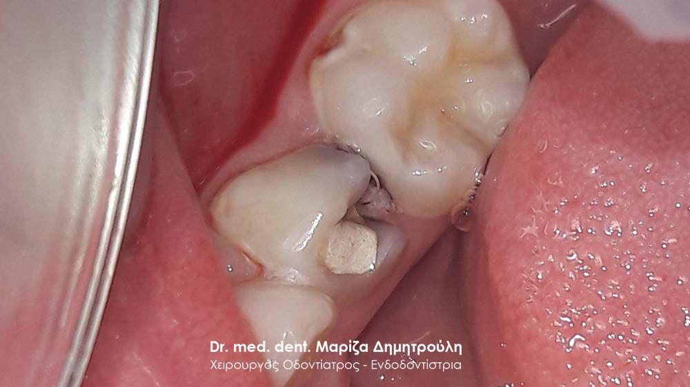
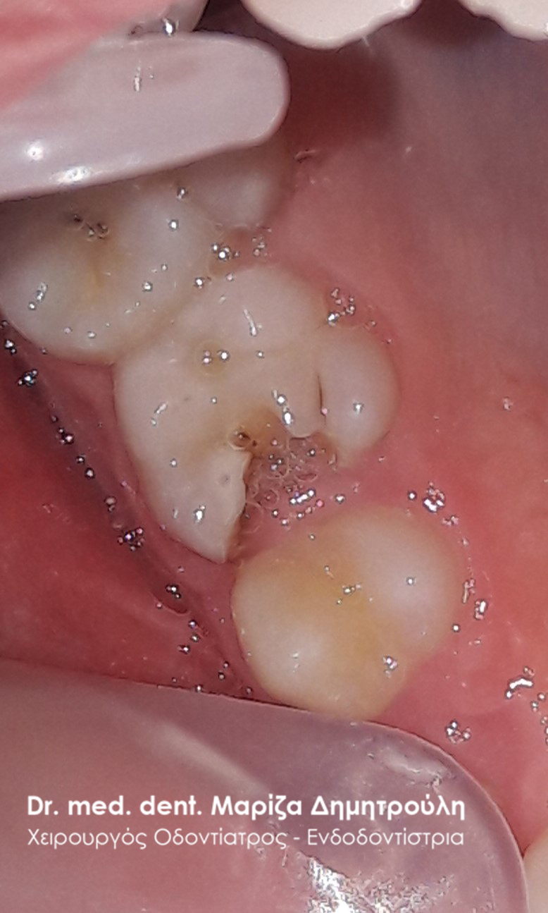
Incident – Pus in a child’s tooth
The little patient complained to his parents that he had a toothache on the left side of his upper jaw. The clinical examination revealed the accumulation of pus in the first molar of the upper jaw, which was evidenced by the swelling in that area. This tooth had an old white filling, which had re-edentified. In this particular case, the tooth could not be kept in the child’s mouth as during the tooth’s derailment, a large discharge of pus was observed and the bleeding from the inside of the tooth could not be stopped.
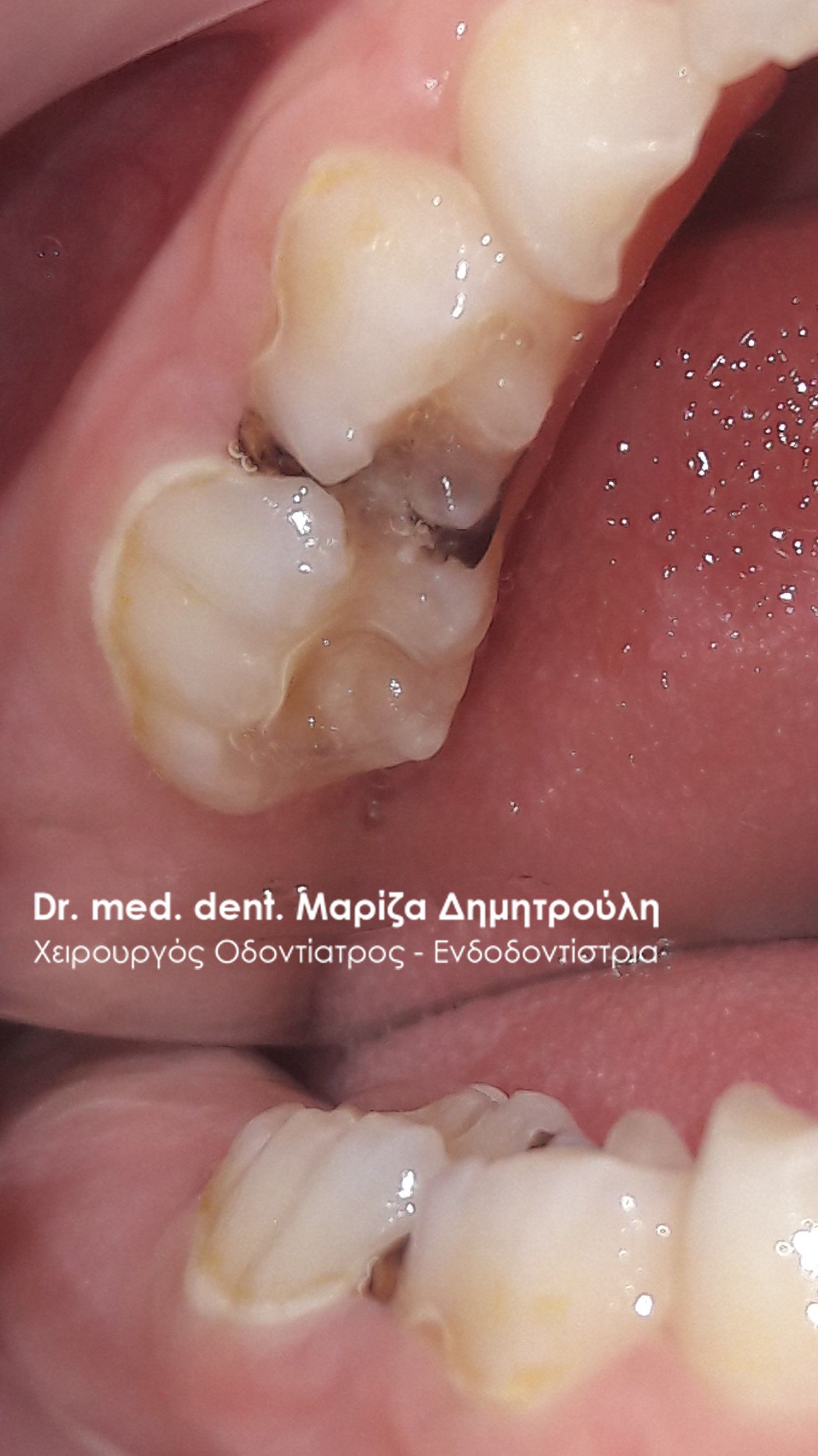
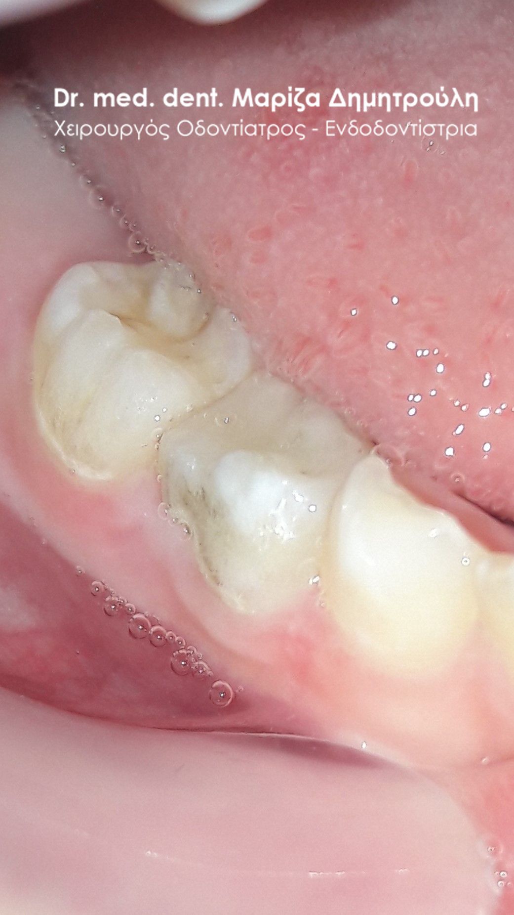
Incident – Children’s dental fillings
The patient had black amalgam fillings, which in the last week caused severe pain symptoms. After administration of local anesthesia and removal of the black fillings, a pulpotomy (due to deep caries) and restoration with a white composite resin filling was performed on one young tooth. A new white filling was placed on the last new tooth without the need for a pulpotomy.
BEFORE
AFTER
Incident – Children’s dental fillings
The patient complains of pain during chewing on the right side of the upper jaw. Clinical examination revealed two small carious cavities, which were restored with white resin filling.
BEFORE

AFTER
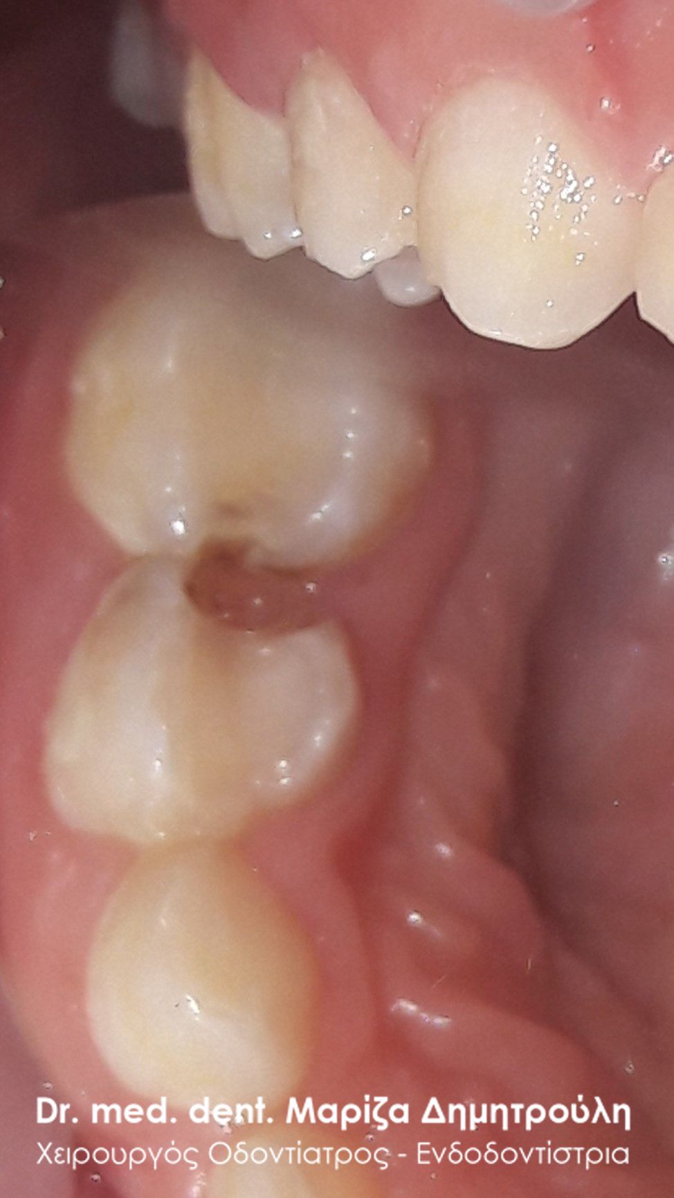
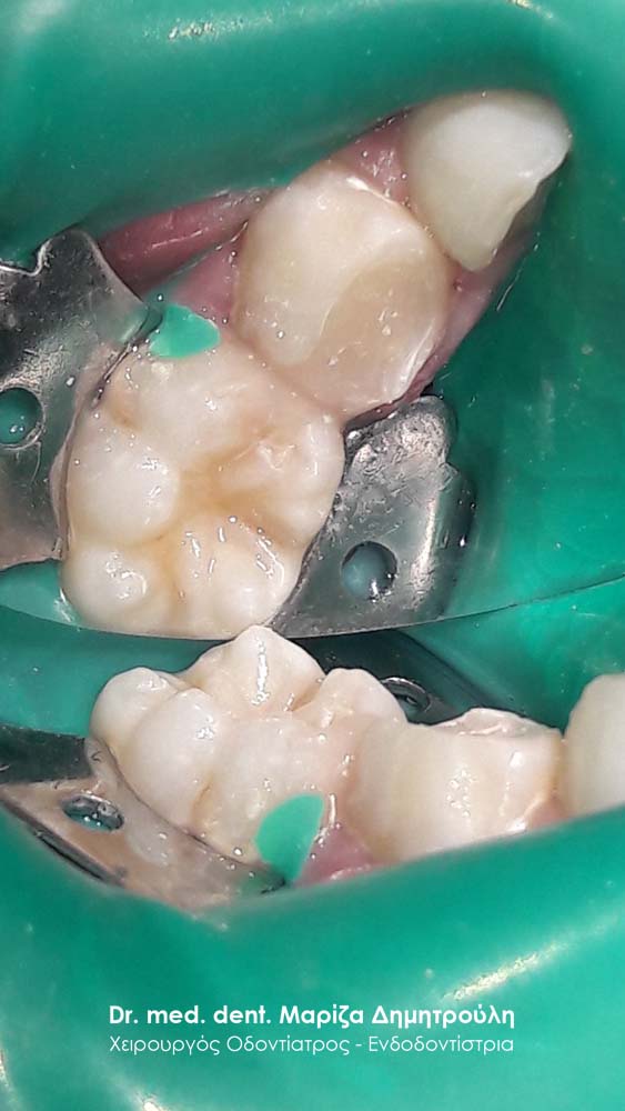
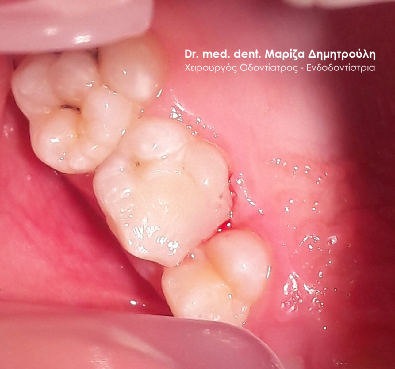
Incident – Children’s dental fillings
The little patient is suffering from pain in the lower left side of the lower jaw. After the clinical examination, the existence of a large carious cavity in the lower left second first molar and a smaller one in the left lower first molar was found.
After administration of local anesthesia, the carious dental tissues were removed and the cavities were restored with white resin filling. on the first permanent molar of the same side, sealanat was placed for preventive reasons.
BEFORE
BEFORE
AFTER
AFTER
Incident – Children’s dental fillings
In this specific case, the original clinical picture of the two teeth, which due to caries were restored with white resin fillings, is absent.
Case – Complete reconstruction of a child’s mouth
The little boy visited the dentist for the first time. The dentist who examined the children at the school noted that there were some carious cavities, which should be restored. In this particular patient, both white fillings were performed on the teeth that were carious, as well as preventive coverings on the chewing surfaces of the permanent molars to protect them from any future carious activity. The sealants in this case were necessary for an additional reason, since the child already had medium caries activity.
Also in the anterior area of the upper jaw, a small fistula was observed in the area of the first left molar, which had strong mobility but did not “fall”. In this case, the extraction of the baby tooth was necessary, because the child had the fistula for at least the last 3 weeks (always according to the mother’s words). With the extraction of the tooth the fistula subsided in the following days and the child was relieved.
Fistula in the area of the first neogill sector
BEFORE
META – Permanent Tooth Sealant
BEFORE
AFTER – Baby tooth seal and permanent tooth sealant
BEFORE
AFTER – Baby tooth seal and permanent tooth sealant
BEFORE
AFTER – Baby tooth seal and permanent tooth sealant
Case – Complete reconstruction of a child’s mouth
The little patient came to the clinic presenting extensive carious lesions on all the back teeth. The mother wished to keep the baby teeth in the girl’s mouth and wanted us to avoid extracting them. Treatment of teeth per quadrant has begun. In some teeth due to deep and extensive caries (which reached the level of the pulp) a pulpotomy was performed. All teeth were restored with white fillings of composite resin.
It is worth noting that in the first years of the child’s life, the mother brushed the teeth with toothpaste that did not contain any fluoride. Fluoride is the shield of the teeth against caries, since the teeth are shielded with fluoride ions and become stronger against any cariogenic microbes. Scientific studies prove that fluoride cannot be harmful to the general health of both a child and an adult, as long as the recommended amount of toothpaste is always used without exaggeration.
BEFORE
AFTER
BEFORE
AFTER
BEFORE
AFTER
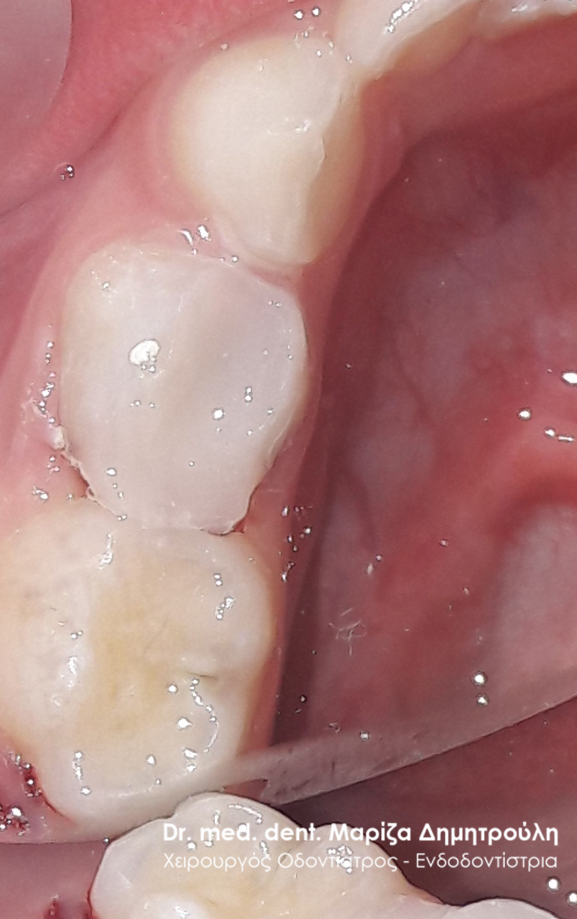
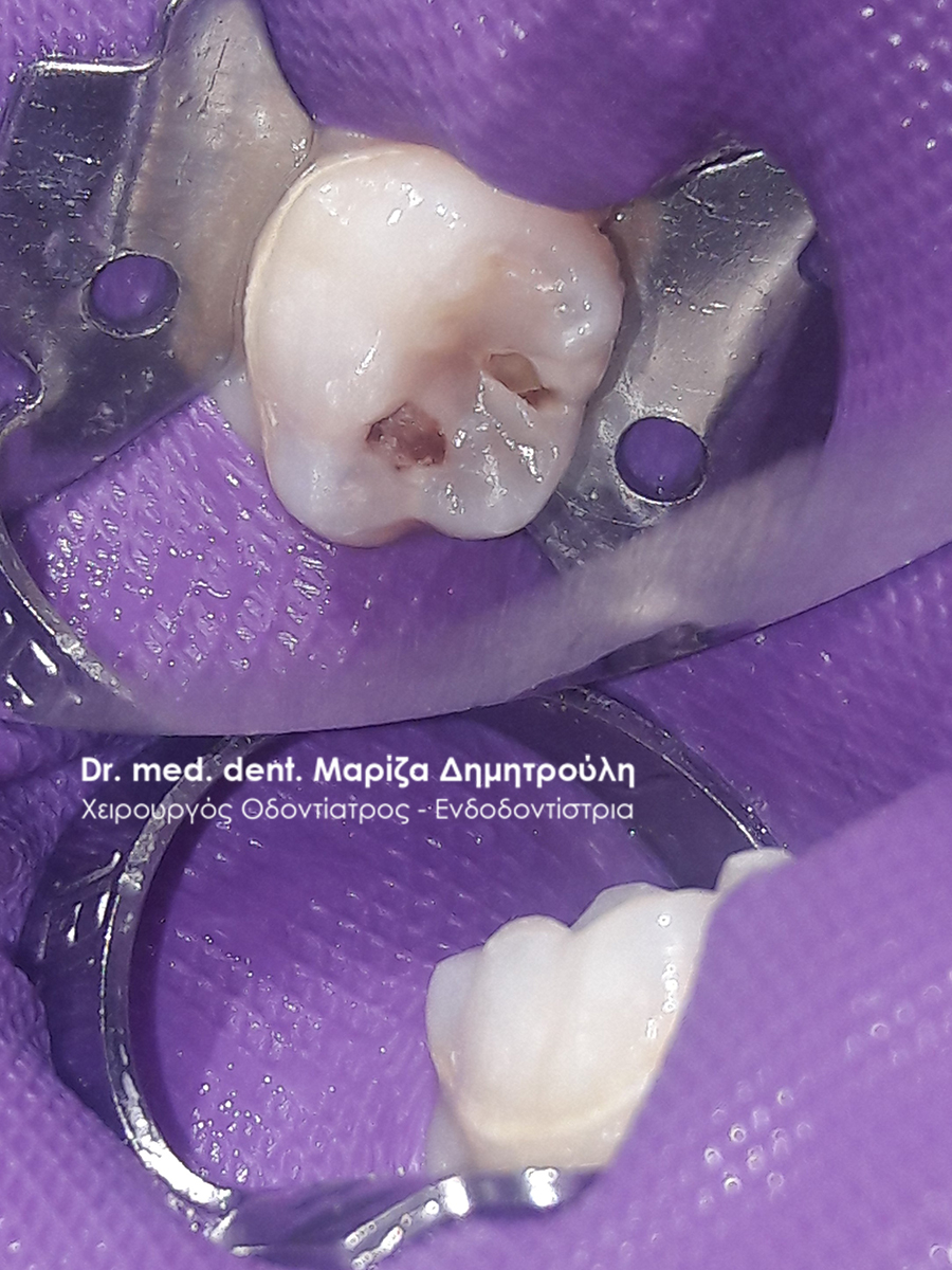
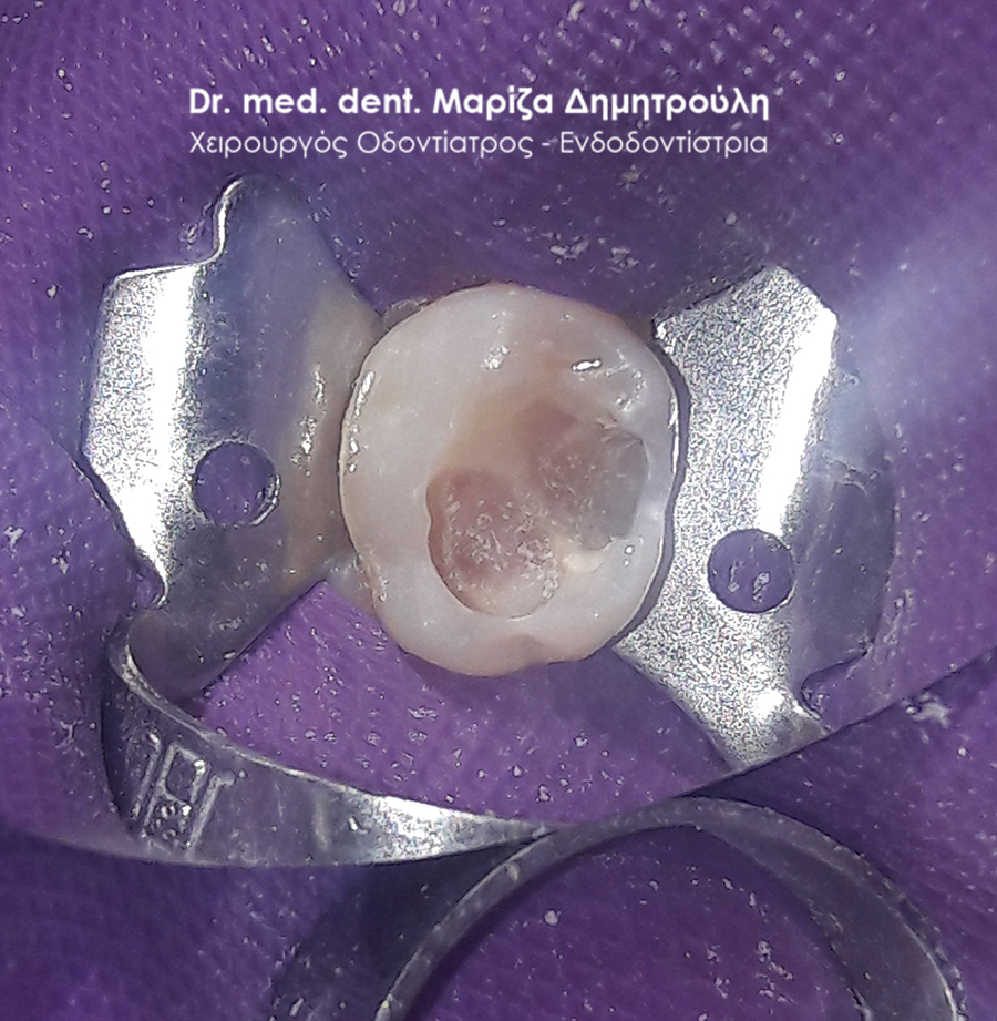
Incident – Pulpotomy (denervation) and sealing of a child’s tooth
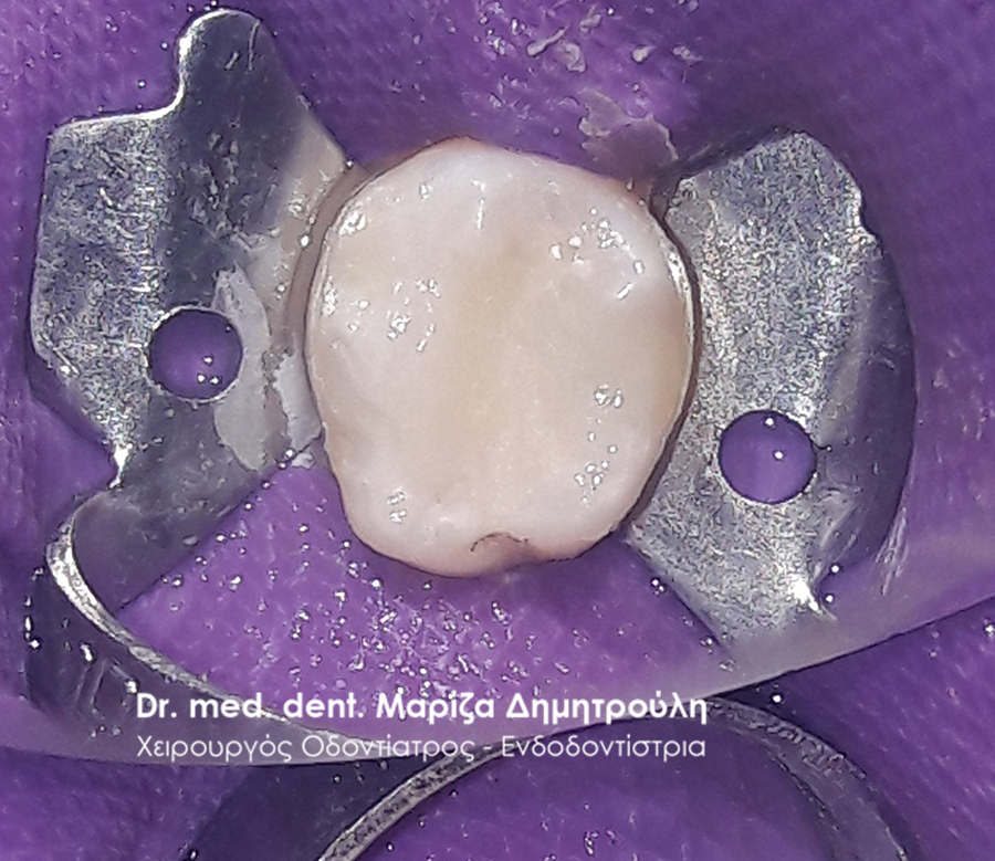
The little patient came to the doctor’s office because of pain in the lower right first molar. After the clinical examination, the presence of a deep carious cavity was established, which was suspected to extend to the nerve of the child’s tooth. After the administration of local anesthesia and the opening of the tooth, the caries was removed. During the derailment of the carious tissues, the nerve of the tooth was exposed and for this reason a pulpotomy was performed. A special medicine was placed in the tooth and the tooth was blocked with a temporary material. At the next session the tooth was asymptomatic and the treatment was completed by restoring the tooth with a white composite resin seal.
BEFORE
AFTER
Incident – Sealant and sealing of a child’s tooth
The patient complains of pain in the upper left side. After the clinical examination it was found that there was a black filling, which appeared that the tooth had decayed after the removal of the amalgam and the caries (under the old black filling) the tooth was restored with a new white filling. A preventive covering of the second permanent molar (sealant) was also performed.
BEFORE
Removing an old filling and cleaning the cavity
AFTER
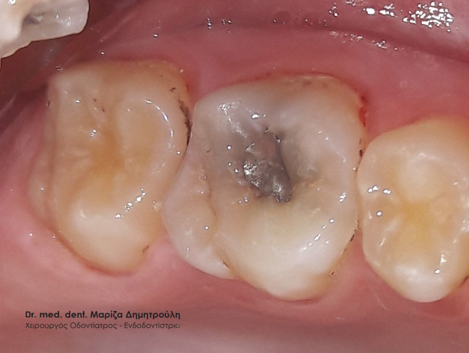
Incident – Pulpotomy (denervation) and tooth filling
The little patient reports pain in the lower right side when he eats. According to the history, a filling was performed on the first lower right molar which “fell out”. After the clinical examination, it was found that there were remnants of the old filling on the tooth, while the rest of the tooth was open and decayed. After the administration of local anesthesia and the placement of the elastic isolator, the carious tissues were removed from the young tooth, a pulpotomy was performed and finally the lesion was restored with a white composite resin seal.
The patient after completing the treatment does not feel pain and the tooth is asymptomatic.
BEFORE
AFTER
Incident – Pulpotomy (denervation) and tooth filling
The patient came to the clinic as the parents noticed a “hole” in one of the upper right teeth. The child’s clinical examination followed and a large carious cavity was found. After local anesthesia was administered, so that the entire dental procedure was painless for the child, the tooth was extracted. All carious dental tissues were then removed, but because the caries extended deep to the level of the nerve of the tooth a pulpotomy was performed. In the next session the tooth was restored with a white composite resin filling.
The patient after completing the treatment does not feel pain and the tooth is asymptomatic.
BEFORE
AFTER
Incident – Child tooth filling
As part of the annual preventive check-up, a small carious cavity was identified, which had not been noticed by the child or the parents. The treatment started with the derailment of the tooth, the caries removal and finished with the restoration of the lesion with a white composite resin filling. During the opening of the tooth, it was found that the adjacent tooth was also affected by caries, more specifically at the point of contact with the “holed” tooth. Similarly, the adjacent tooth was also restored with a white filling, which is evident if one looks more closely at the right photo.
BEFORE
AFTER
Incident – Children’s dental fillings
In this specific case, only the final image of the restoration of the two children’s teeth with white resin fillings is available. In the first child’s molar, a pulpotomy of the tooth was performed, due to the deep extent of caries at the level of the nerve of the tooth.
BEFORE
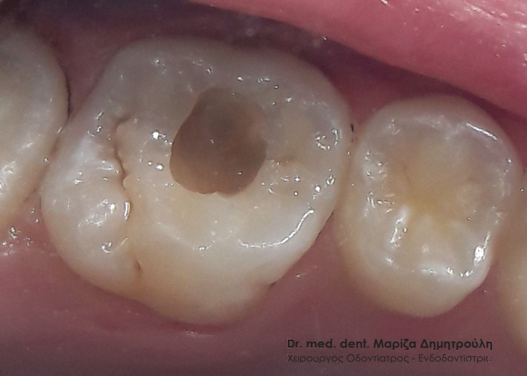
Incident – Child tooth filling
The little patient visited the clinic for the restoration of his decayed childhood tooth. The reconstruction was performed with a white composite resin seal using the isolator.
BEFORE
Tooth cavity clean after caries removal
AFTER
Incident – Children’s dental fillings
The patient complained of pain when chewing on his upper right side. After the clinical examination, the existence of 2 teeth with deep and extensive caries was established. After the caries was removed, white composite resin fillings were made.
BEFORE
AFTER
Incident – Children’s dental fillings
A 9-year-old girl reports pain in the upper left side of her mouth when chewing. After the clinical examination, the existence of 2 carious teeth was found. In the first permanent left molar, the presence of caries is evident, the extent of which proved to be quite deep when the tooth was drilled. The existence of a carious cavity of medium depth was observed in the second newborn (child) molar.
After the caries was removed from the two teeth, white composite resin fillings were made.
BEFORE
AFTER
Incident – Children’s dental fillings
A 6-year-old girl was diagnosed by the dentist, who visited the school, with 2 decayed teeth. After removing the carious tissues from the 2 children’s back teeth, white composite resin fillings were made.
BEFORE
AFTER
Incident – Child tooth filling
A 9-year-old boy complained of pain in the left side of the lower jaw. After the clinical examination, a carious cavity was found, which was restored with a white composite resin filling.
BEFORE
AFTER
Incident – Child tooth filling
8-year-old girl had pain during chewing on the left side of the upper jaw. The clinical examination revealed the existence of a deep caries in one of the back baby teeth. After the tooth was derailed and the caries was removed, the dental deficit was restored with white composite resin filling.
BEFORE
AFTER
Incident – Child tooth filling
During the annual dental checkup, a small carious cavity was found in the first left mandibular permanent molar. In consultation with the child’s parents, it was decided to restore the tooth, firstly because a hole had been caused in the tooth and secondly to stop the active caries activity in the child’s mouth which is likely to be transmitted to the rest of the teeth. The restoration was performed with a white resin seal.
BEFORE
AFTER
Incident – Child tooth filling
The young patient reports intermittent mild chewing discomfort on the left side of the mandible. Clinical examination revealed the presence of a small caries in the first permanent molar on the left side of the mandible. It was decided to treat the tooth with a white filling, since the caries had already created a small hole in the tooth. As is well known, tooth decay must be treated immediately if it has visibly damaged the tooth. A small hole heals much more quickly and painlessly compared to a large carious cavity.
BEFORE

AFTER
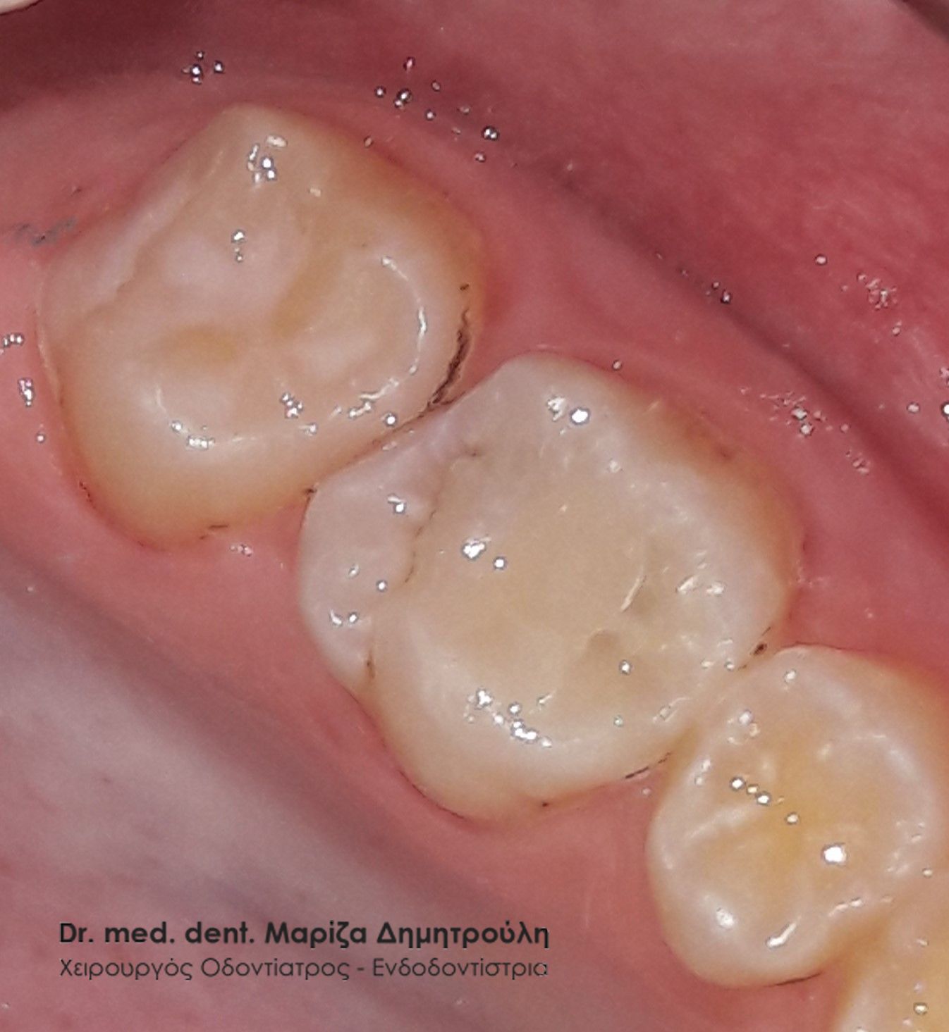
Incident – Child tooth filling
This particular patient came to the doctor’s office, as he had pain on the left side of the lower jaw while chewing. After the clinical examination, a small carious cavity was found at the border of the last two lower teeth, which was restored with white resin filling.
BEFORE
AFTER
Widget After Content

Dr. med. dent. Mariza Dimitrouli
Dr. med. dent. Mariza Dimitrouli is a specialist at the University of Hannover, Germany (MHI) and also holds a PhD from the same university. She also worked in a dental clinic in Berlin. The title of Endodontist held by Dr. M. Dimitrouli is renewed every six years by the German Endodontics Society. She has a private practice in Thessaloniki, Thermi.