Case – Full Restoration of Dental Fillings
The patient had old fillings and his desire was the total restoration of his mouth and the replacement of his old fillings with new white composite resin fillings.
BEFORE
META
Cervical seal – original image
Nervical sealing – final image
BEFORE
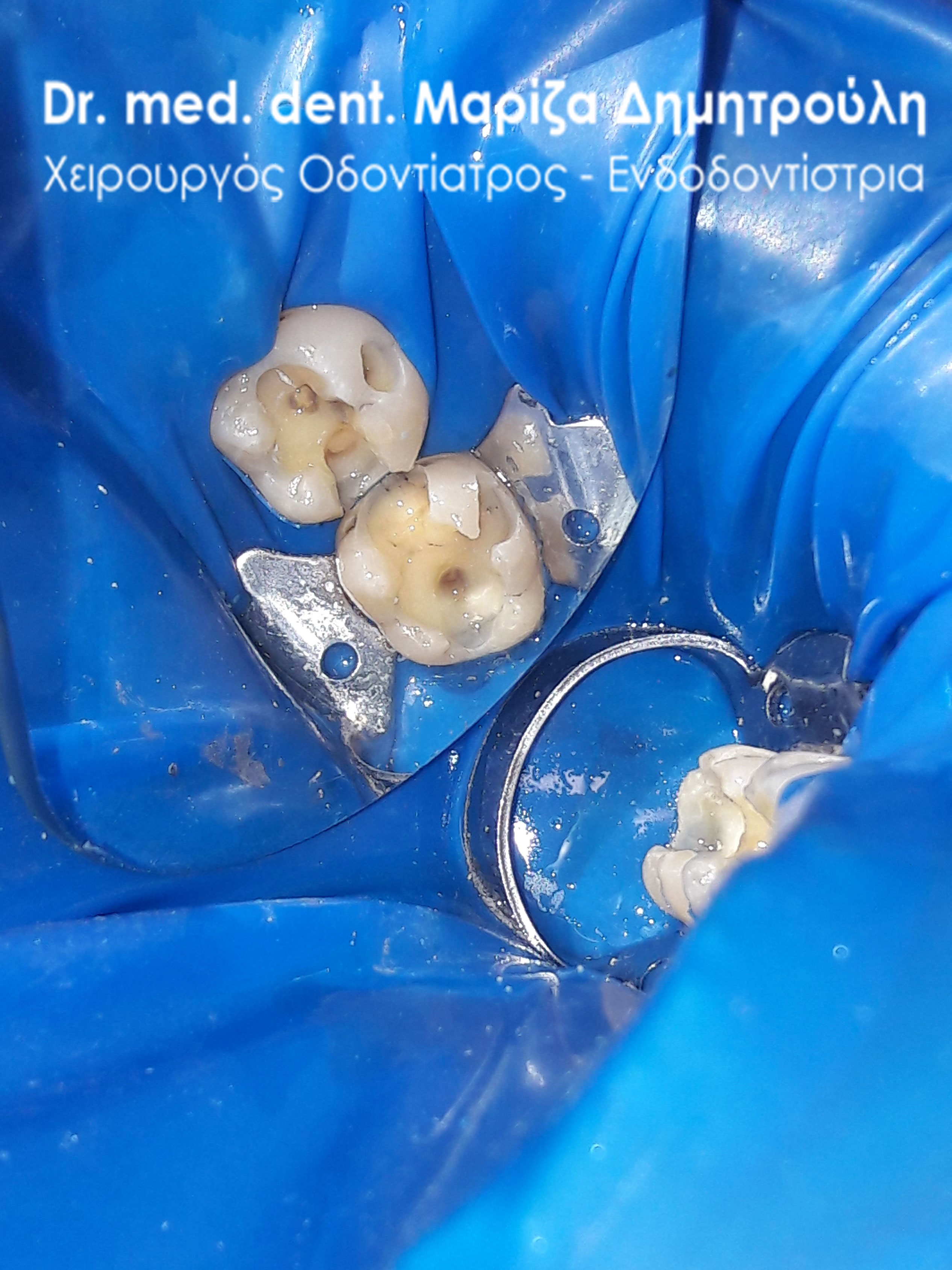
Image of teeth after removal of black fillings
META
META
Case – Full Restoration of Dental Fillings
The patient came to the clinic with the intention of replacing some old fillings, which occasionally gave her symptoms of pain. as the photos show the teeth were restored with new white fillings.
BEFORE
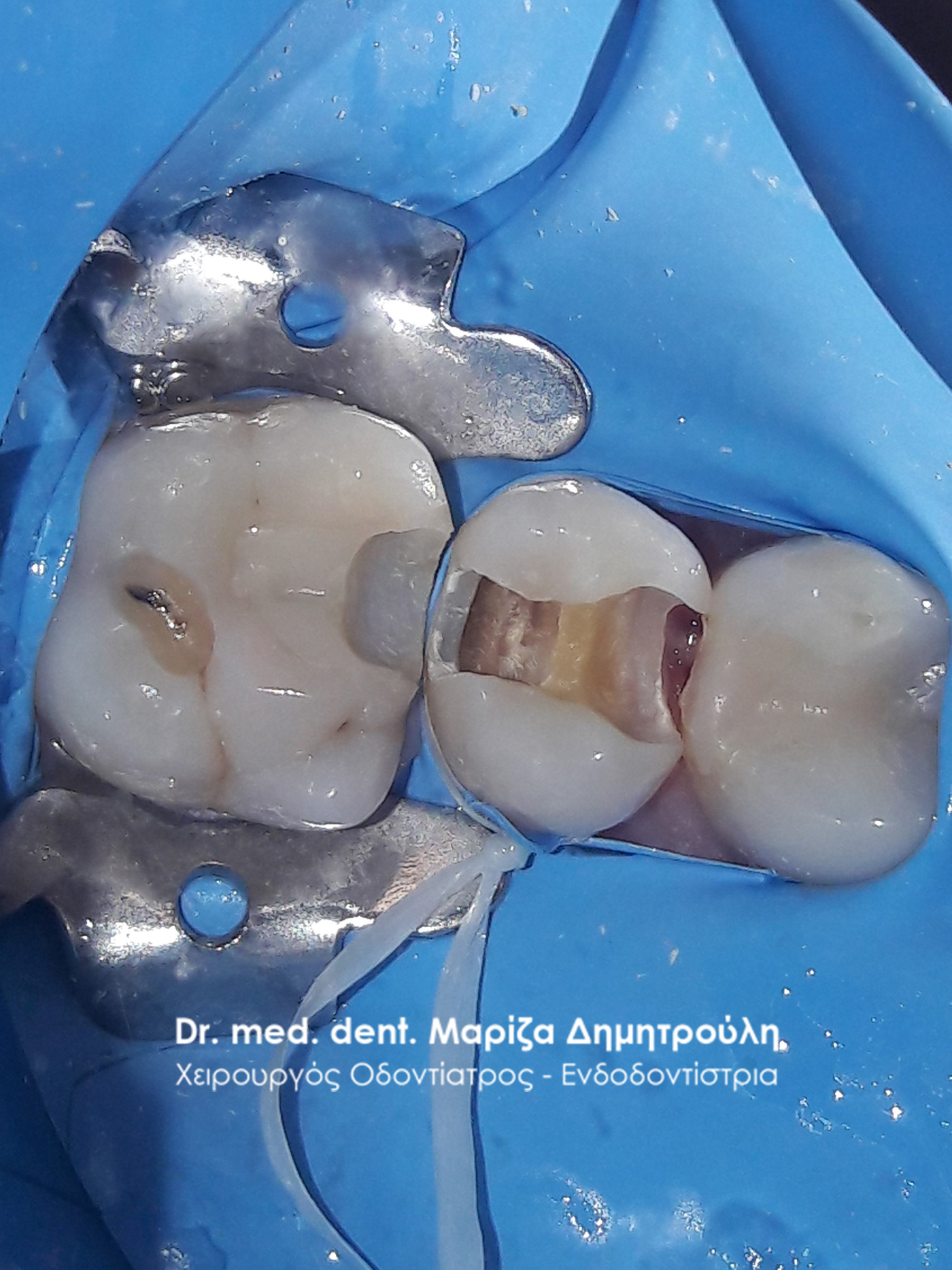
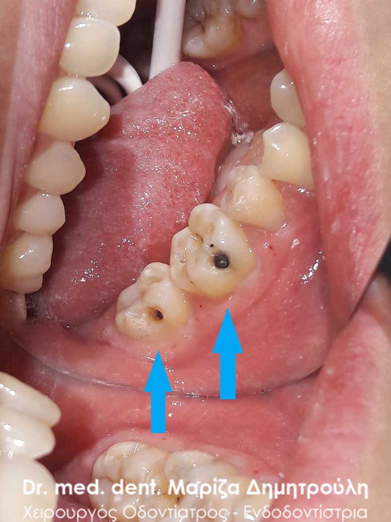
Image of caries cavity after tooth extraction
META
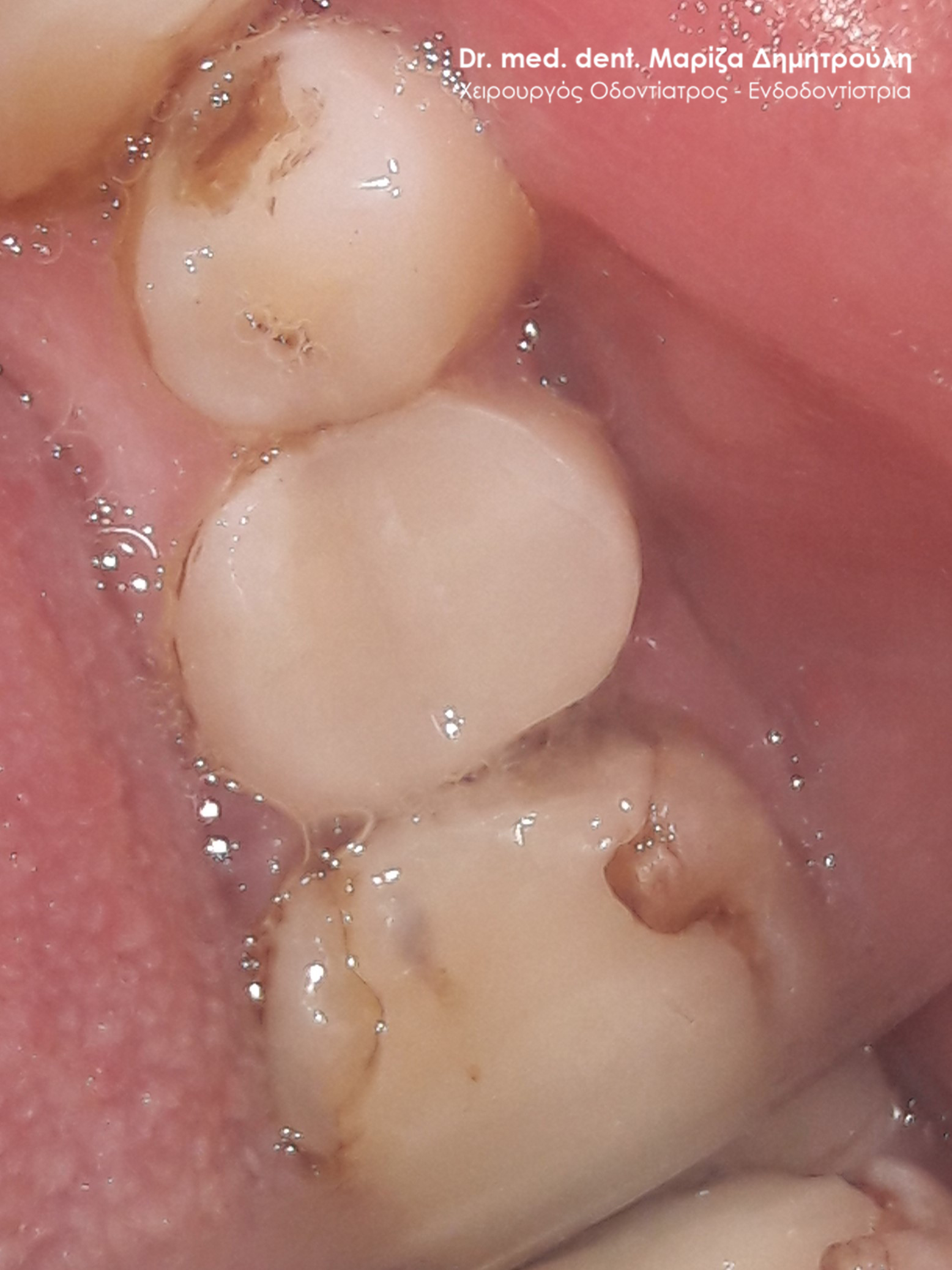
Original image of lower left molars
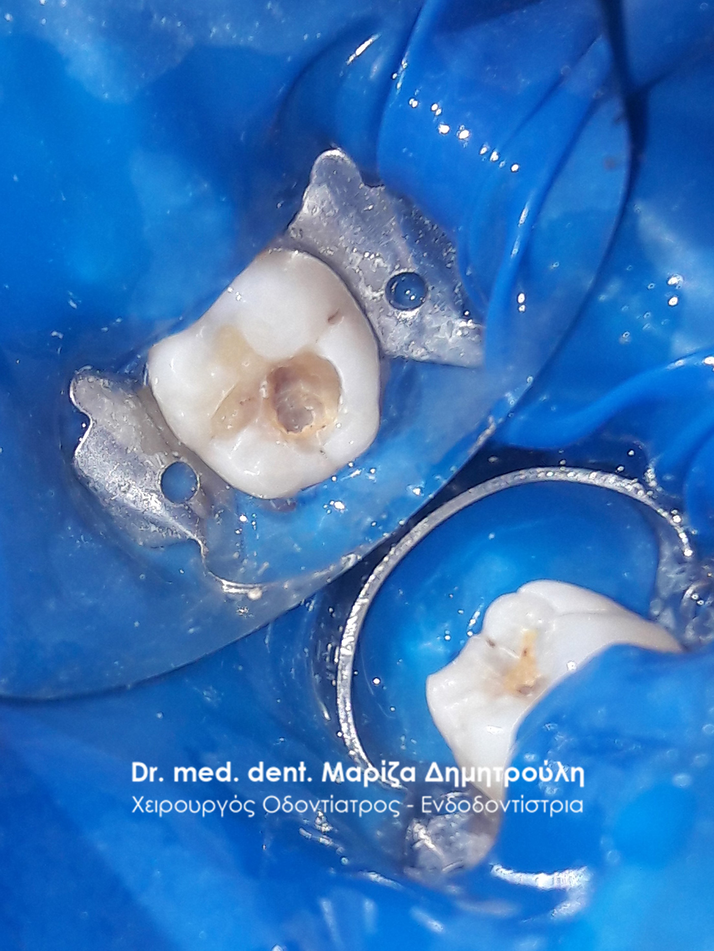
Image of 1st lower left molar after its eruption
Final image of 1st lower left molar
Final image of 2nd lower left molar
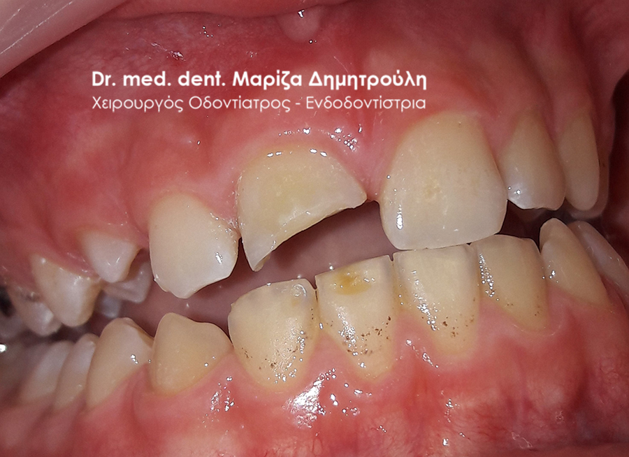
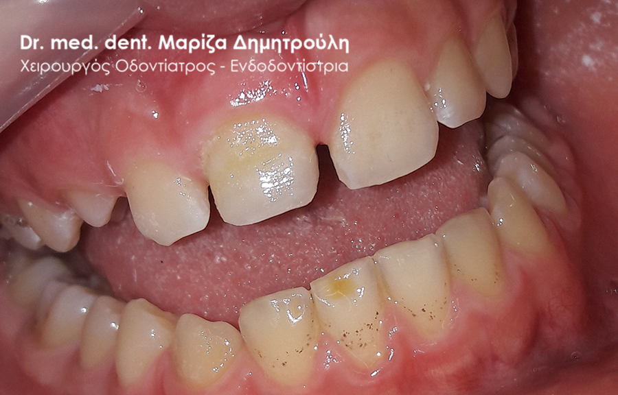
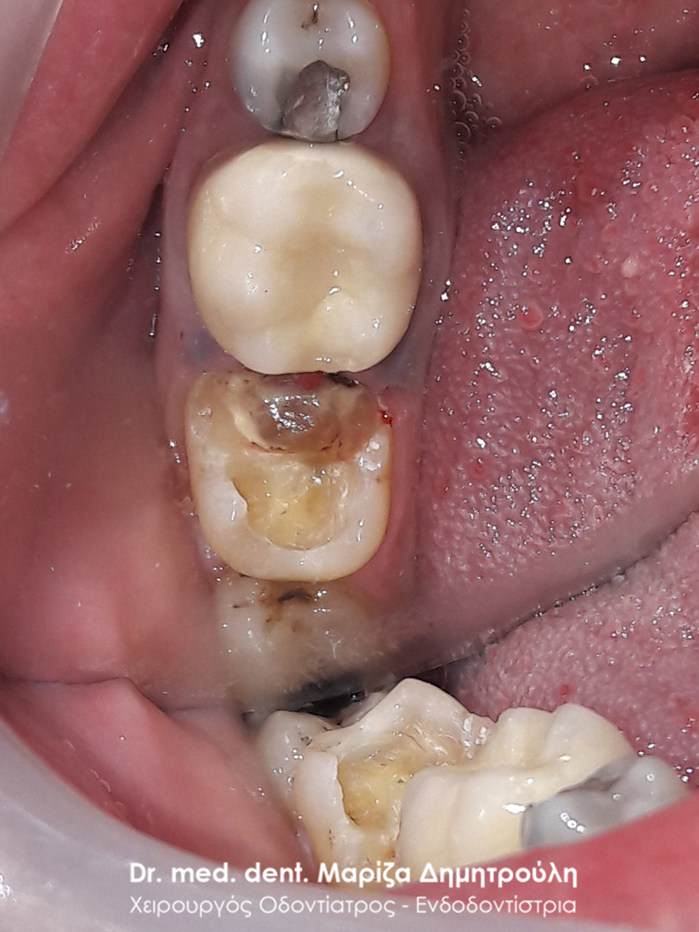
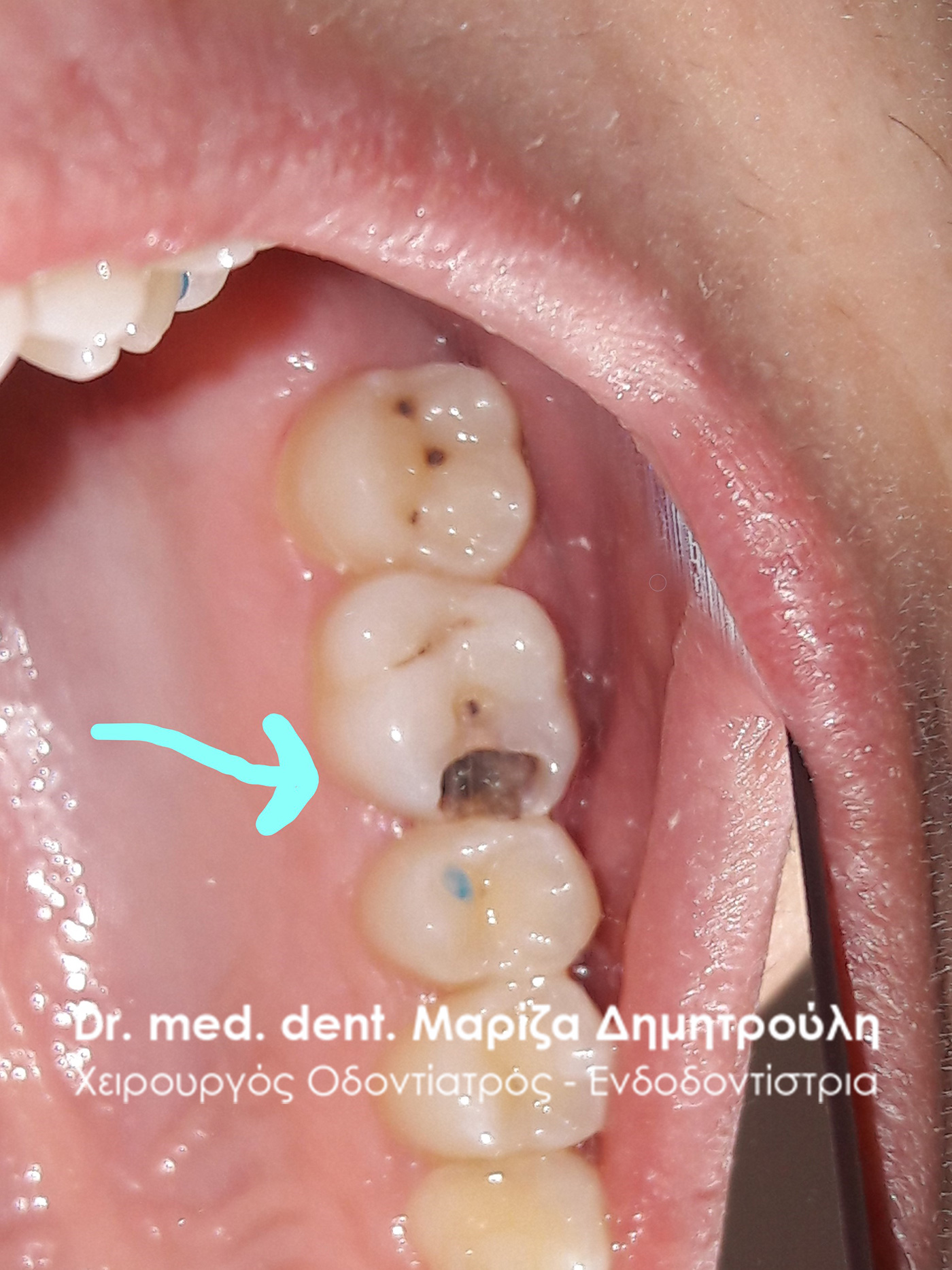
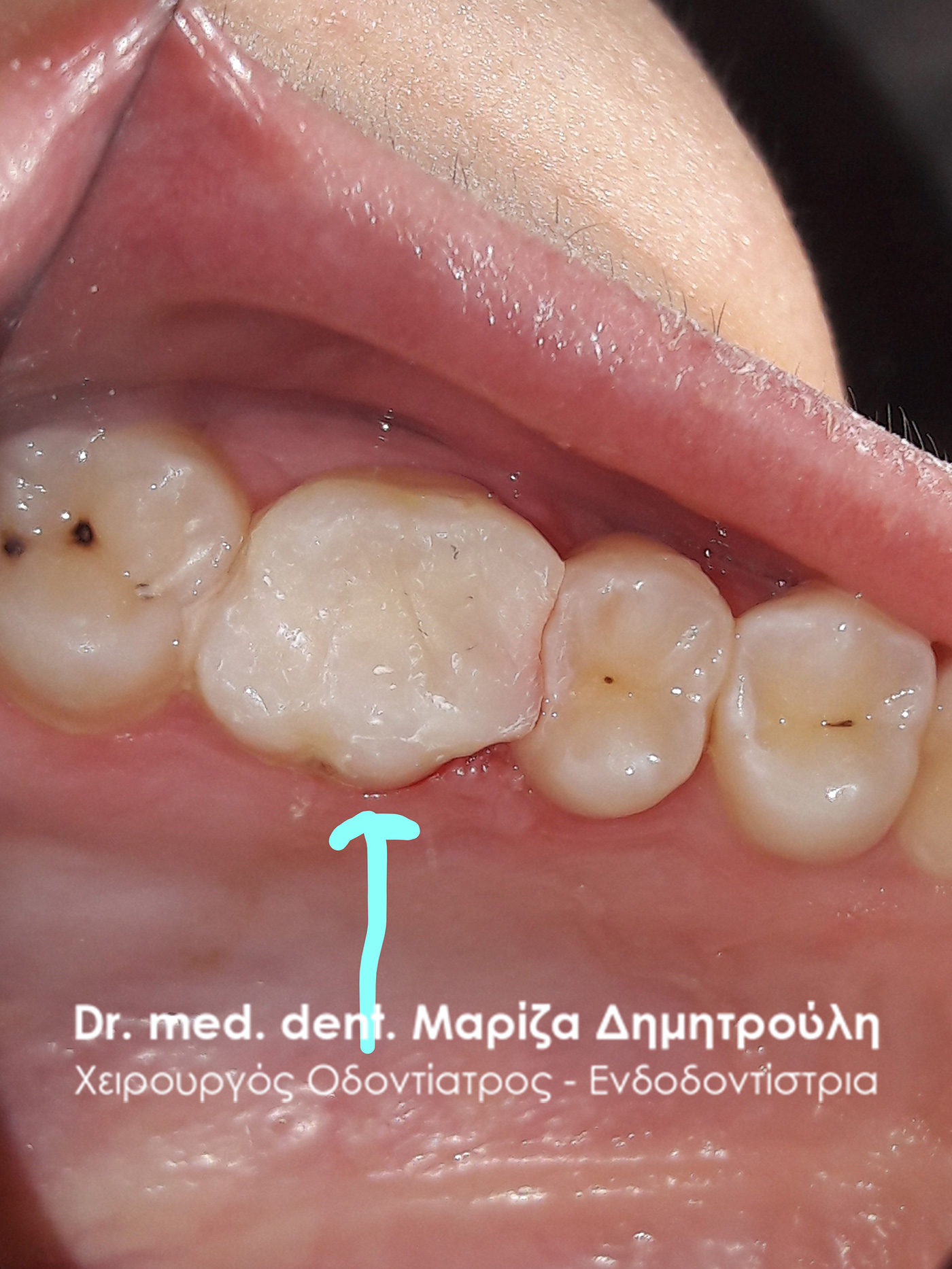
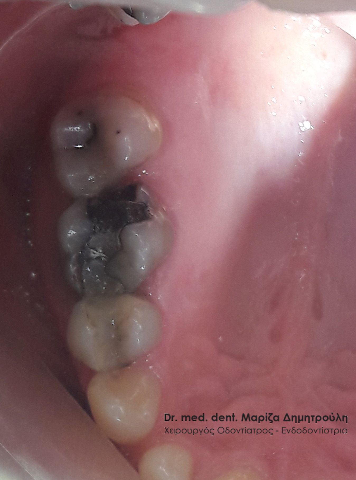
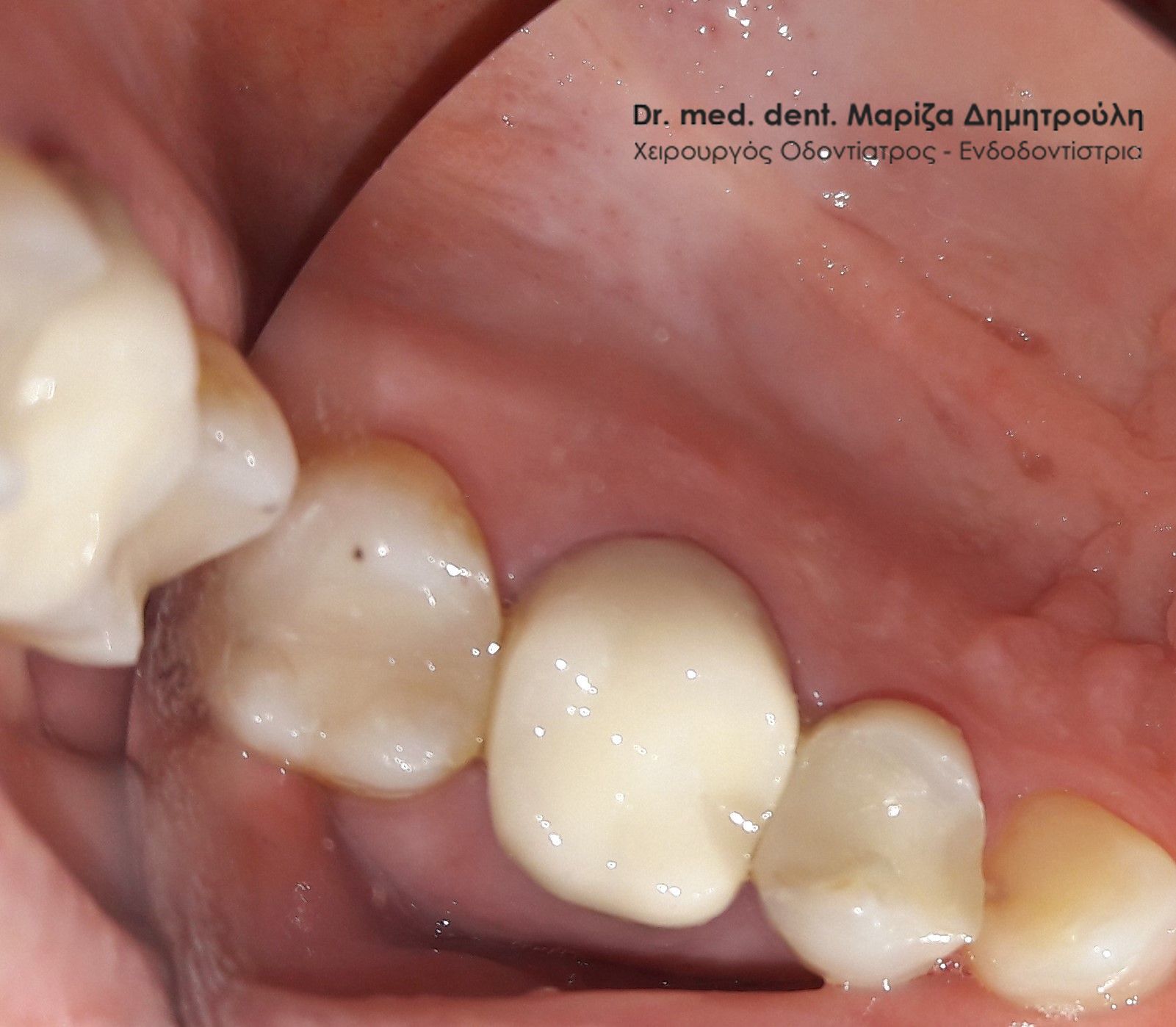
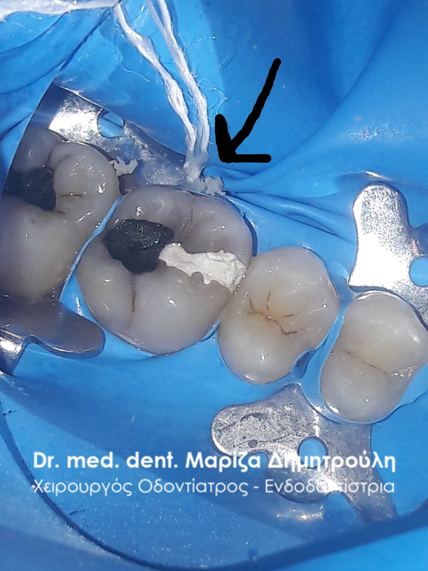
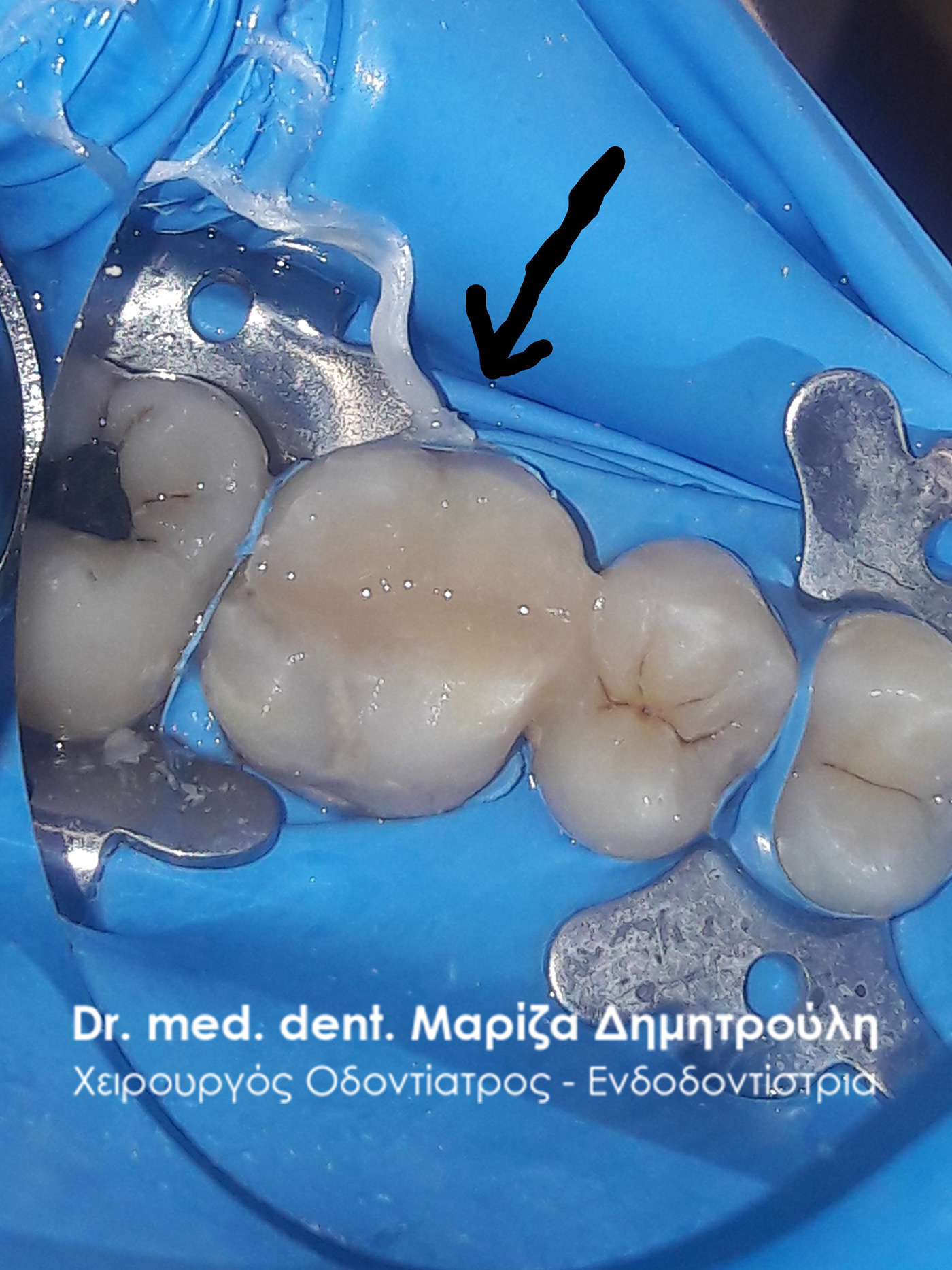
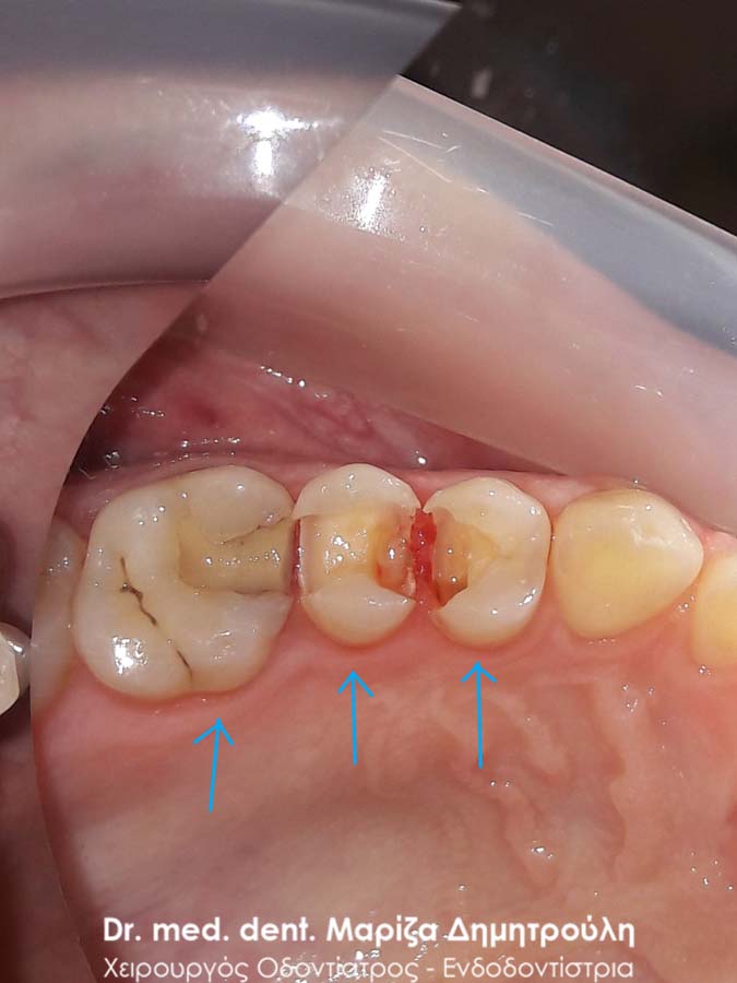
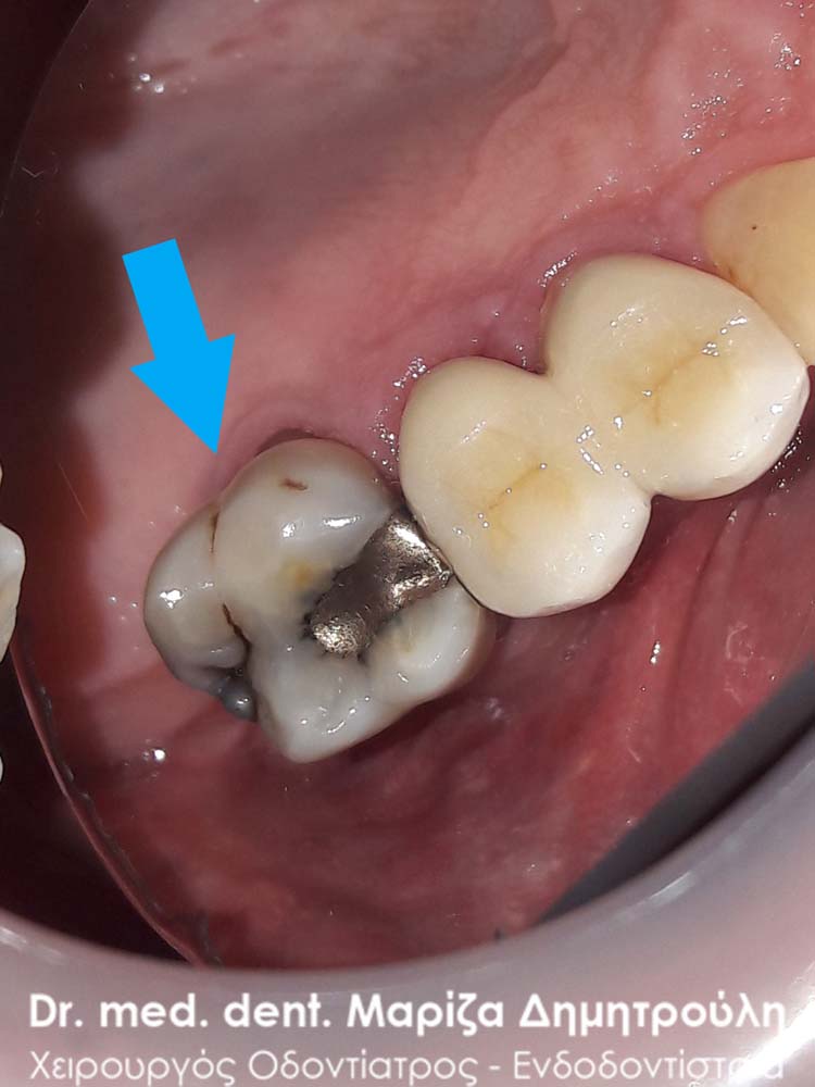
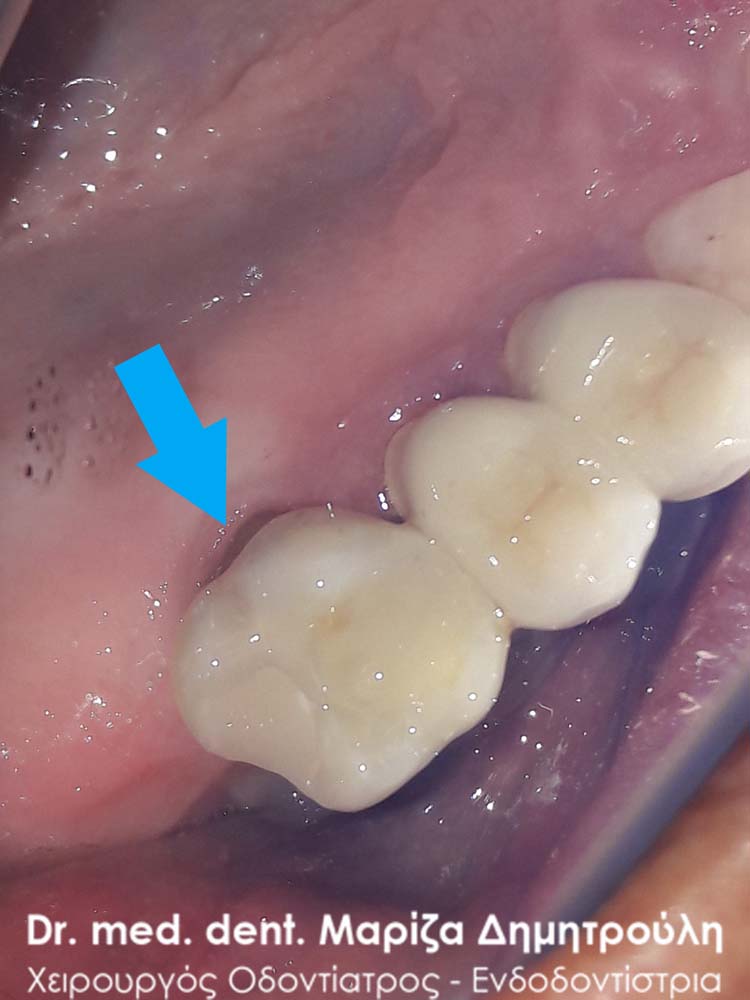
Incident – Change of black tooth fillings
The patient wished to replace the black amalgam fillings with white fillings, because they bothered her both aesthetically and functionally, since food was collected between the two teeth. Replaced old seals with new white seals.
BEFORE
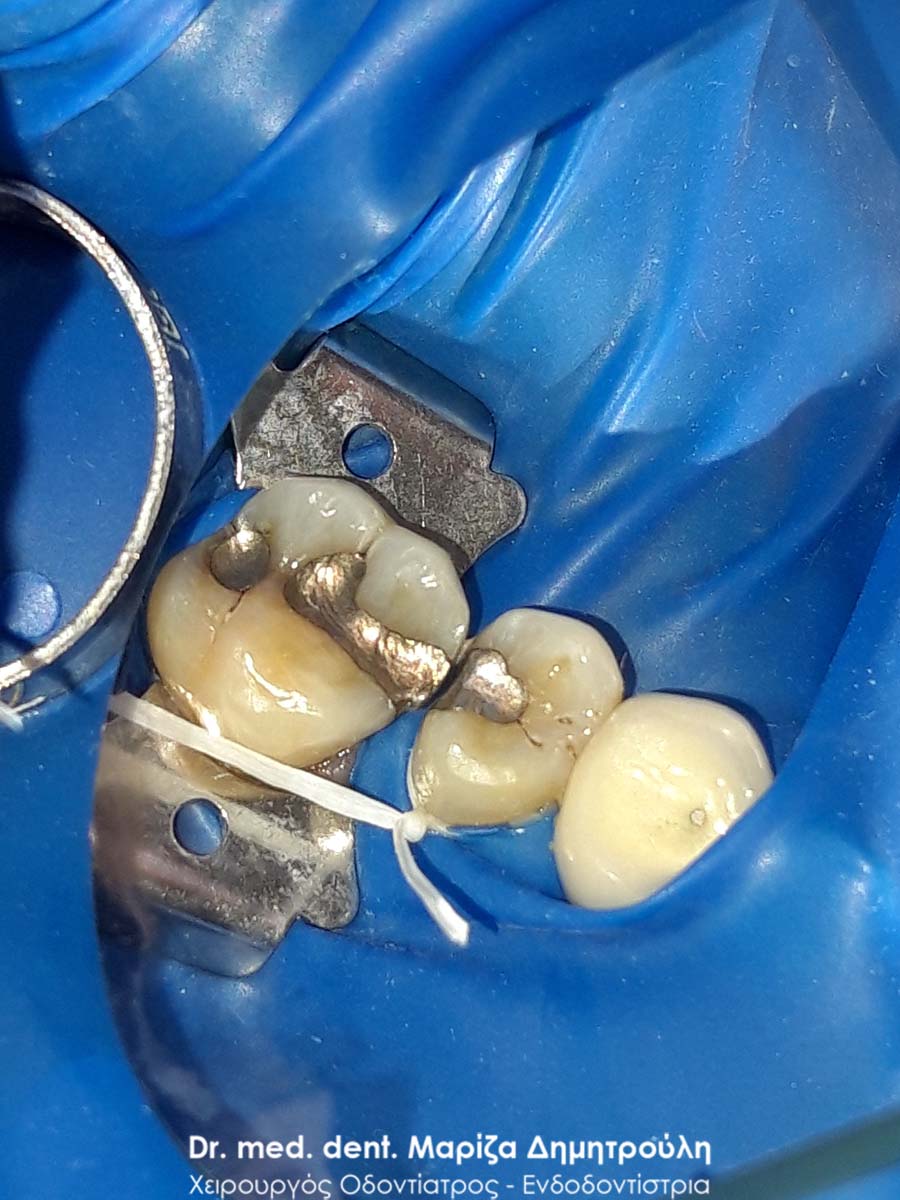
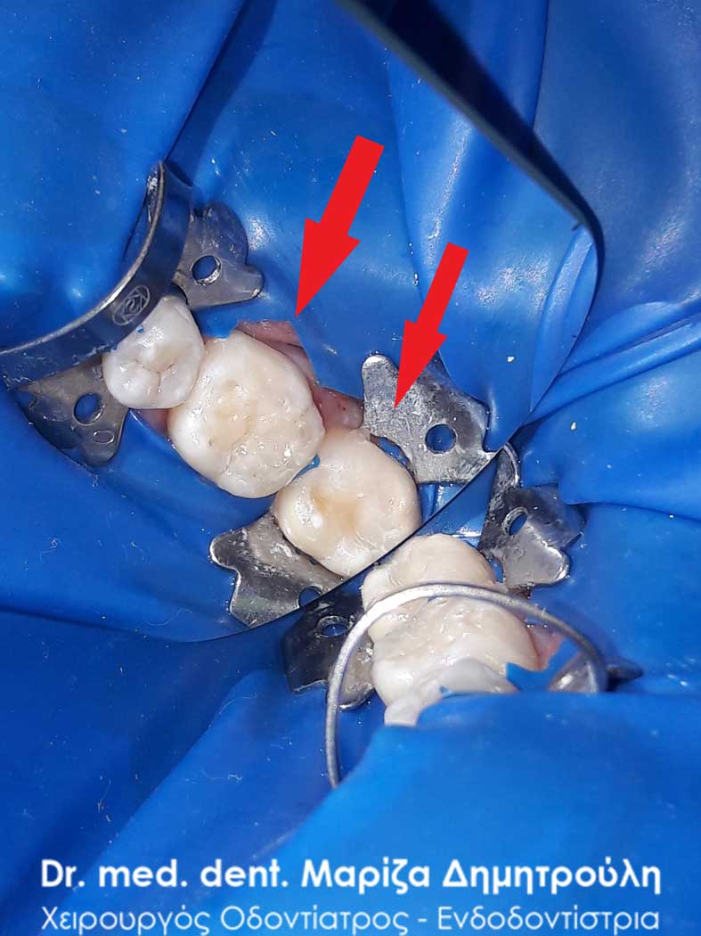
Image of teeth using a rubber spacer
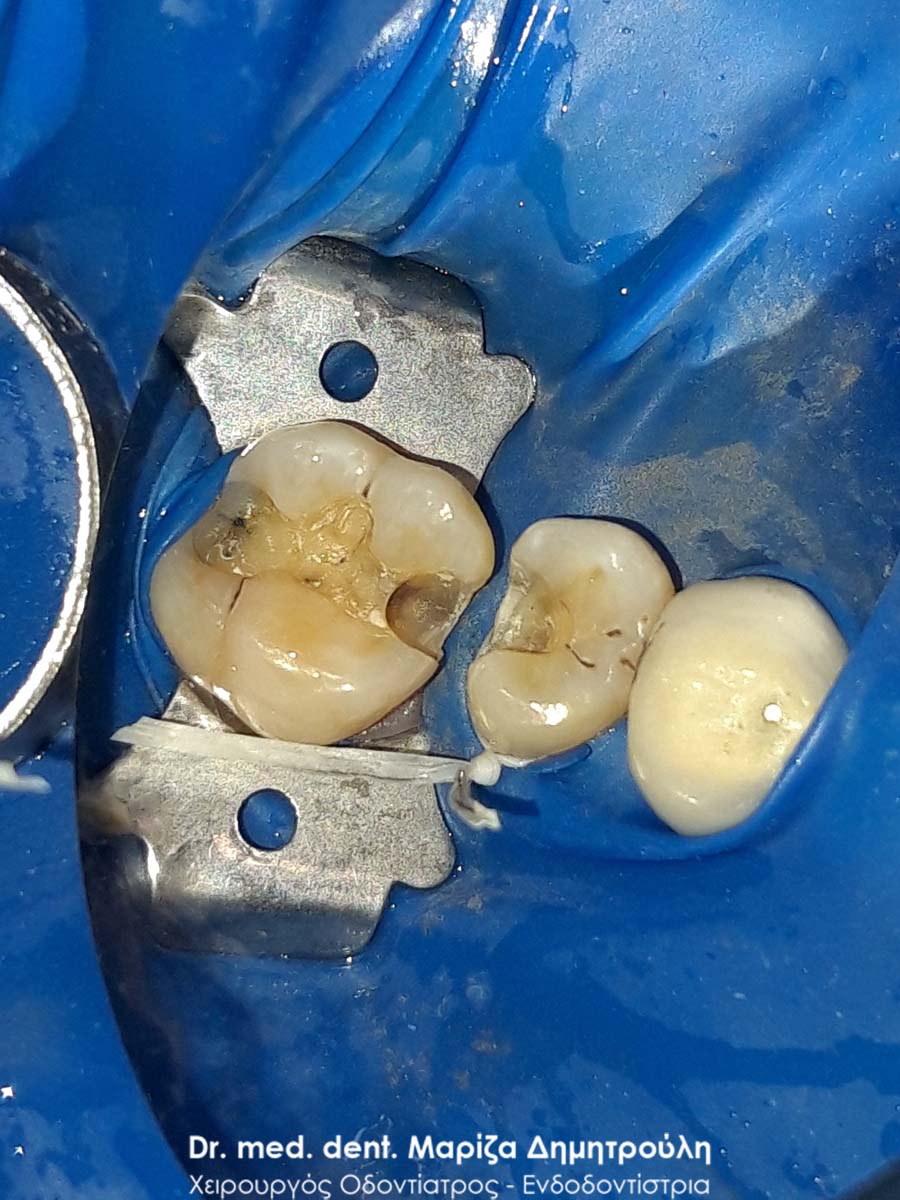
Image of teeth after removal of amalgam fillings
AFTER
AFTER
Case – Front Teeth Fillings
The patient came to the doctor’s office for the preventive annual check-up. After the clinical examination, it was established that the four upper front teeth were caries. The replacement of the old fillings with new white fillings was planned and carried out.
BEFORE
META
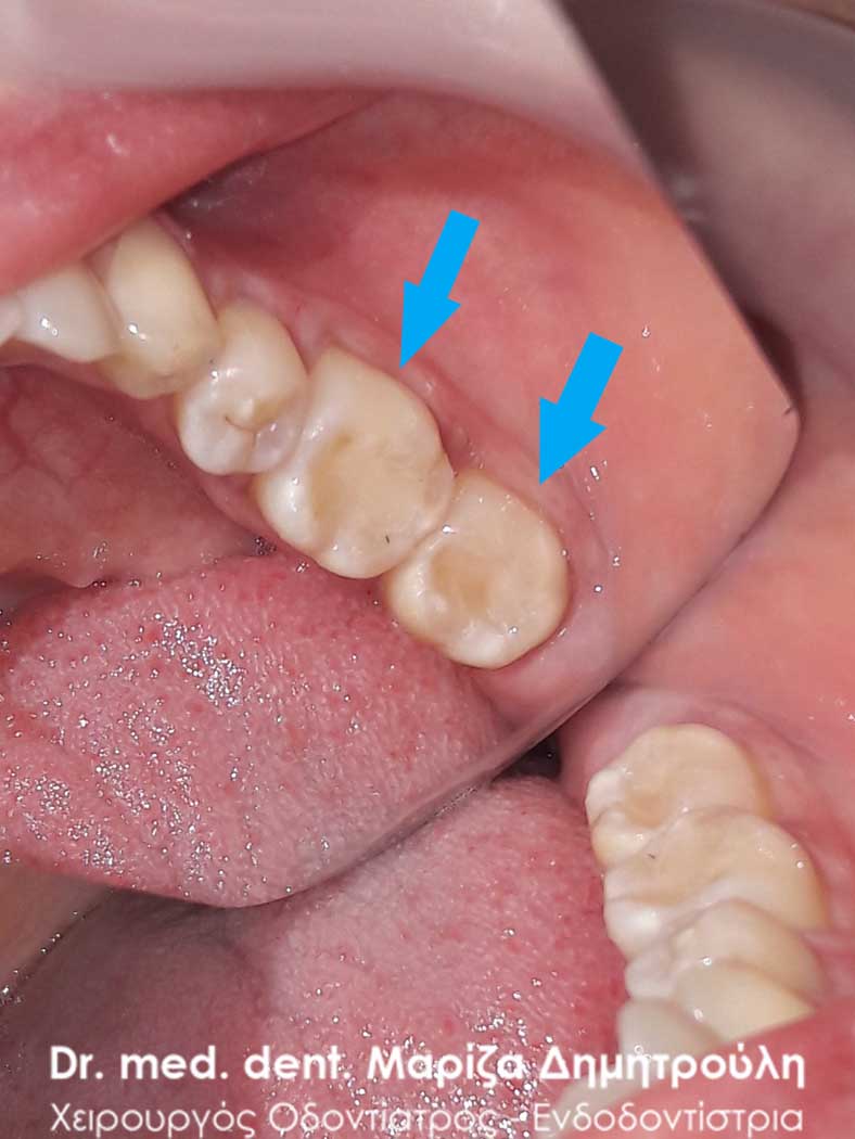
Case – Front Teeth Fillings
The patient visited the clinic for the purpose of replacing the four old fillings on the upper front teeth. After removing the old composite resin fillings and removing the caries (underneath the old fillings) the teeth were restored with new white fillings.
BEFORE
META
Incident – Front tooth filling
In this particular patient, denervation of the upper right central sector was performed in the clinic. During the last denervation appointment, he dislodged the white resin filling that had been performed by the referring dentist. The treatment had to be carried out immediately due to the position of the tooth in a prominent part of the mouth.
The following restoration was performed within 15 minutes (due to time pressure of the office schedule) as a temporary solution to the patient’s problem. In the future, the tooth will have to be restored with a fiberglass shaft and the placement of a tooth holder, which due to the patient’s young age will have to be postponed until the age of 18.
BEFORE
META
Incident – White tooth filling
The patient wanted the restoration of a cavity that had been bothering him lately while chewing. The carious cavity was located on the lower second right premolar and the white filling was performed with the composite resin material.
BEFORE
META
Incident – White fillings on two teeth
As part of the annual dental checkup, two small carious teeth on the right side of the upper jaw were found in the patient. The old white fillings on the two upper right molars had been re-caried. After the administration of local anesthesia, the old white fillings of the teeth were removed, the decay was removed and two teeth were restored with new white resin fillings.
BEFORE
META
Incident – White fillings on two teeth
In the present case, the original image of the two teeth before their derailment began is absent. Only the intermediate image showing the extent of the dental deficit in the two molars of the upper left side and the final restoration image of the two teeth are available.
The patient had felt a mild pain while chewing food for the last few days. After the clinical examination of the mouth, the existence of carious cavities in the two upper left molars was established. It was decided with the patient’s consent to replace the fillings. In the first molar, only half the extent of the old filling was corrected, as it was deemed unnecessary to remove the entire old filling.
BEFORE
META
Incident – Cervical tooth filling
The patient wanted the restoration of the cervical lesion, which he presented in the lower right second premolar. The treatment was performed with white composite resin filling.
BEFORE
META
Περιστατικό – Tooth Filling
Η ασθενής επισκέφτηκε το ιατρείο ως επείγον περιστατικό με την επιθυμία της άμεσης και γρήγορης αποκατάστασης του σπασμένου δοντιού της που την πονούσε. Υπό τις προαναφερθέντες συνθήκες η θεραπεία επιλογής ήταν η ανασύσταση του δοντιού λευκό σφράγισμα σύνθετης ρητίνης. Η θεραπεία ολοκληρώθηκε εντός 20 λεπτών και η ασθενής μπορούσε να επιστρέψει άμεσα στη δουλειά της.
BEFORE
META
Περιστατικό – Tooth Filling
Στα πλαίσια του ετήσιου οδοντιατρικού ελέγχου εντοπίστηκε ένα τερηδονισμένο δόντι, το οποίο δεν είχε γίνει αντιληπτό από τον ασθενή. Προτού ξεκινήσει η διαδικασία τροχίσματος του δοντιού, ο ασθενής ενημερώθηκε οτι η τερηδόνα πιθανόν να εκτείνεται βαθιά (στο ύψος του νεύρου του δοντιού), γεγονός που επιβεβαιώθηκε κατά τη διάνοιξη της κοιλότητας (2η φωτογραφία). Η αποκατάσταση του δοντιού πραγματοποιήθηκε με λευκό σφράγισμα ρητίνης.
BEFORE
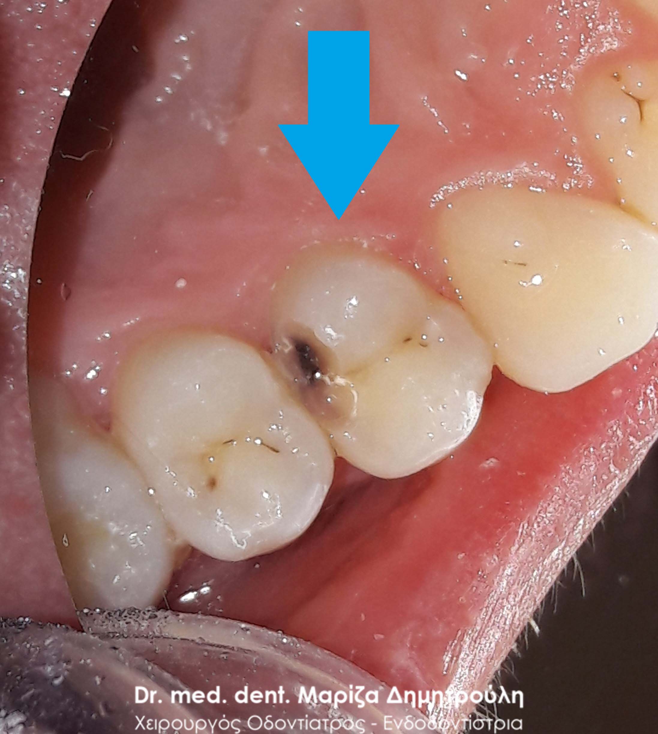
Image of deep carious tooth cavity
AFTER
Περιστατικό – Tooth Filling
Ο ασθενής μετά το πέρας της ενδοδοντικής θεραπείας του δοντιού προτίμησε (για οικονομικούς λόγους) την αποκατάσταση του με λευκό σφράγισμα σύνθετης ρητίνης έναντι της τοποθέτησης θήκης δοντιού, η οποία σαφώς προστατεύει το δόντι με μεγαλύτερη ασφάλεια. Στο συγκεκριμένο δόντι επειδή απουσίαζε μεγάλο τμήμα οδοντικού ιστού δεν ήταν δυνατόν ένα απλό σφράγισμα ρητίνης να αποδώσει επαρκές σημείο επαφής με το παρακείμενο δόντι. Ο ασθενής ενημερώθηκε και δεν τον ενοχλούσε.
Στην προκειμένη περίπτωση οι οδοντίατροι θα πρέπει να ενημερώνουν τον ασθενή οτι υπάρχει μεγάλος κίνδυνος θραύσης τόσο του σφραγίσματος όσο και του απονευρωμένου δοντιού. Η τοποθέτηση θήκης σε απονευρωμένα δόντια με μεγάλο έλλειμμα οδοντικής ουσίας ενδείκνυται για προστασία από τυχόν κατάγματα δοντιού.
BEFORE
META
Περιστατικό – Tooth Filling
Ο ασθενής συνηδειτοποίησε οτι έσπασε ένα κομμάτι από το παλιό σφράγισμα αμαλγάματος που είχε στο δεξιό κάτω πρώτο γομφίο. Η κλινική εξέταση αποκάλυψε οτι μαζί με κομμάτι του αμαλγάματος έσπασε επιπρόσθετα και κομμάτι του δοντιού. Η πρώτη φωτογραφία δείχνει την εικόνα του δοντιού αμέσως μετά την αφαίρεση του εναπομείναντος αμαλγάματος και τον εκτροχισμό των τερηδονισμένων οδοντικών ιστών. Στον ασθενή εξηγήθηκε οτι το έλλειμμα ήταν μεγάλο και για την καλύτερη προστασία του δοντιού θα έπρεπε να κατασκεαστεί στεφάνη / θήκη δοντιού. Ο ασθενής προτίμησε για οικονομικούς λόγους την αποκατάσταση του δοντιού με λευκό σφράγισμα σύνθετης ρητίνης.
BEFORE
META
Περιστατικό – Tooth Filling
Στα πλαίσια του ετήσιου οδοντιατρικού ελέγχου διαπιστώθηκε μία μικρής έκτασης τερηδονική κοιλότητα στον κάτω δεύτερο γομφίο. Η αποκατάσταση πραγματοποιήθηκε με λευκό σφράγισμα σύνθετης ρητίνης. Σε γενικές γραμμές συστήνεται η τερηδόνα να προλαμβάνεται σε πρώιμο στάδιο προτού δράσει για μεγάλο χρονικό διάστημα σε ένα δόντι και προκαλέσει την εκτεταμένη απώλεια οδοντικού ιστού. Στην προκειμένη περίπτωση ισχύει “όσο νωρίτερα τόσο καλύτερα”.
BEFORE
META
Περιστατικό – Tooth Filling
Ο ασθενής προσήλθε με πόνο στο ιατρείο στην αριστερή πλευρά της άνω γνάθου. Μετά την κλινική εξέταση διαπιστώθηκε η ύπαρξη ενός παλιού μαύρου σφραγίσματος αμαλγάματος στον αριστερό δεύτερο γομφίο (προτελευταίο δόντι), το οποίο είχε επανατερηδονιστεί και προκαλούσε πόνο στον ασθενή κατά τη μάσηση. Μετά τη χορήγηση τοπικής αναισθησίας αφαιρέθηκε το παλιό σφράγισμα αμαλγάματος, απομακρύνθηκαν οι τερηδονισμένοι οδοντικοί ιστοί και το δόντι αποκαταστάθηκε με νέο λευκό σφράγισμα σύνθετης ρητίνης.
BEFORE
META
Περιστατικό – Tooth Filling
Η ασθενής προσήλθε στο ιατρείο με ήπιες ενοχλήσεις στην αριστερή πλευρά της κάτω γνάθου. Μετά την κλινική εξέταση διαπιστώθηκε τερηδονική κοιλότητα μικρής έκτασης, αλλά με υποψία οτι εκτεινόταν σε αρκετό βάθος κοντά στο νεύρο του δοντιού. Μετά τον εκτροχισμό του δοντιού και τη διάνοιξη της κοιλότητας, επιβεβαιώθηκαν οι υποψίες καθώς η τερηδόνα εκτεινόταν κοντά στο ύψος του νεύρου του δοντιού. Ακολούθησε η απομάκρυνση της τερηδόνας και η τοποθέτηση ειδικού υλικού με αναγεννητικές και επουλωτικές ιδιότητες για την προστασία του πολφού (νεύρου) του δοντιού.
BEFORE
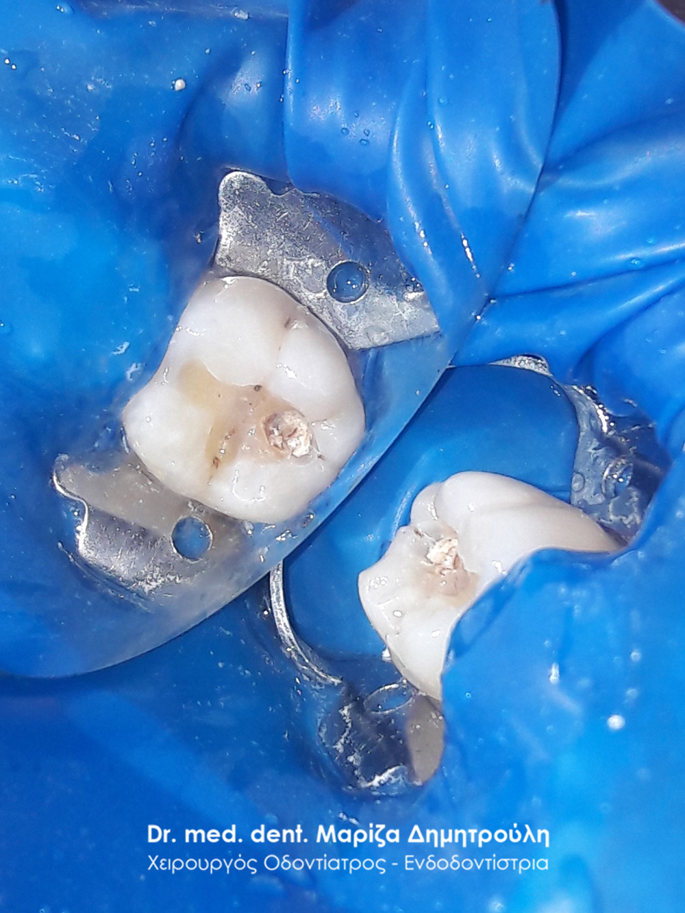
Image of tooth immediately after cavity opening
Placing material to protect the tooth’s nerve
META
Περιστατικό – Tooth Filling
Η ασθενής παραπονιόταν για έναν ενοχλητικό πόνο στην αριστερή πλευρά της άνω γνάθου. Μετά την κλινική εξέταση διαπιστώθηκε μια βαθιά τερηδονική κοιλότητα στον πρώτο αριστερό γομφίο της άνω γνάθου. Χορηγήθηκε τοπική αναισθησία και πραγματοποιήθηκε η διάνοιξη της κοιλότητας και η απομάκρυνση των τερηδονισμένων οδοντικών ιστών. Το έλλειμμα του δοντιού αποκαταστάθηκε με λευκό σφράγισμα σύνθετης ρητίνης.
BEFORE
The extent of caries on the tooth after opening the cavity
META
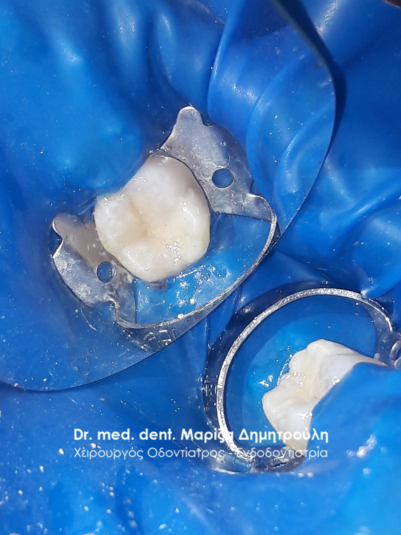
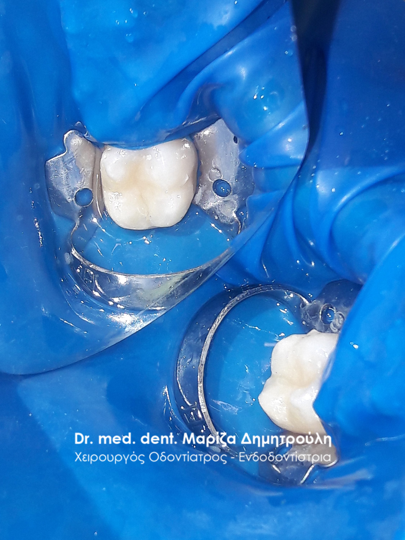
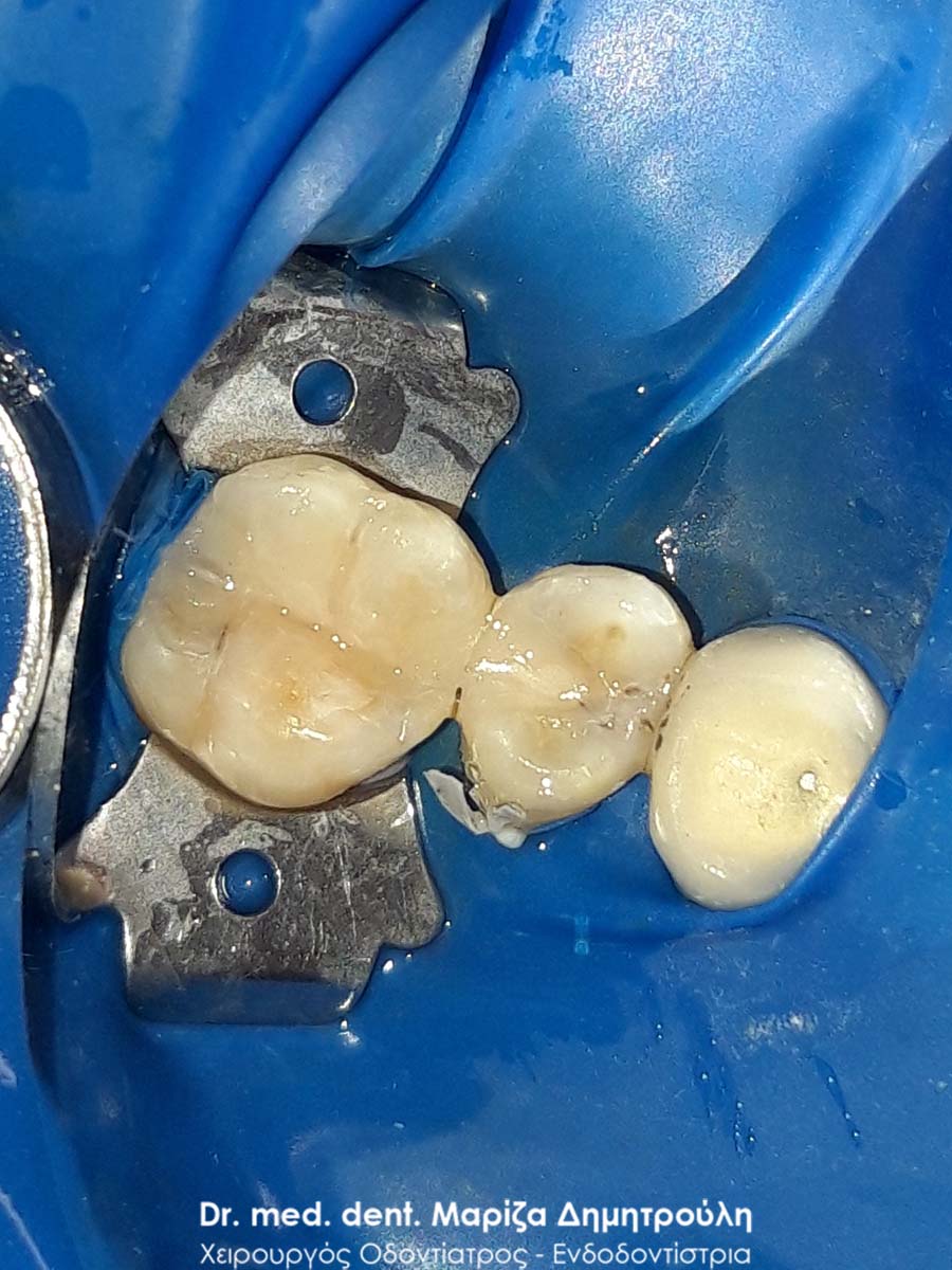
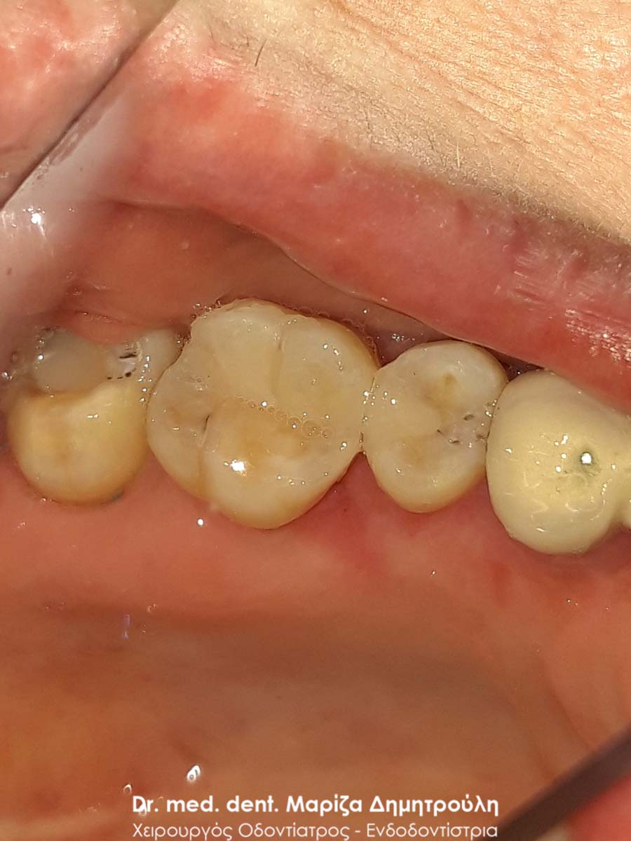
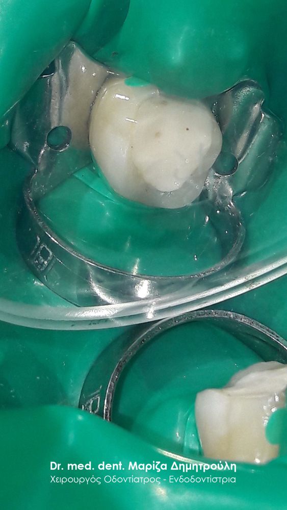
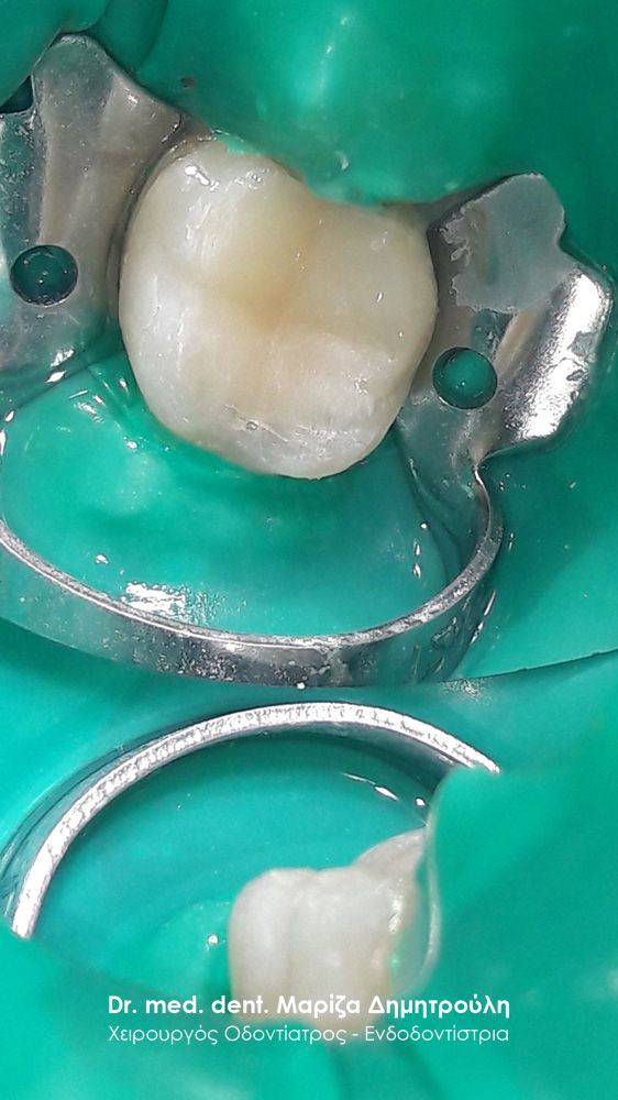
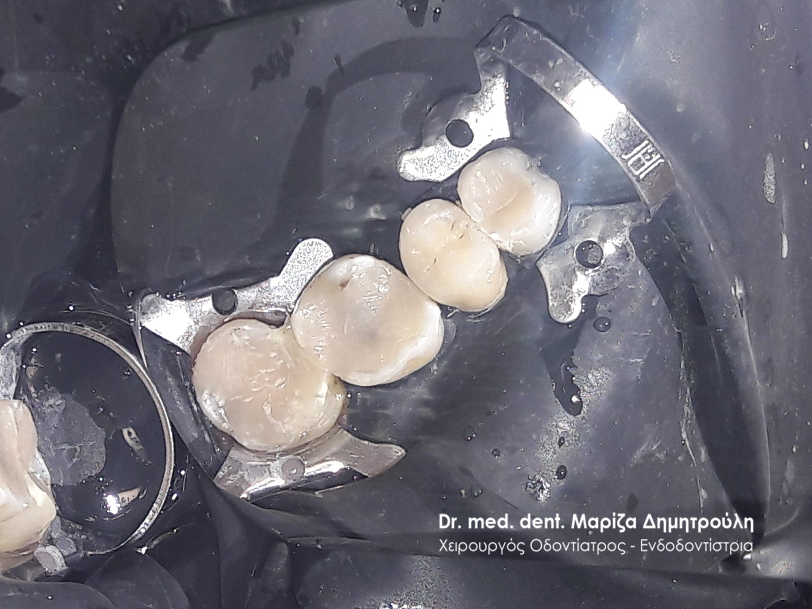
Case – Dental Fillings and Prosthetics
The patient came to the clinic with the desire to restore her teeth. After the clinical examination it was found on the upper left side that 3 teeth needed immediate dental treatment. As can be seen in the photos, there were old resin fillings (white fillings) in the first premolar and the second molar, which had been re-carious. After the old fillings were derailed, the underlying caries was removed and these 2 teeth were restored with new white fillings. The first molar had an extensive old amalgam filling and in addition a piece of the natural tooth had broken off. Due to the extensive damage to the tooth, it was decided to restore the tooth with a zirconium crown.
BEFORE
META
Περιστατικό – Tooth Filling
Ο ασθενής ανέφερε πόνο στην πάνω δεξιά πλευρά. Μετά την κλινική εξέταση διαπιστώθηκε μια μεσαίου βαθμού τερηδονική αλλοίωση στο δεξί πάνω πρώτο γομφίο. Αποφασίστηκε η αποκατάσταση του δοντιού με λευκό σφράγισμα.
BEFORE
META
Περιστατικό – Tooth Filling
Ο ασθενής ανέφερε έναν ήπιο περιοδικό πόνο κατά τη μάσηση, που τον εντόπιζε στην άνω δεξιά πλευρά του στόματος του. Μετά την κλινική εξέταση διαπιστώθηκε μία μικρή τερηδονική κοιλότητα, η οποία και αποκαταστάθηκε με λευκό σφράγισμα σύνθετης ρητίνης.
BEFORE
META
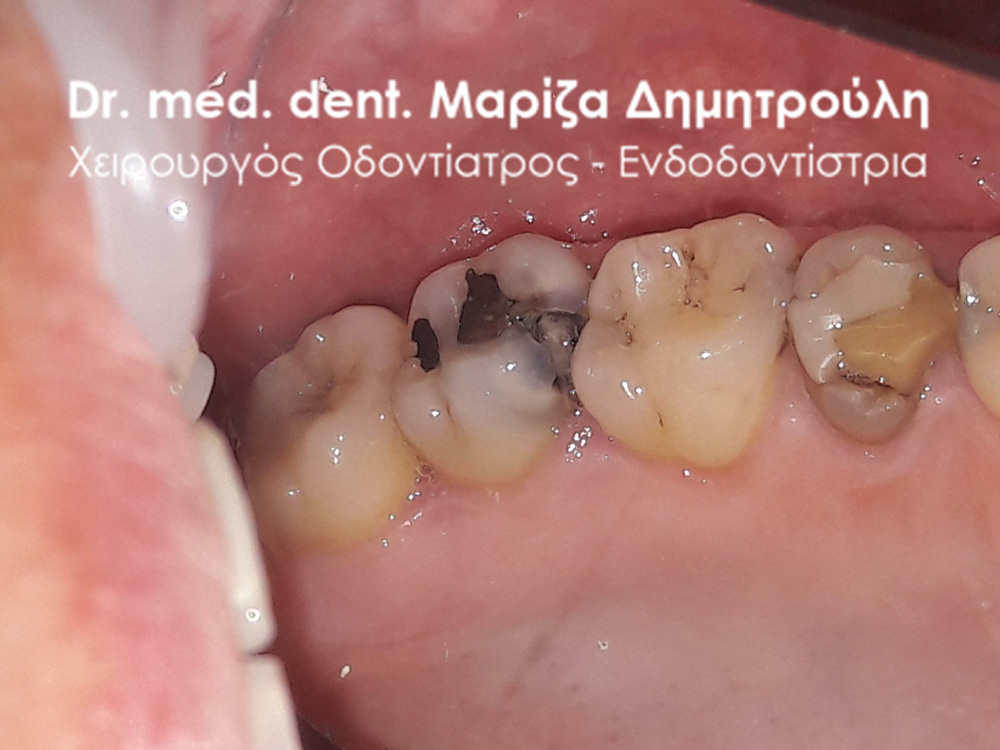
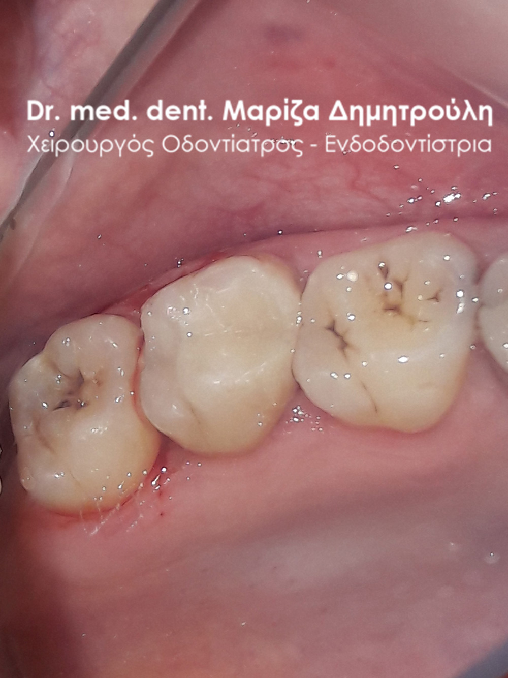
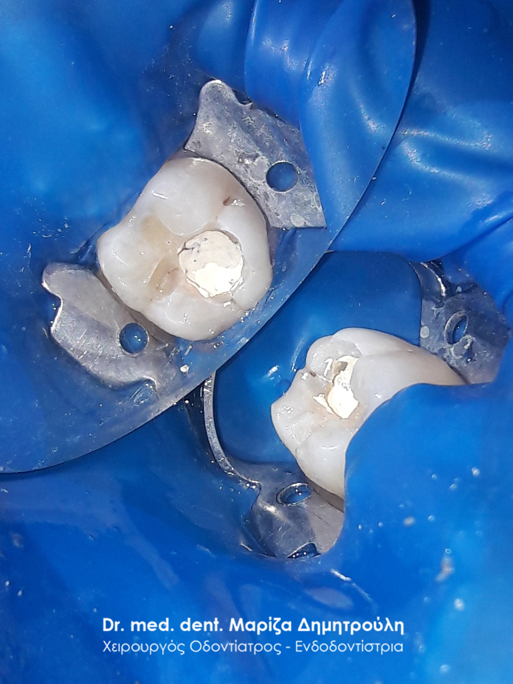
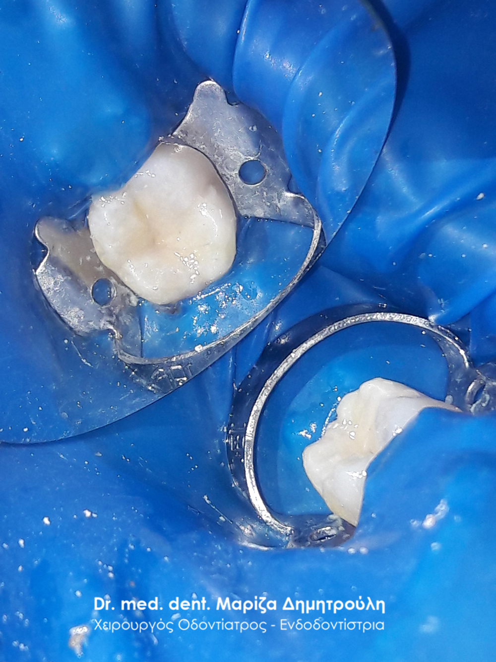
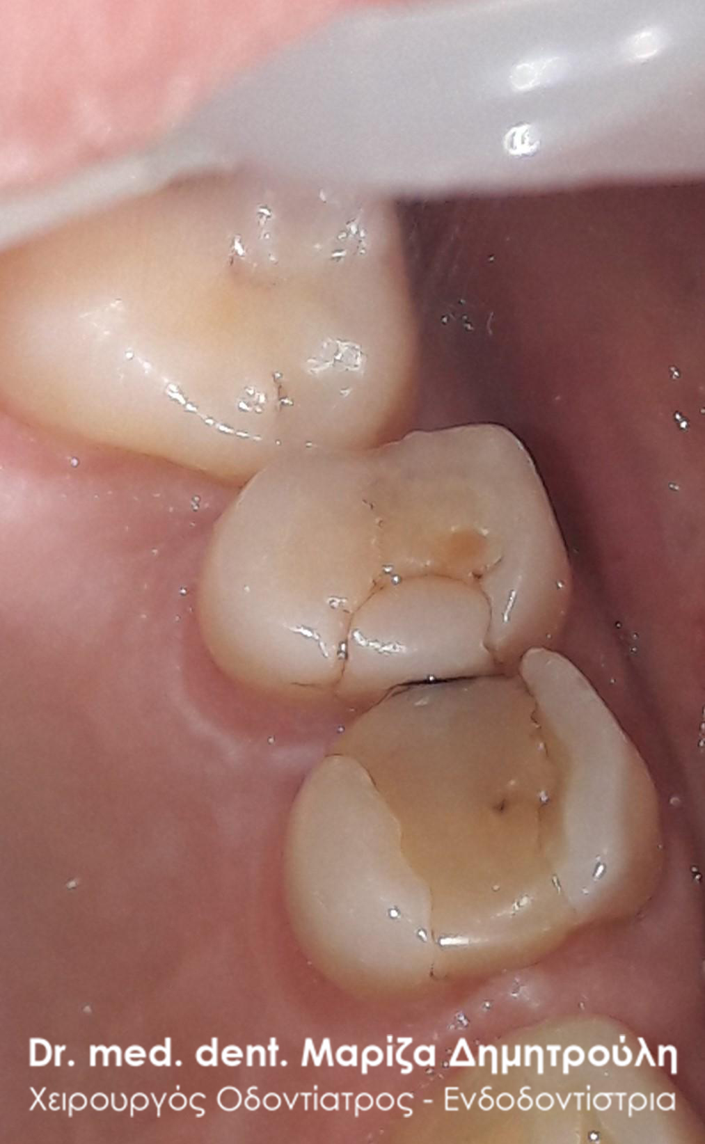
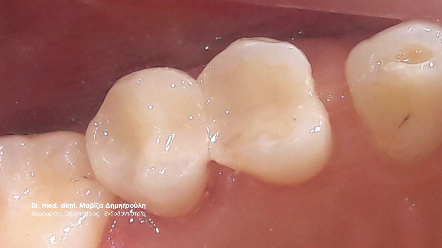
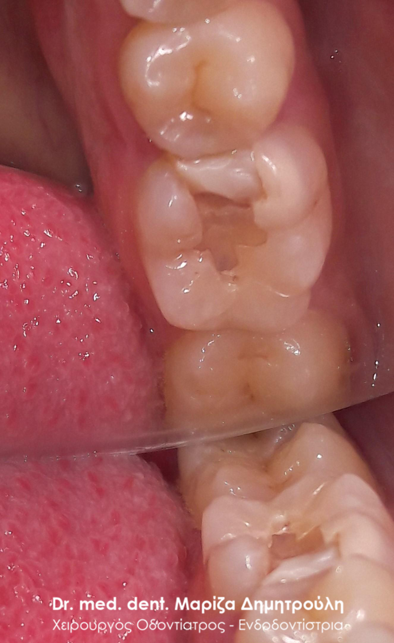
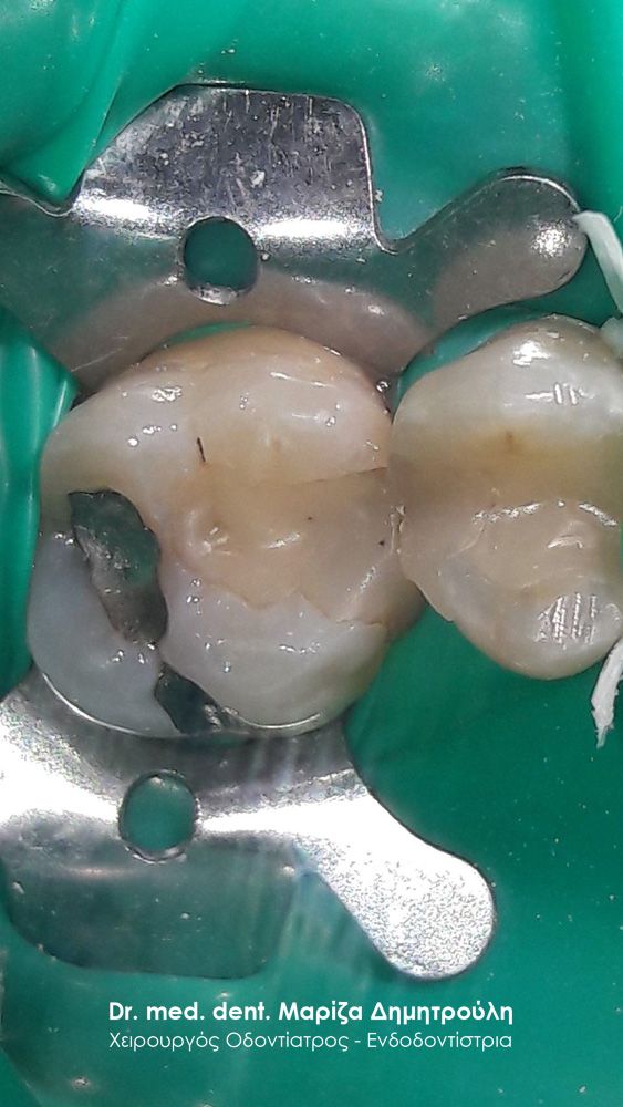
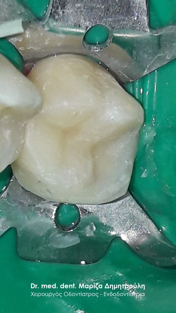
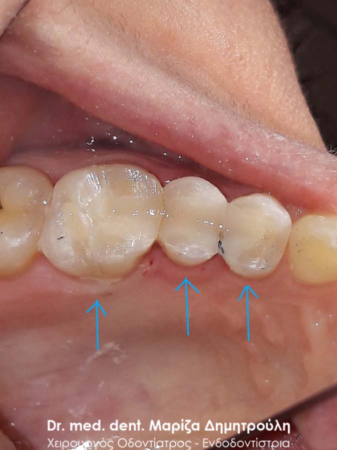
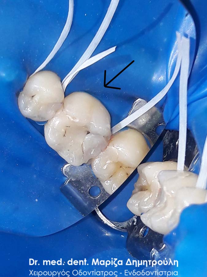
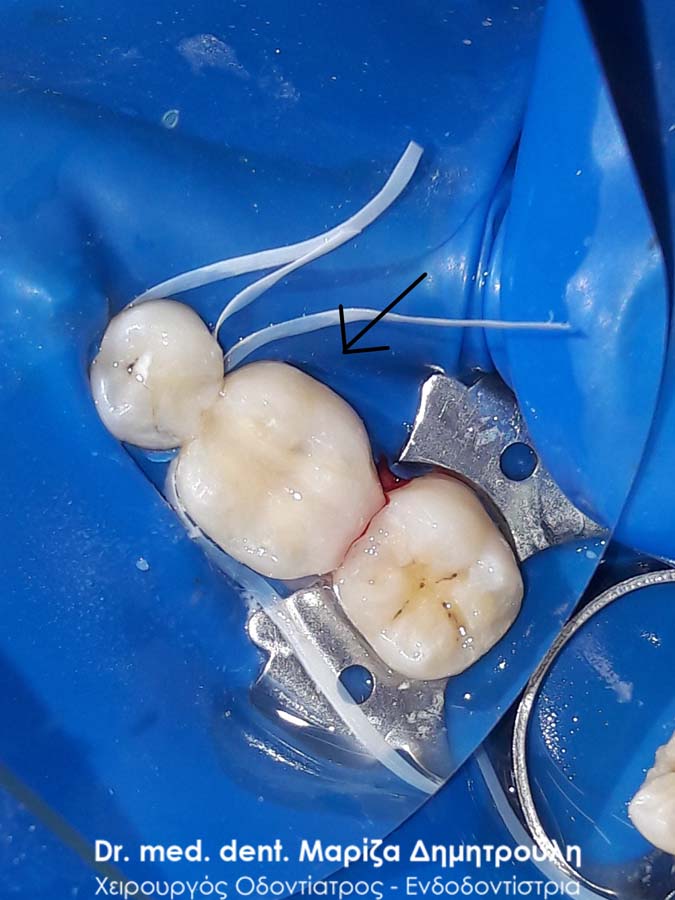
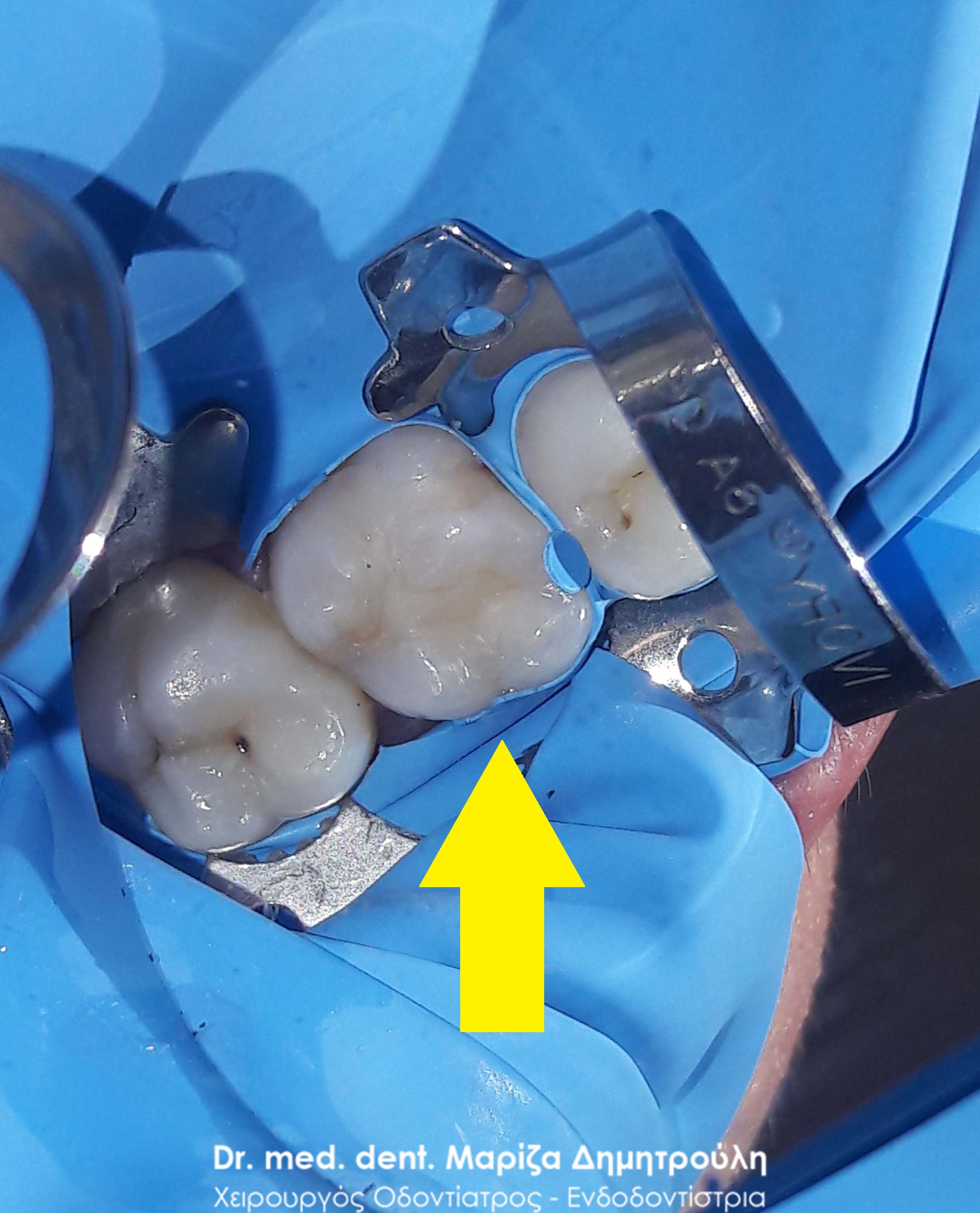
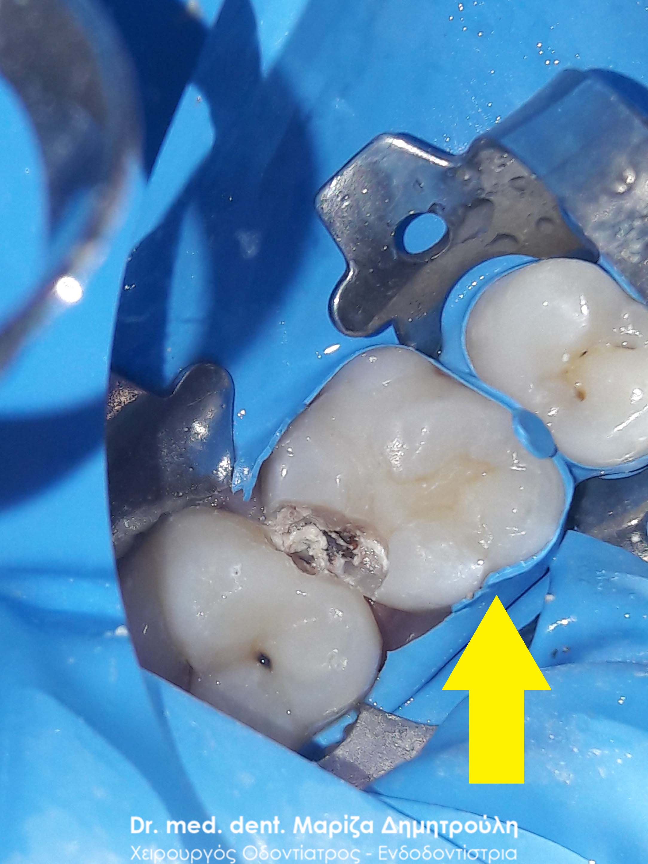
Incident – Replacement of fillings on 2 teeth
As part of the annual dental checkup, some old fillings were found, which required their replacement since their margins were imperfect and did not have a hermetic contact with the tooth. The original left photo also shows the strong yellow tint of the old fillings. After removing the old blockages and cleaning the caries, the teeth were restored with new white fillings of composite resin.
BEFORE
META
Incident – White Tooth Seal
BEFORE
META
Incident – White Tooth Seal
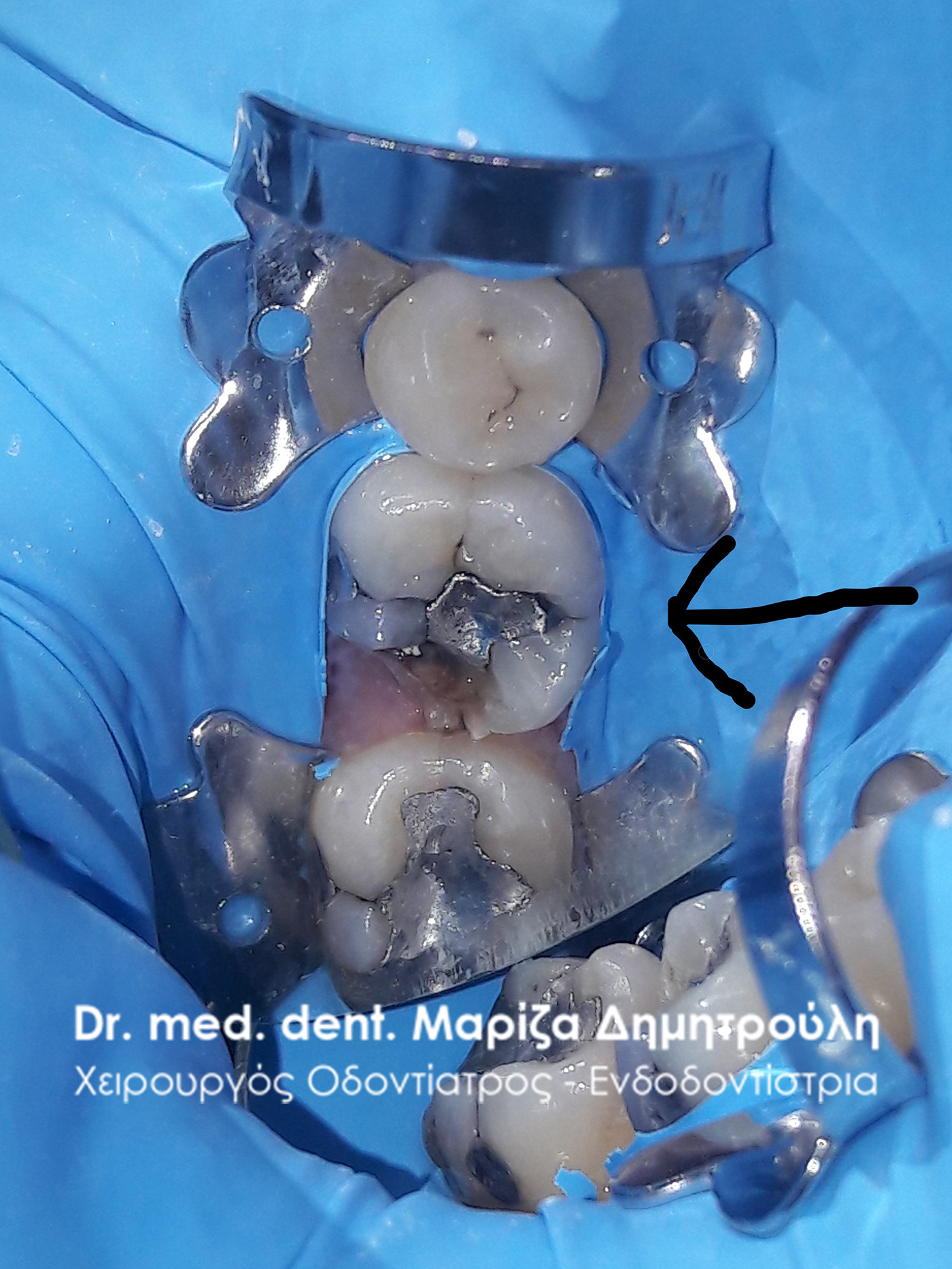
The lower right second molar was restored one month ago with a resin seal. The patient visited the clinic because her tooth was constantly hurting after the filling was created. After administration of local anesthesia, the old white filling was ground and it was found that the filling material extended to the level of the nerve of the tooth. This event caused the patient’s pain symptoms. A special filling material with soothing and healing properties was placed on the tooth. At a second appointment, since the patient was no longer in pain, the tooth was restored with the new final white composite resin filling.
BEFORE

META
Incident – Tooth Filling Replacement
The patient had an old filling, which hurt either when she chewed or spontaneously in the absence of any stimulus. It was decided to remove the old filling and temporarily restore the tooth with a special occlusive material, which has soothing properties. Since the patient’s symptoms subsided, the final restoration of the tooth was carried out at a second appointment with a white composite resin filling.
BEFORE
META
Περιστατικό – Tooth Filling
Η ασθενής περιοδικά αισθανόταν μία μικρή ενόχληση κάθε φορά που έτρωγε στην αριστερή πλευρά της άνω γνάθου. Η εξέταση αποκάλυψε την ύπαρξη μιας μικρής τερηδόνας στο δεύτερο γομφίο της άνω γνάθου. Μετά τον εκτροχισμό και την αφαίρεση της τερηδόνας το δόντι αποκαταστάθηκε με λευκό σφράγισμα ρητίνης.
BEFORE
META
Incident – Replace Seals
The patient came to the clinic with the desire to restore the old fillings in 2 teeth. The patient reported that the one filling that was on the first upper left molar hurt her periodically without the pain being intense and unbearable. As the photos testify, after the removal of the old filling, caries was found, which caused the pain when chewing. After the removal of the carious dental tissues, the tooth was restored with a white composite resin filling.
Regarding the second filling on the lower right first molar, it was a temporary restoration of the tooth after endodontic treatment (denervation), which had to be replaced with a final composite resin filling (white filling).
BEFORE
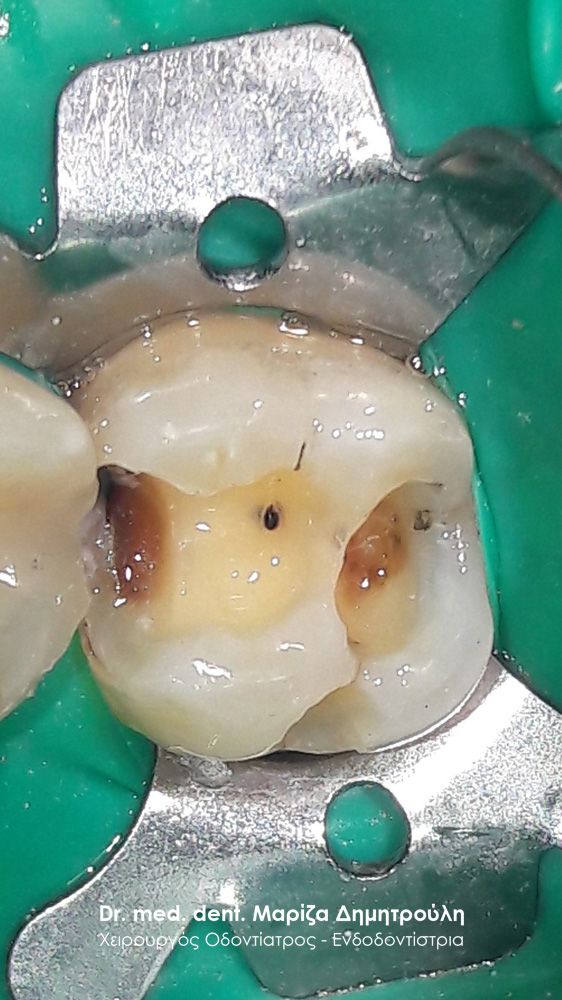
Image of the tooth after removing the old filling
META
BEFORE
META
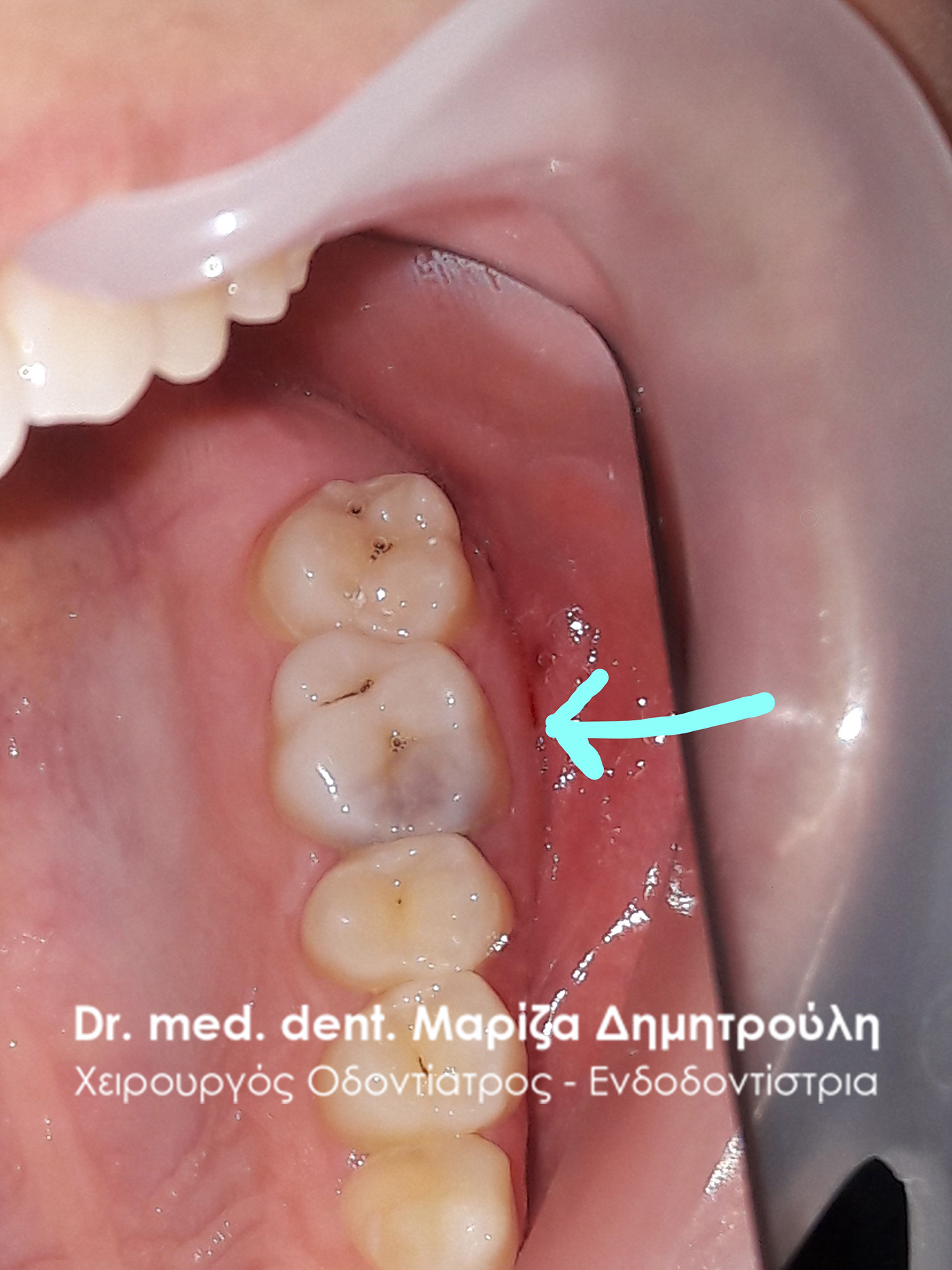
Incident – White Tooth Fillings
As part of the annual standard dental checkup, small carious cavities were found in the patient’s mouth, which were restored with white composite resin fillings.
BEFORE
META
META
Incident – White Tooth Fillings
The patient had 2 fillings replaced 2 months ago (on the 1st molar and on the 1st premolar) on the left side of the upper jaw. She stated that she has been suffering from severe pain ever since. After the clinical examination, the presence of caries in the 2nd molar and the wisdom tooth was established. It was decided to restore the decayed teeth with white composite resin fillings. As for the teeth with sensitivity, they were replaced with a temporary soothing filling material. At a subsequent appointment, the final restoration of these teeth was carried out with white resin fillings.
BEFORE
META
META
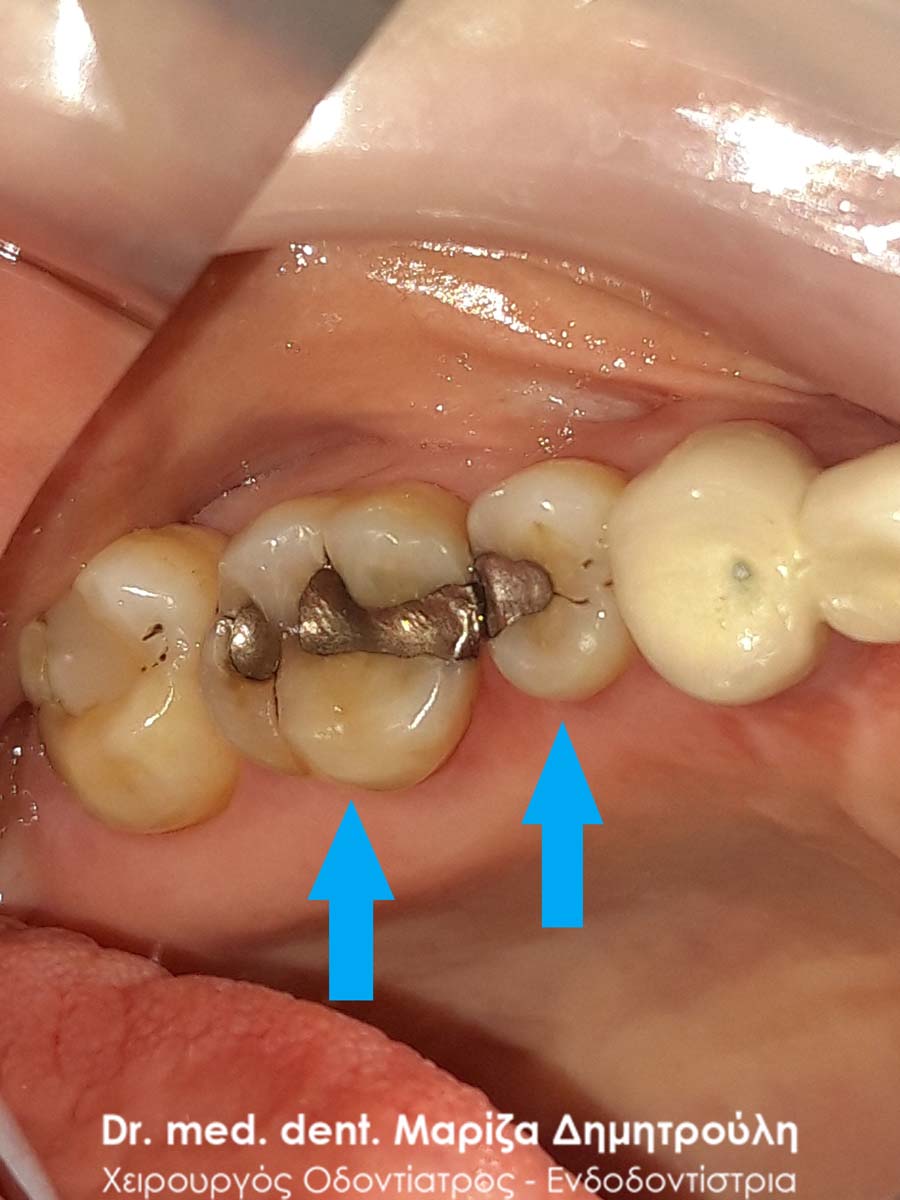
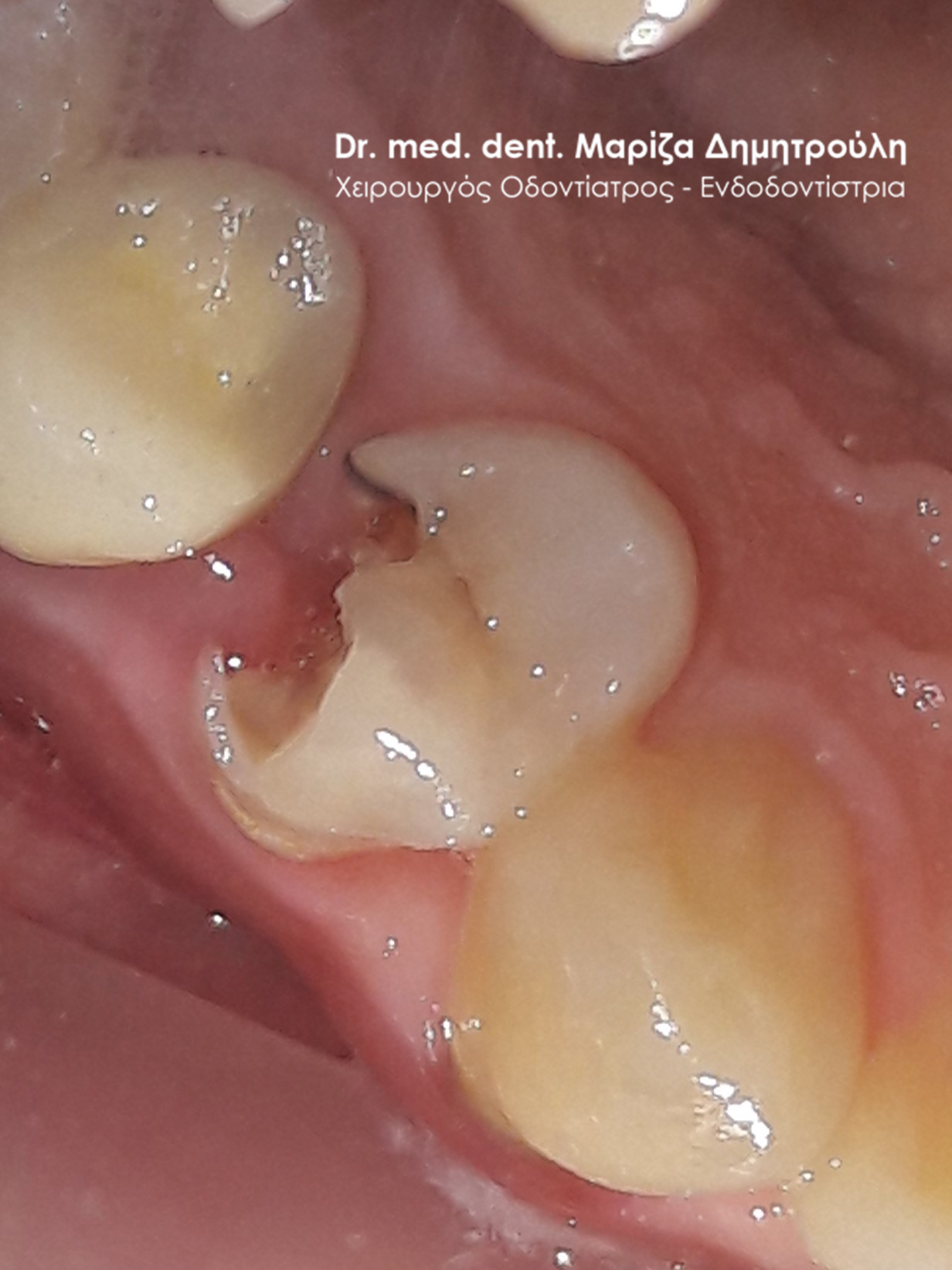
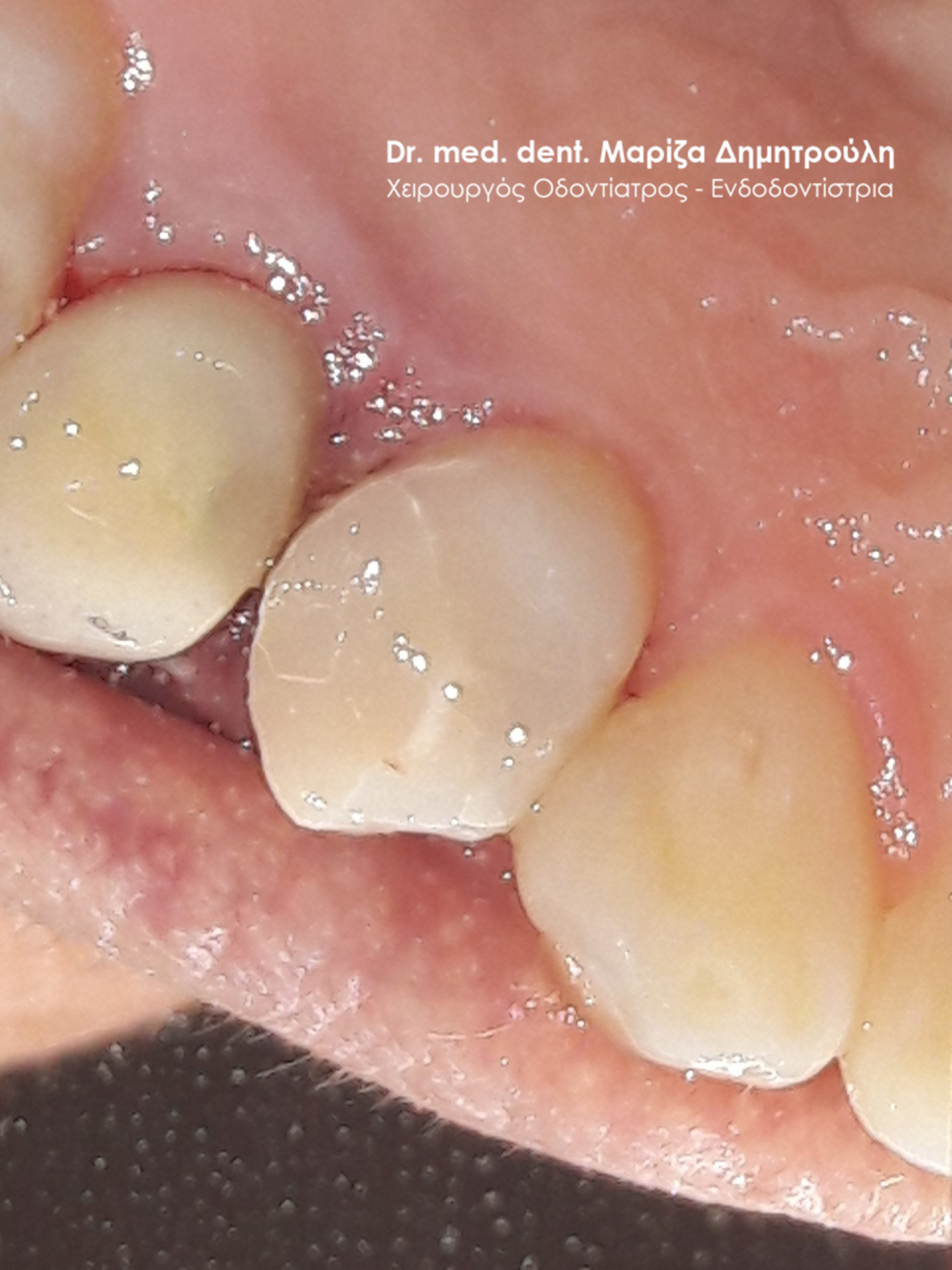
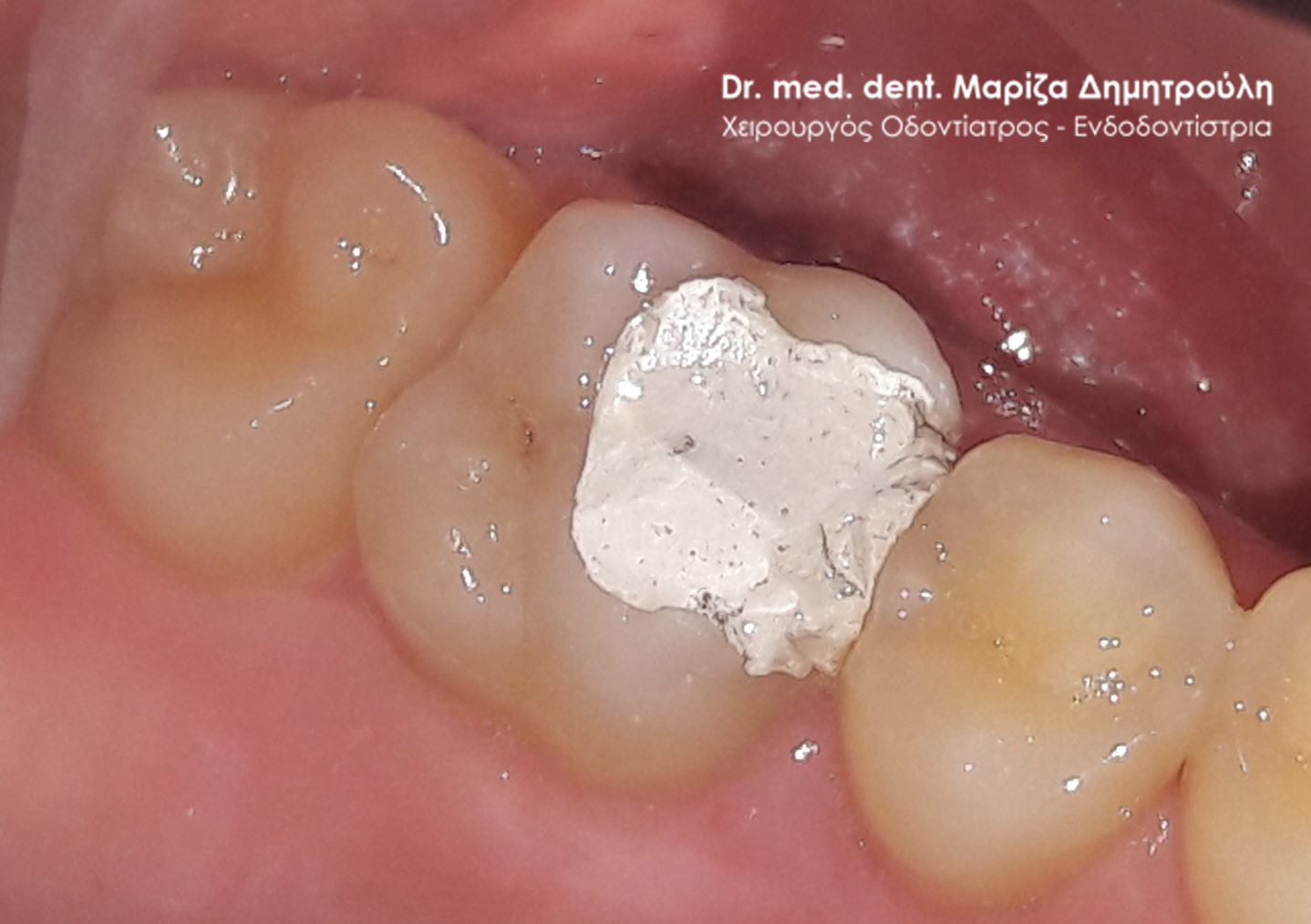
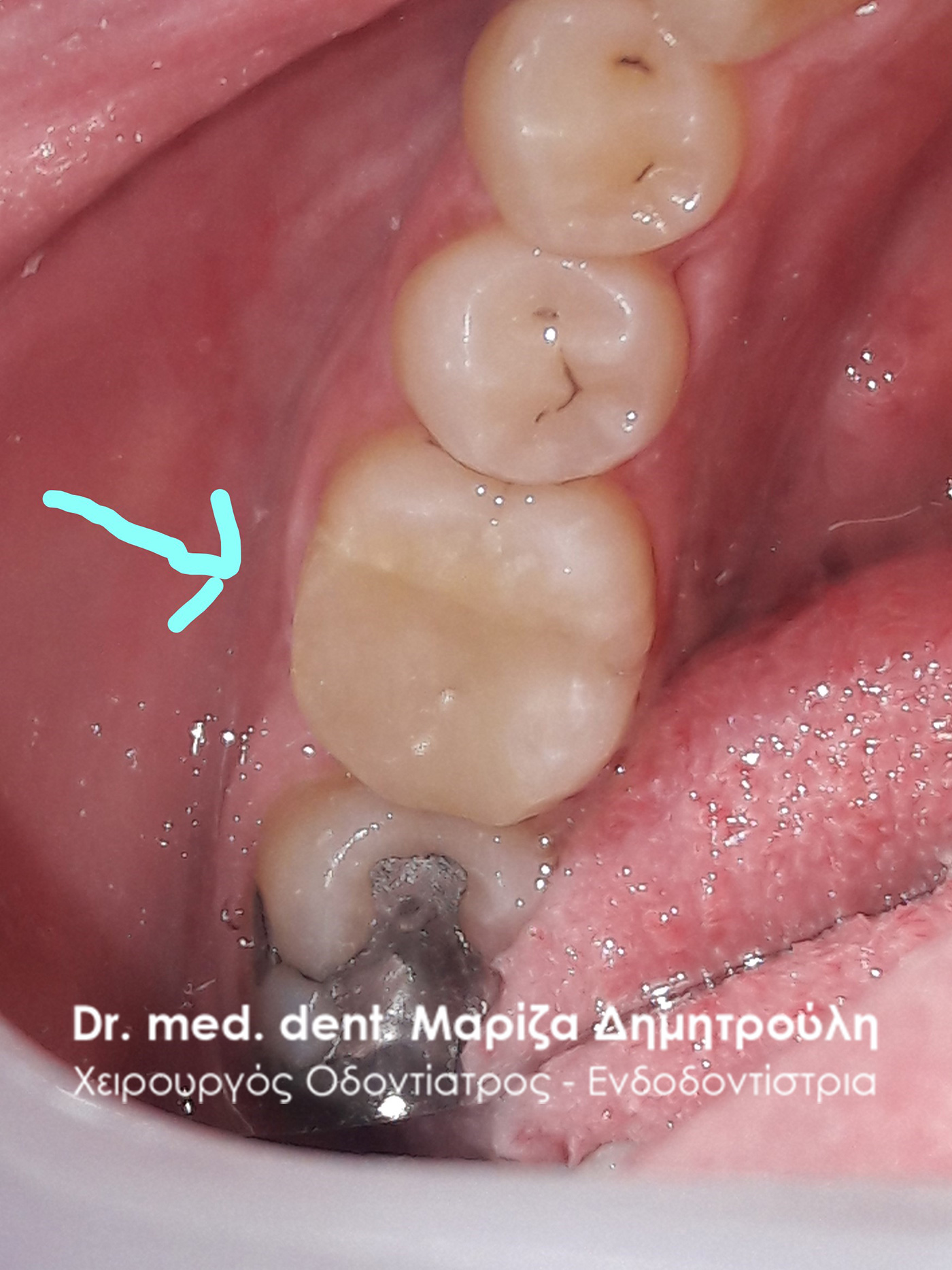
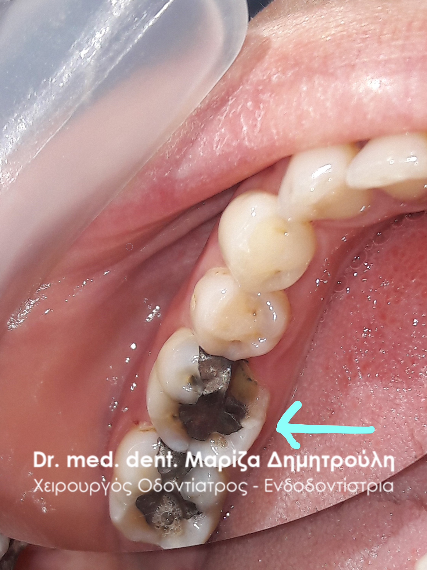
Incident – Tooth Filling Replacement
The patient visited the office as her lower right first molar was periodically painful. After clinical examination, an old large amalgam filling was found, the margins of which were not in contact with the tooth and gaps had formed between the black filling and tooth walls. It was decided to replace the old seal with a new white composite resin seal.
The middle photo shows the tooth deficit after removal of the amalgam filling and carious dental tissues. The photo on the right shows the final restoration of the tooth.
BEFORE
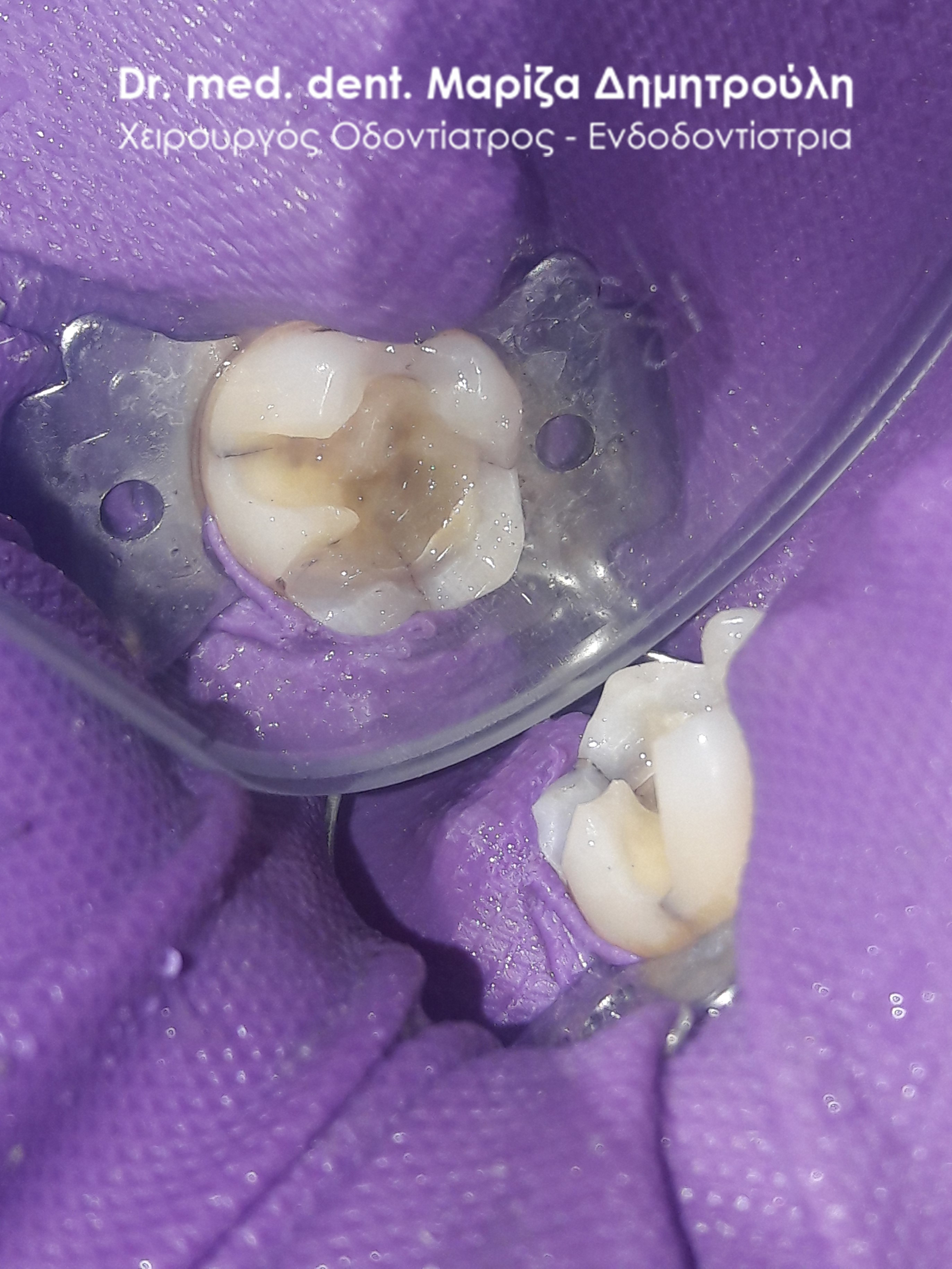
Image of tooth after removal of old filling and caries
META
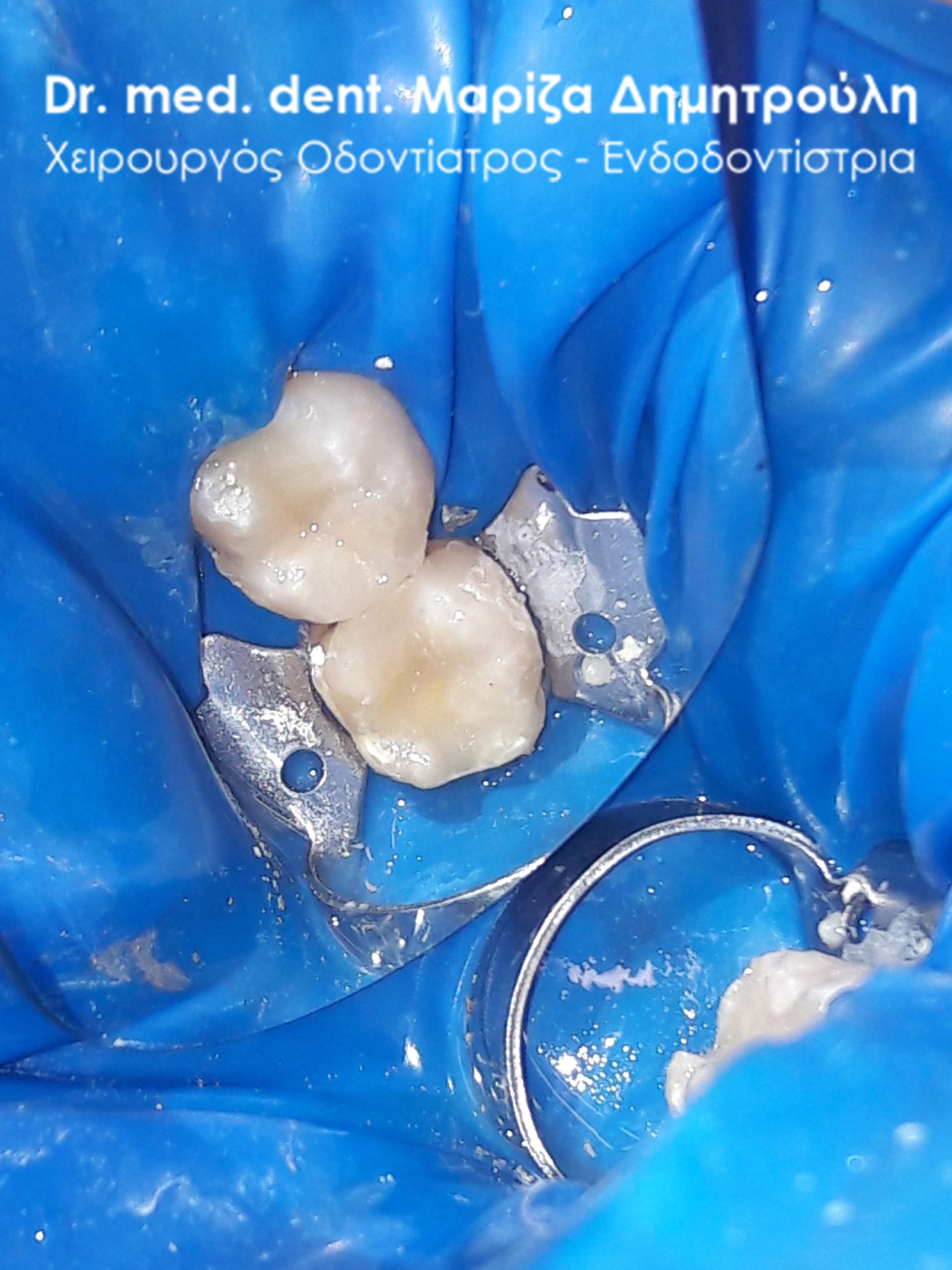
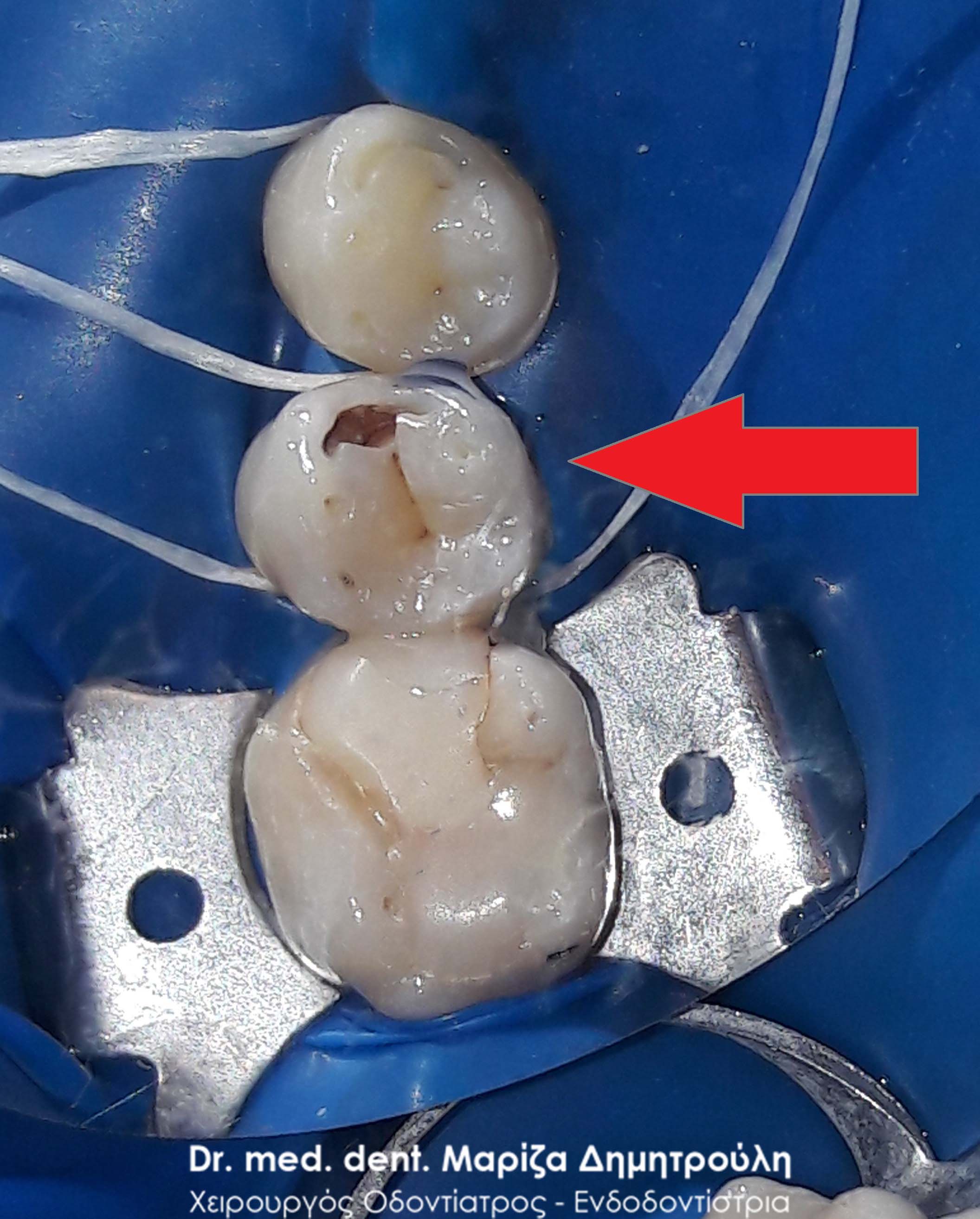
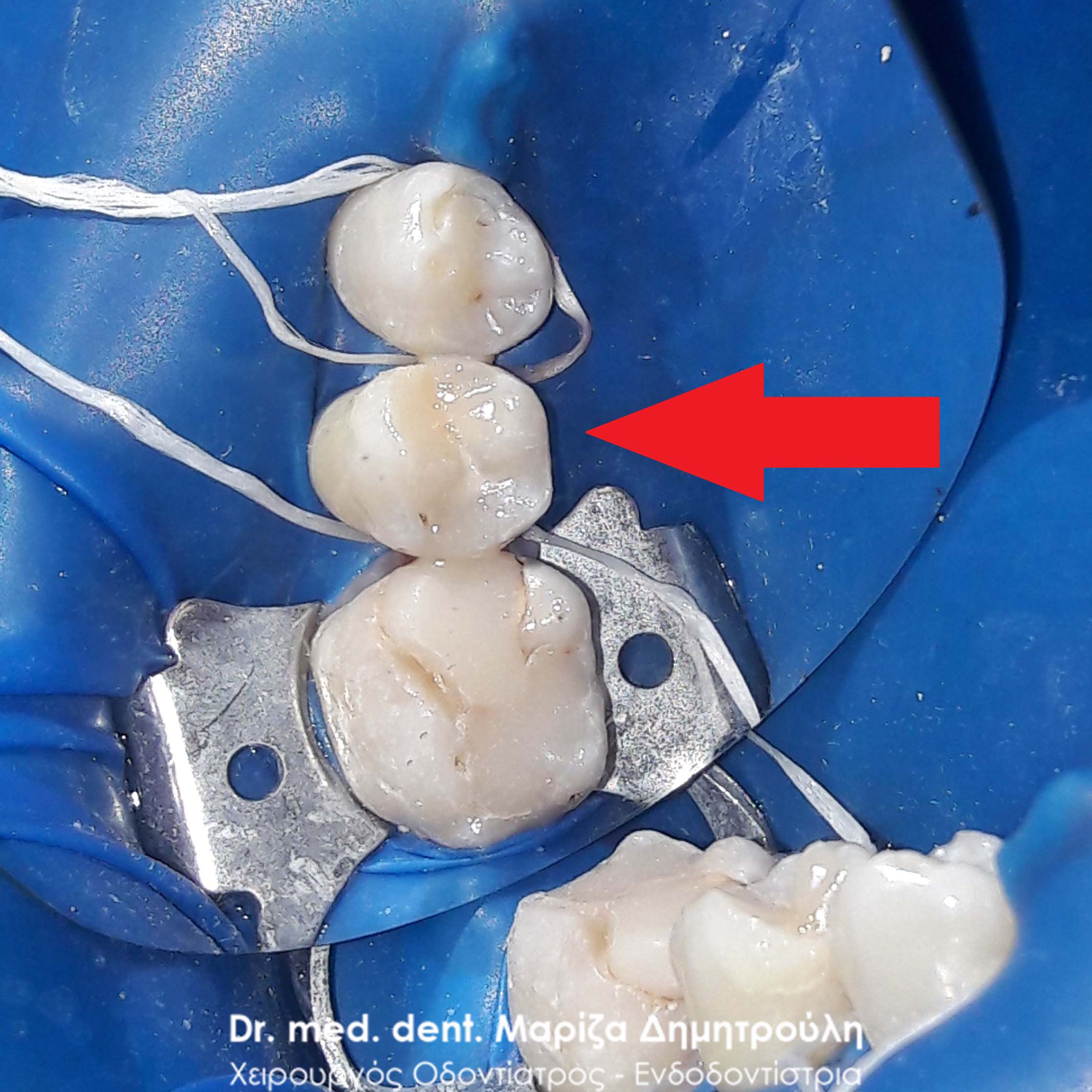
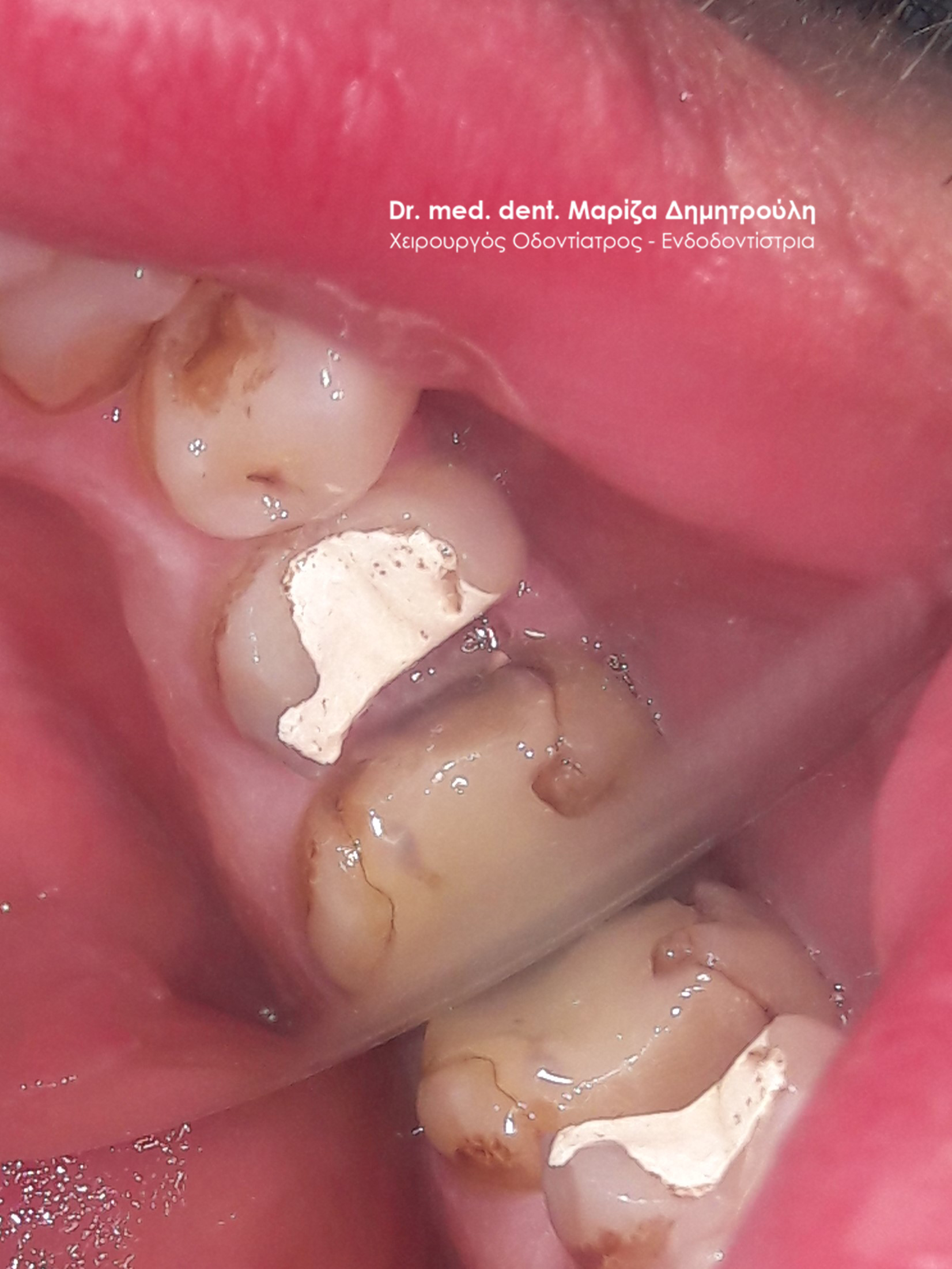
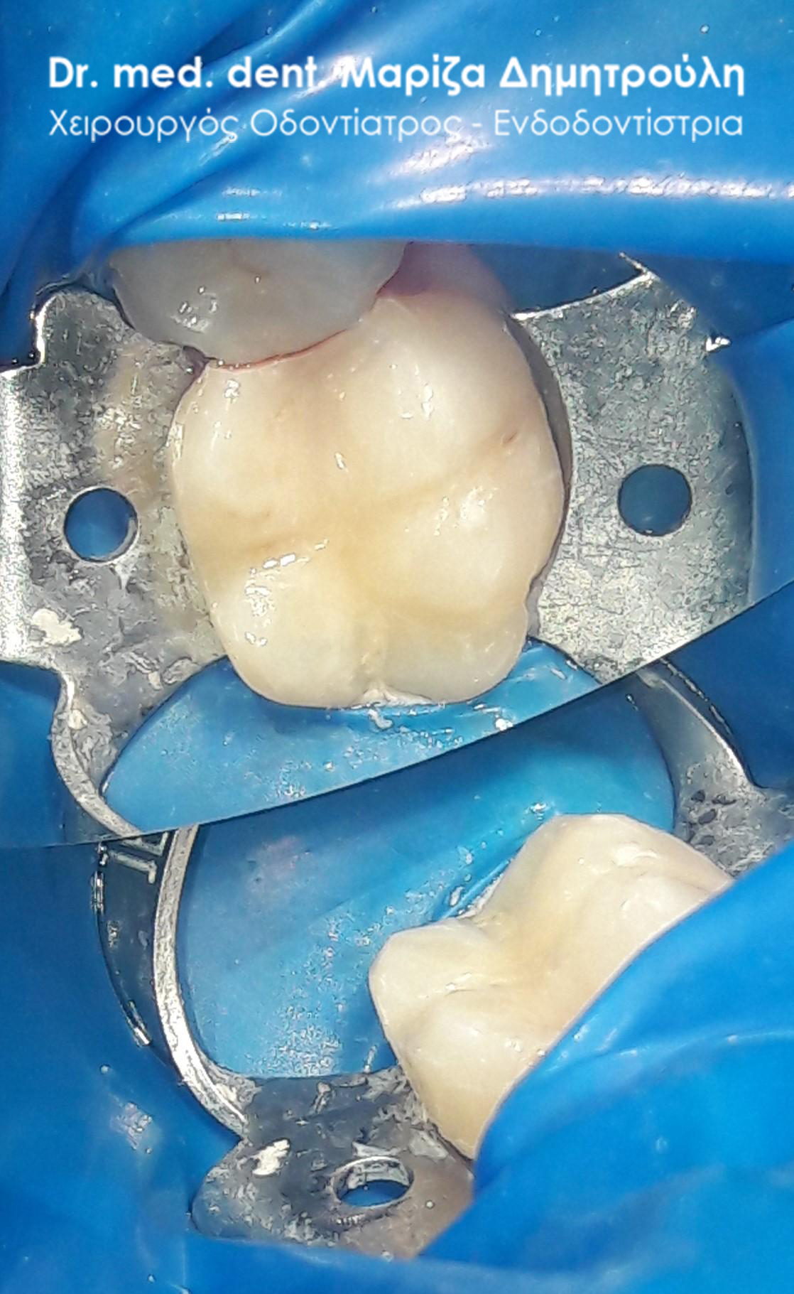
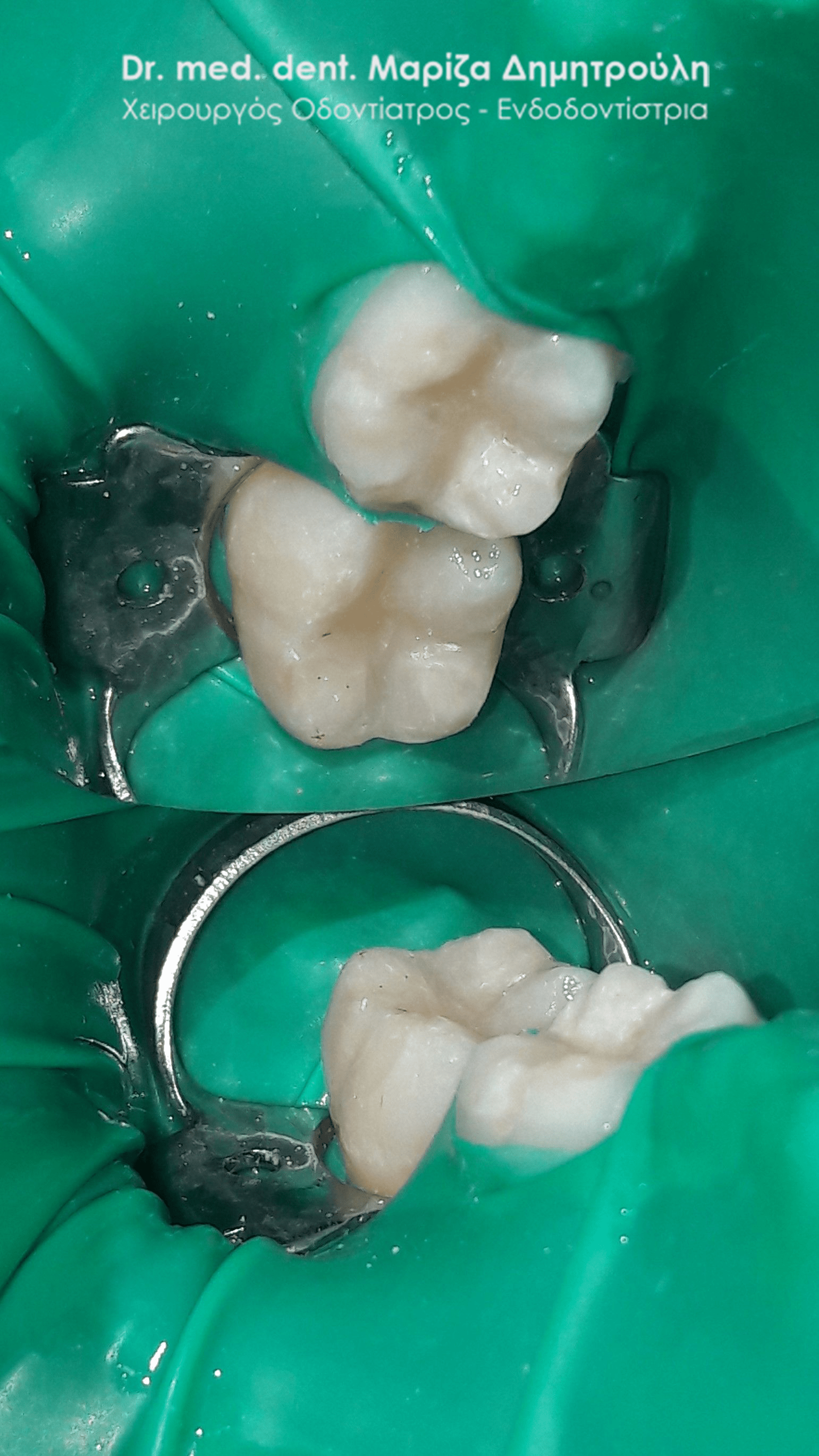
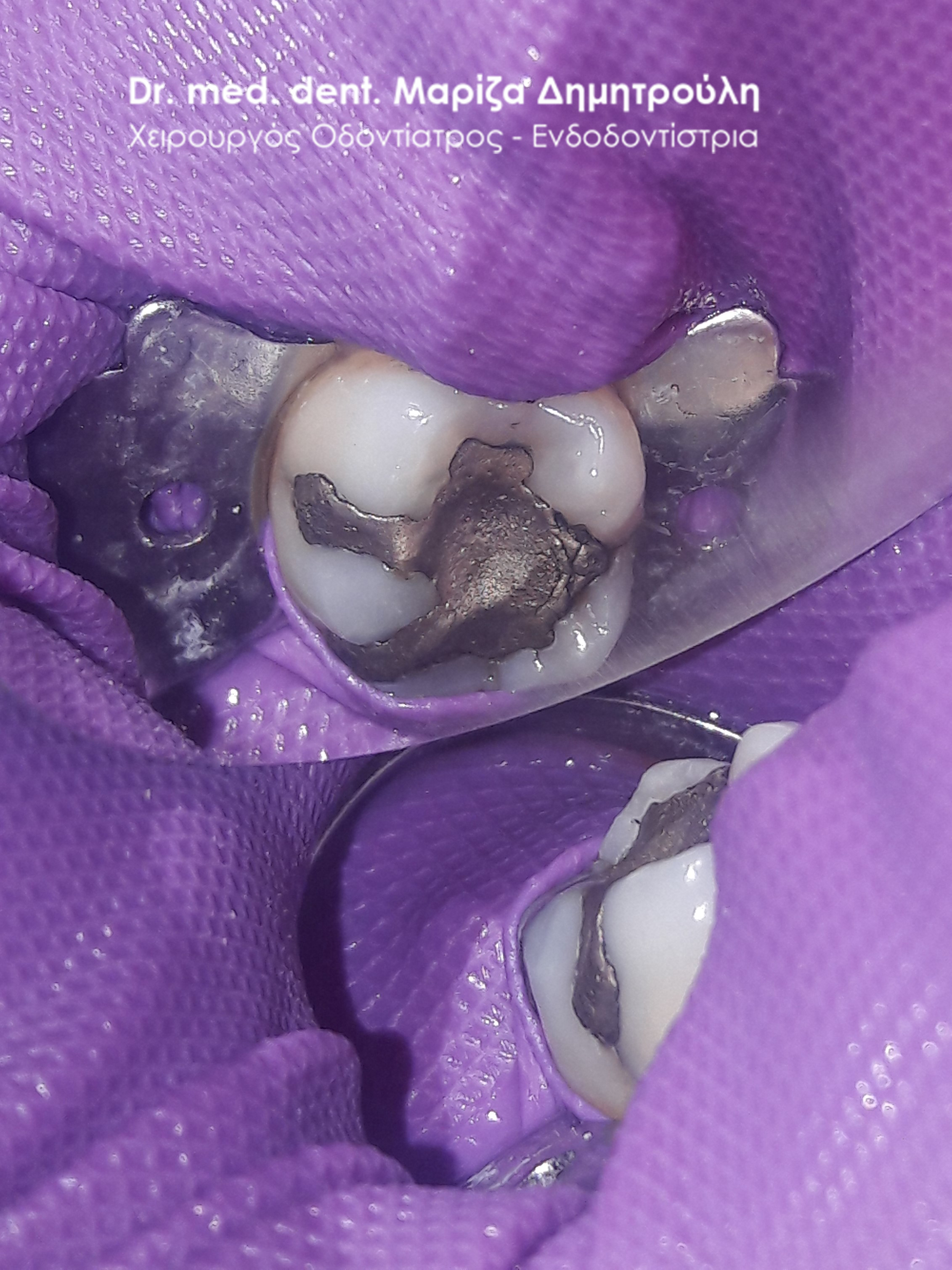
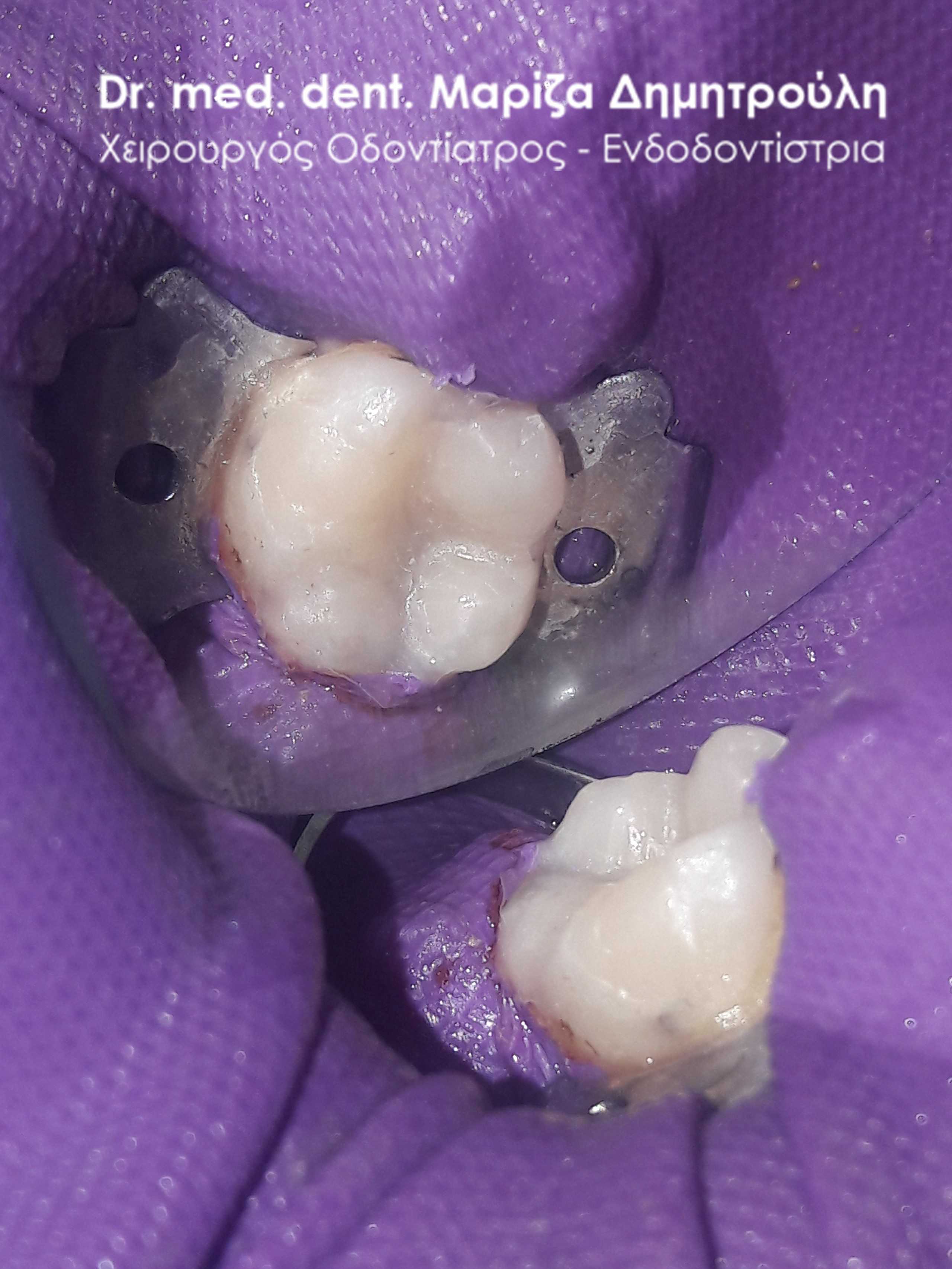
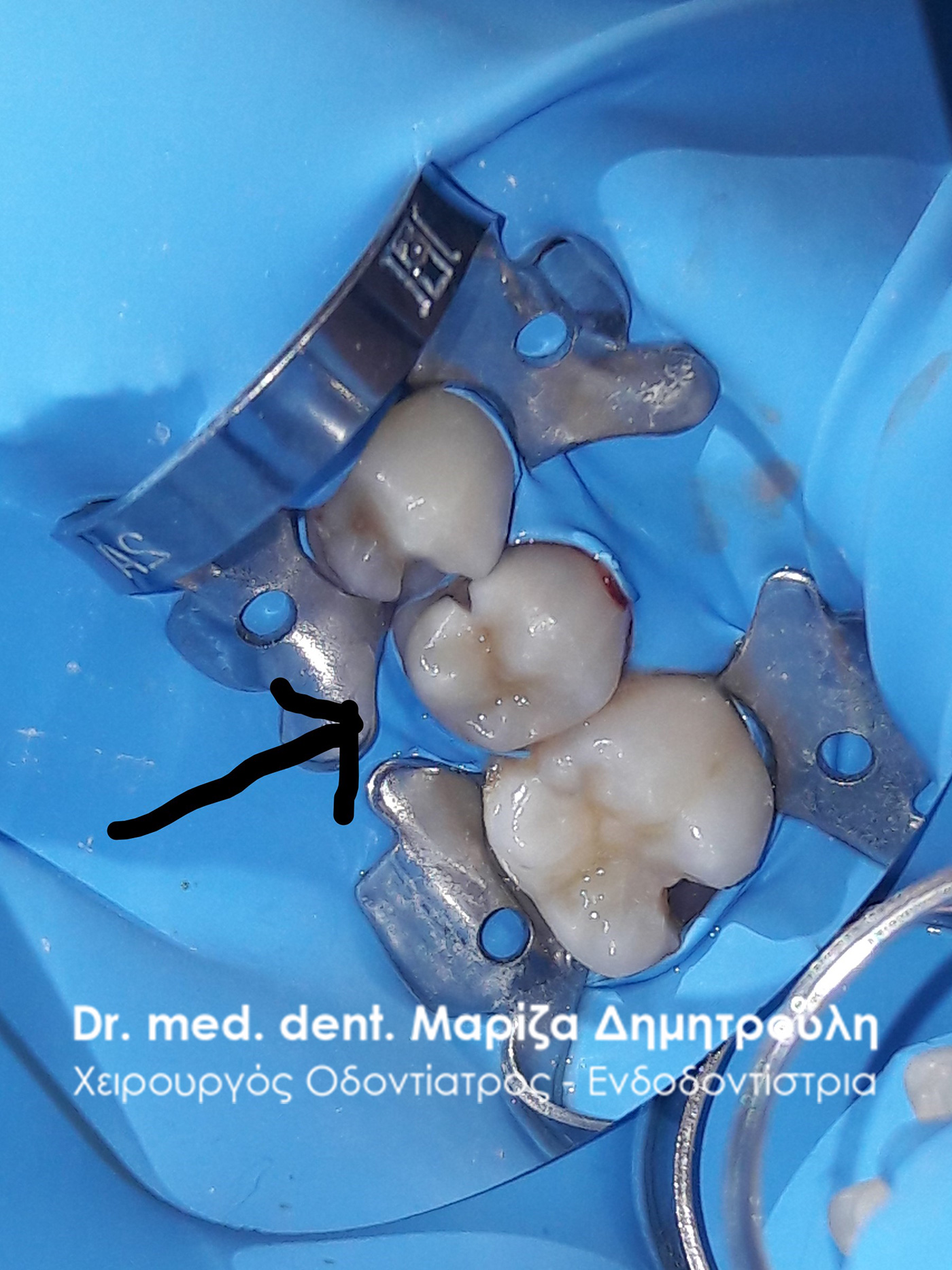
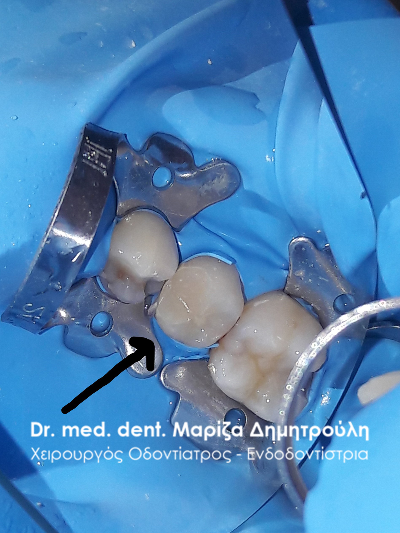
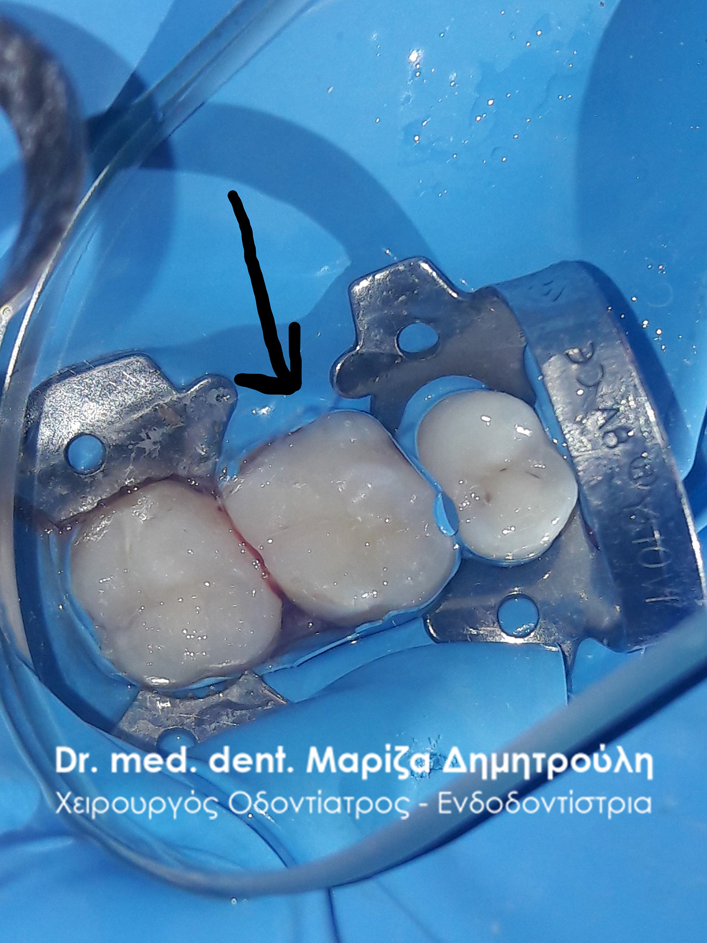
Incident – Replacing Dental Fillings
The patient wanted to replace the old black amalgam fillings withwhite composite resin fillings.
The treatment was performed using a rubber isolator as defined by global dental protocols for the removal of black fillings. Dentists would do well to use isolators when removing amalgam fillings so that the patient inhales as little as possible and does not ingest the mercury that is released during the process.
The treatment plan for the first molar (tooth with a very white filling) was under discussion, hence the restoration of the tooth with a temporary filling.
BEFORE
META
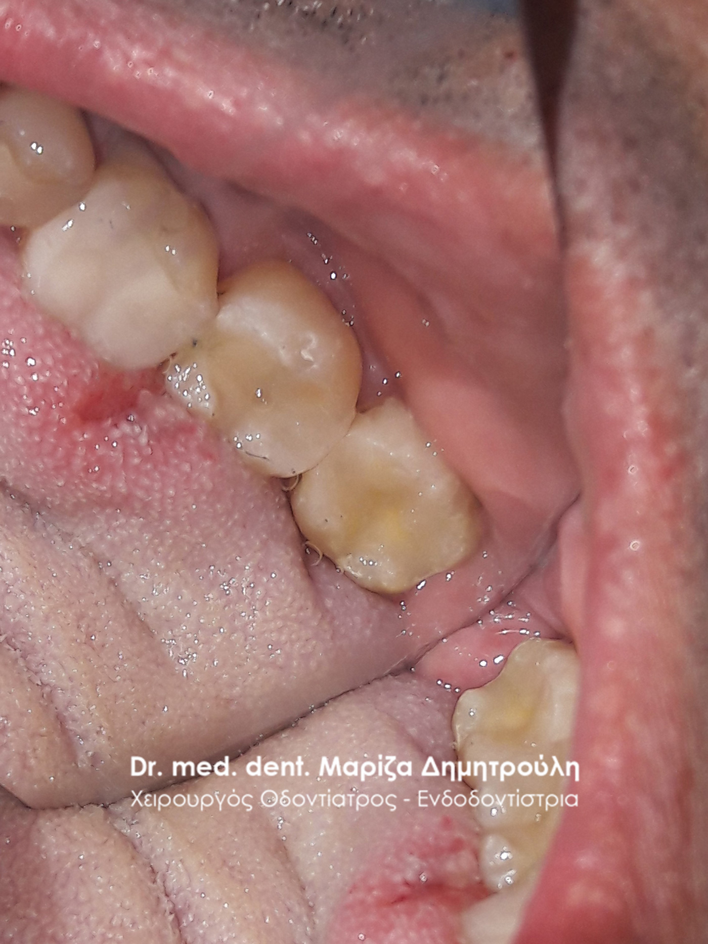
Incident – Replacing Dental Fillings
The patient reports a periodic pain in the left side of the upper jaw.
After the clinical examination, three old white fillings were found, the borders of which in some were re-carious and in others were not in contact with the tooth and gaps had formed between the filling and the dental walls. It was decided to replace the old fillings with new white composite resin fillings.
BEFORE
META
Incident – Replacing Dental Fillings
The patient wished to replace the black amalgam fillings on the right first molar and the second molar with white composite resin fillings. The treatment was performed using a rubber isolator as defined by global dental protocols for the removal of black fillings. When dentists use isolators when removing amalgam fillings, the patient inhales very little and does not ingest the mercury that is released during the process.
BEFORE
META
Incident – Tooth denervation and filling
The patient visited the office to repair a self-perceived “hole”. After the clinical examination, a carious lesion was found on the left first molar. Even though the cavity was not wide, the caries extended to a great depth at the level of the pulp, i.e. the nerve of the tooth. The procedure of denervation of the tooth began and after its completion a white composite resin filling was performed. The restoration of the tooth was performed with a filling and not with a tooth holder, because the tooth deficit was not so great that a crown was required to protect it.
BEFORE
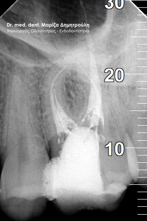
Root Canal Treatment
Restoration of a tooth with a temporary seal after the end of denervation
Final white stamp
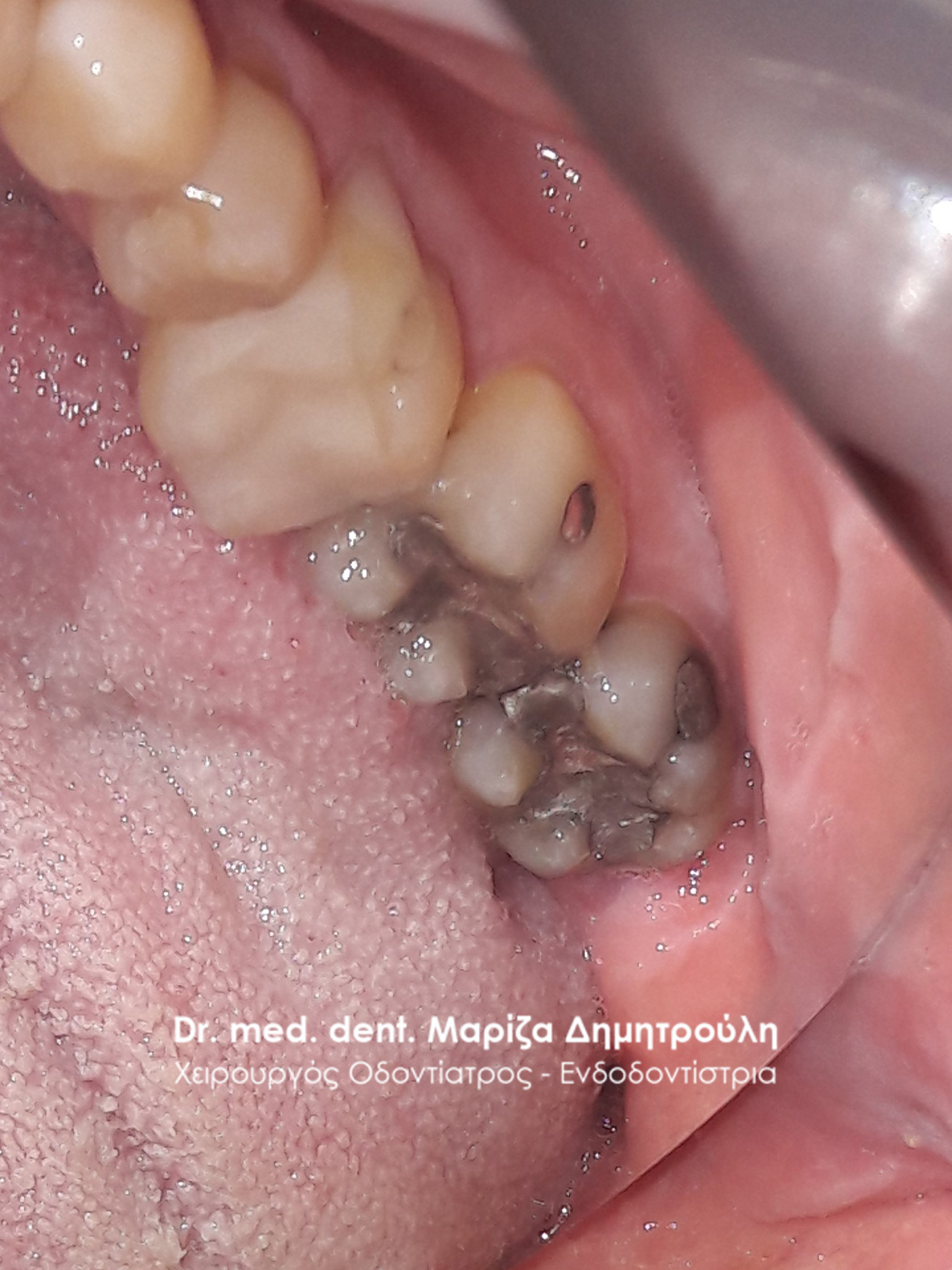
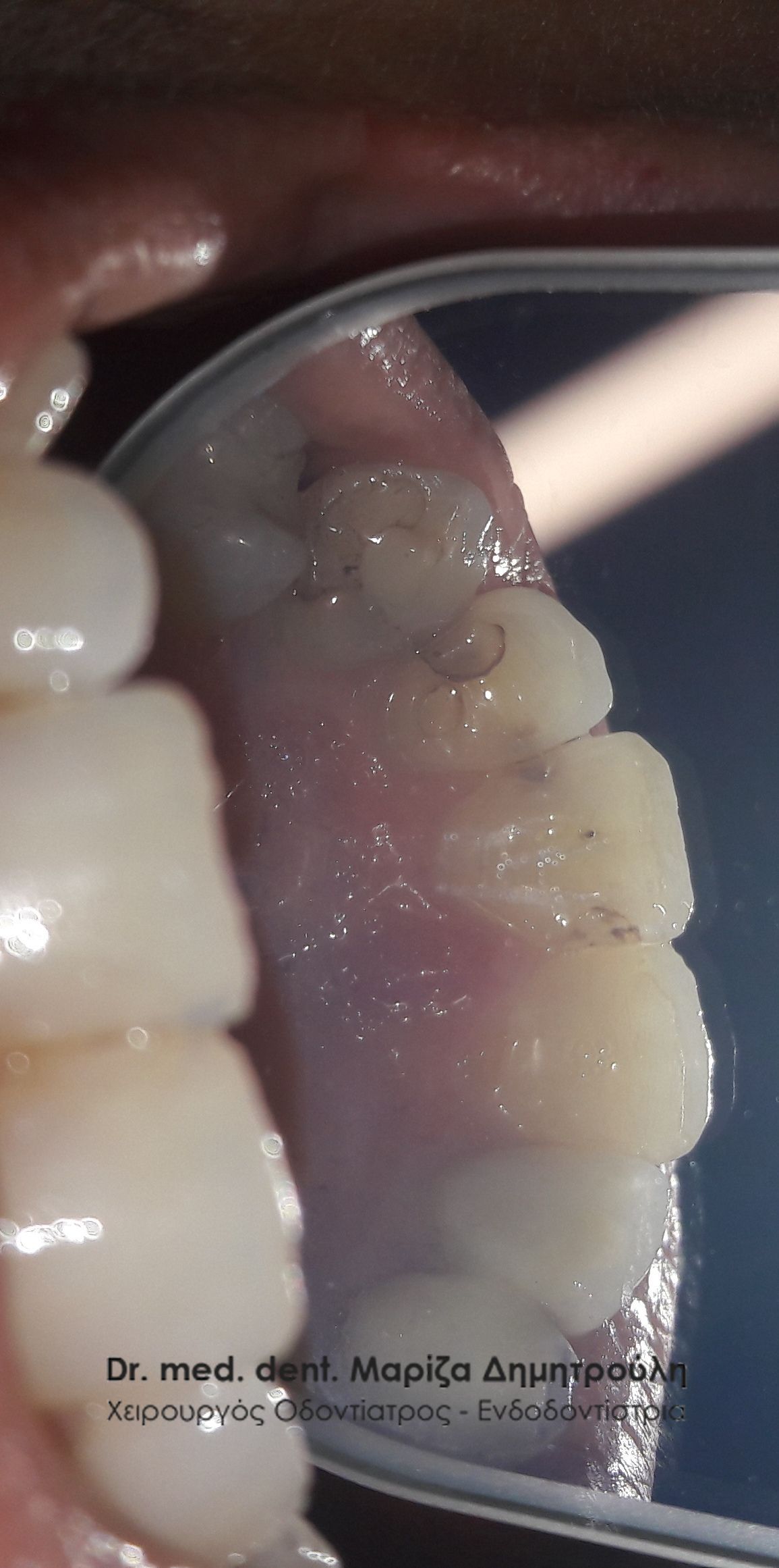
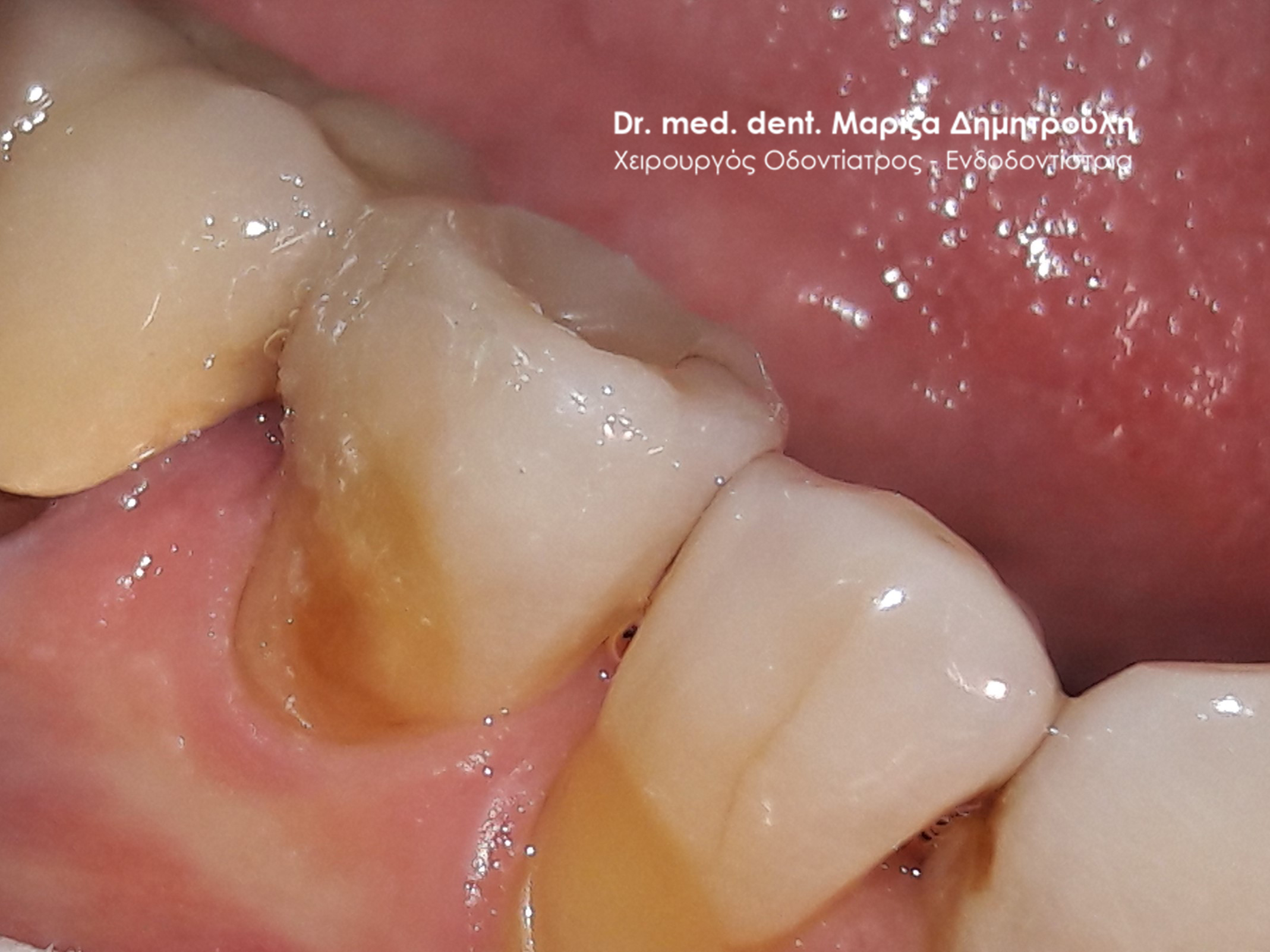
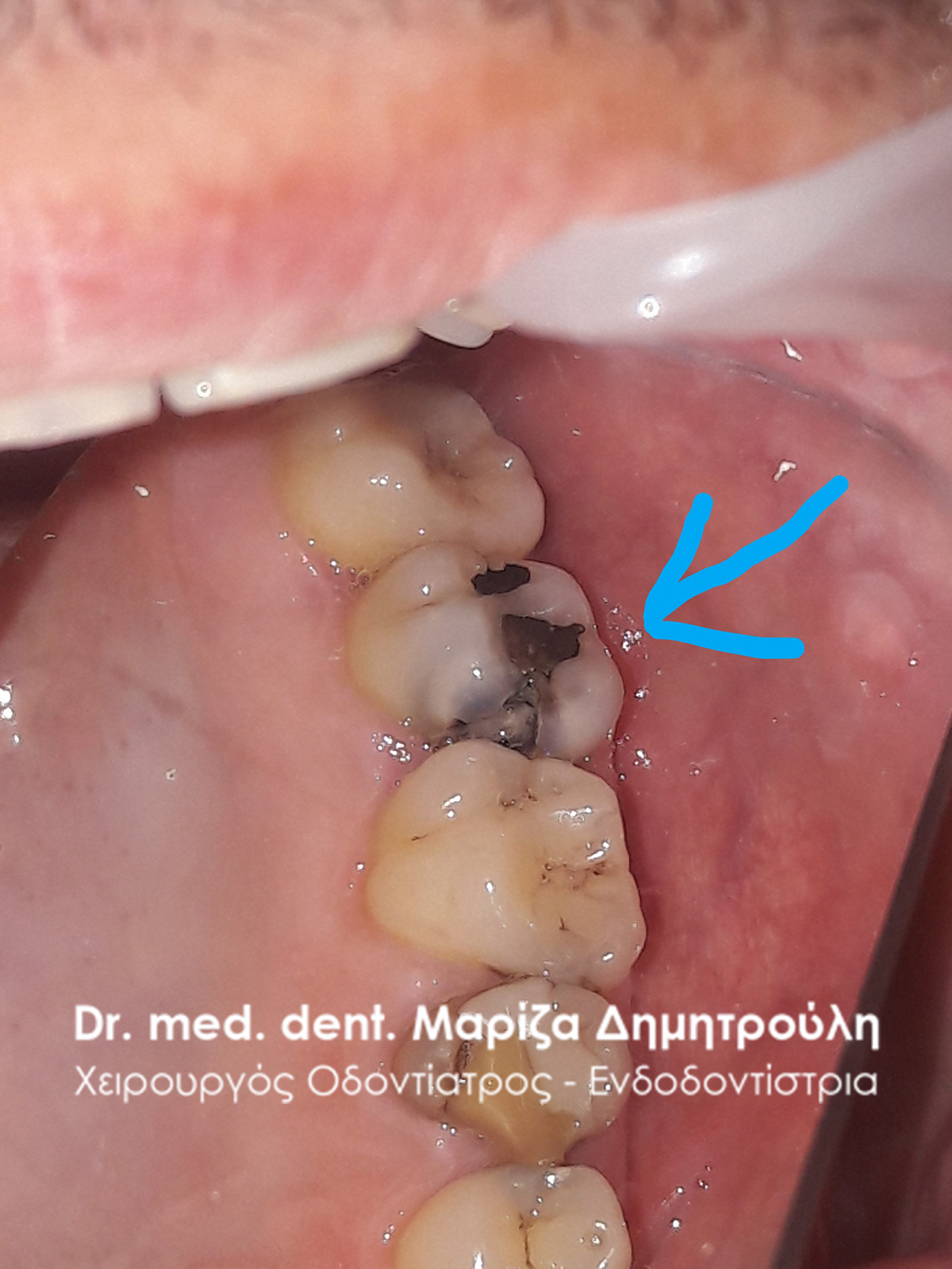
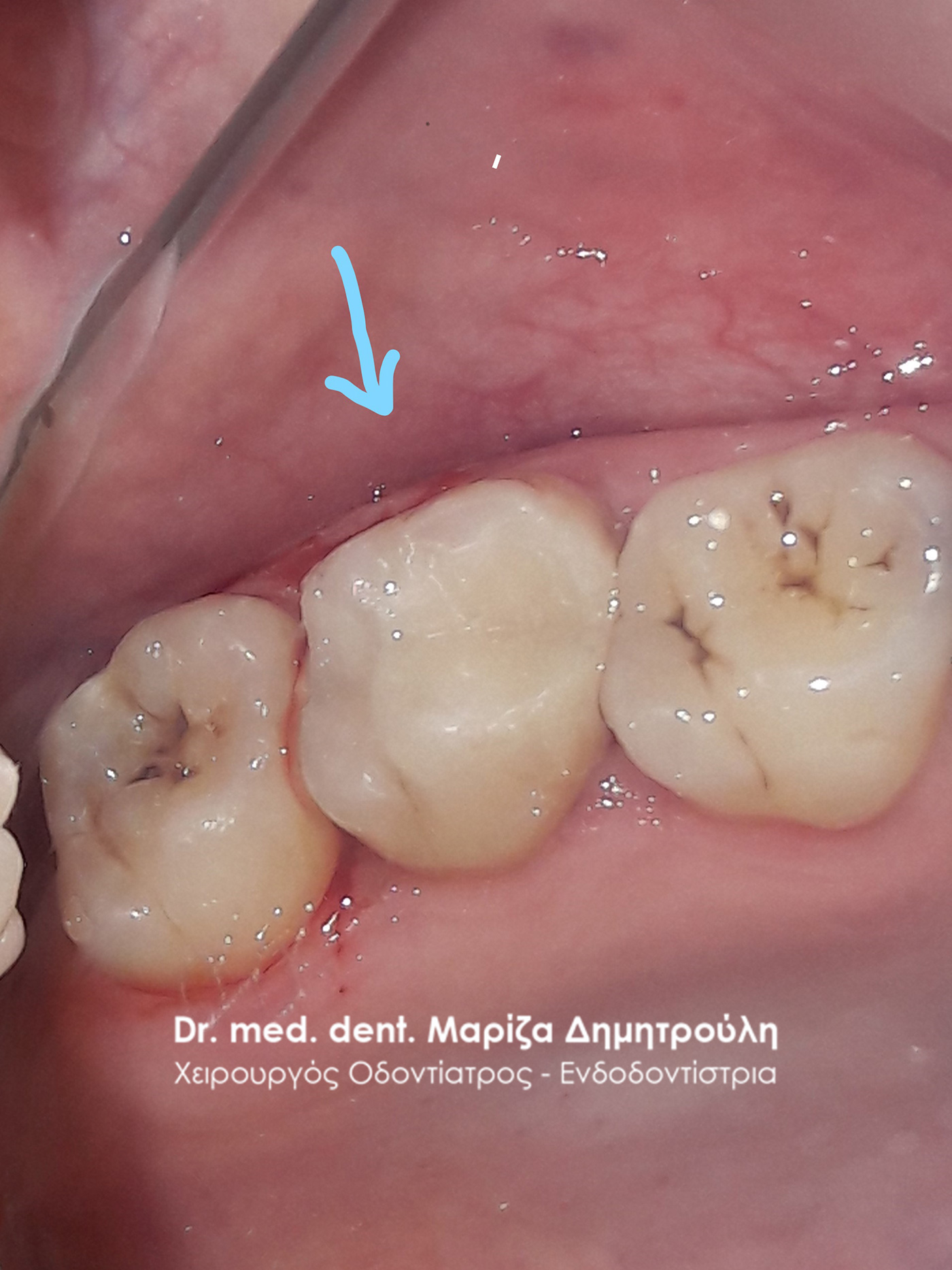
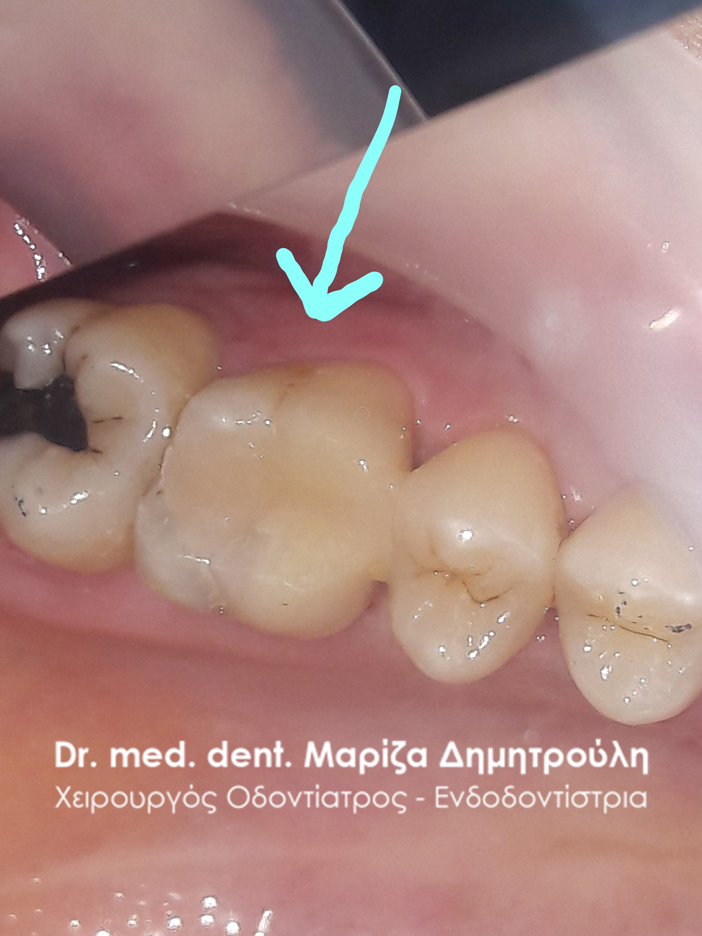
Case – Replacing a black amalgam filling with a white tooth filling
The patient noticed that an old black filling on the first molar on the right side of the mandible broke. After the administration of local anesthesia, a rubber isolator was placed to make the procedure of removing the black amalgam filling safer. The rubber isolator reduces the risk of swallowing fragments and mercury vapor released during the removal of an amalgam filling. Once the old filling was removed, the tooth was restored with a new white resin filling.
BEFORE
META
Case – Replacing a black amalgam filling with a white tooth filling
The patient reports pain during chewing on the right side of the lower jaw. After clinical examination of the area, it was found that a piece of the first molar on the right side of the lower jaw, which had previously been restored with a black amalgam filling, had broken off. It is common for teeth with black fillings after years to have a piece of the tooth break off, while the amalgam filling remains intact in place. This was followed by administering a local anesthetic to the area of the broken tooth and removing both the old amalgam filling and the decay exposed under the black filling. The procedure was completed by restoring the tooth with a new white resin filling.
BEFORE
META
Incident – Correction of white tooth filling
The patient had a piece of the old white resin filling on his mandibular right first molar break off. Since the seal was large and the patient was not in pain, it was decided to correct and restore only the missing part of the seal. Initially, a rubber isolator was placed to prevent saliva from coming into contact with the restoration site of the tooth. The broken part of the seal was then restored with a new resin seal.
BEFORE
META
META
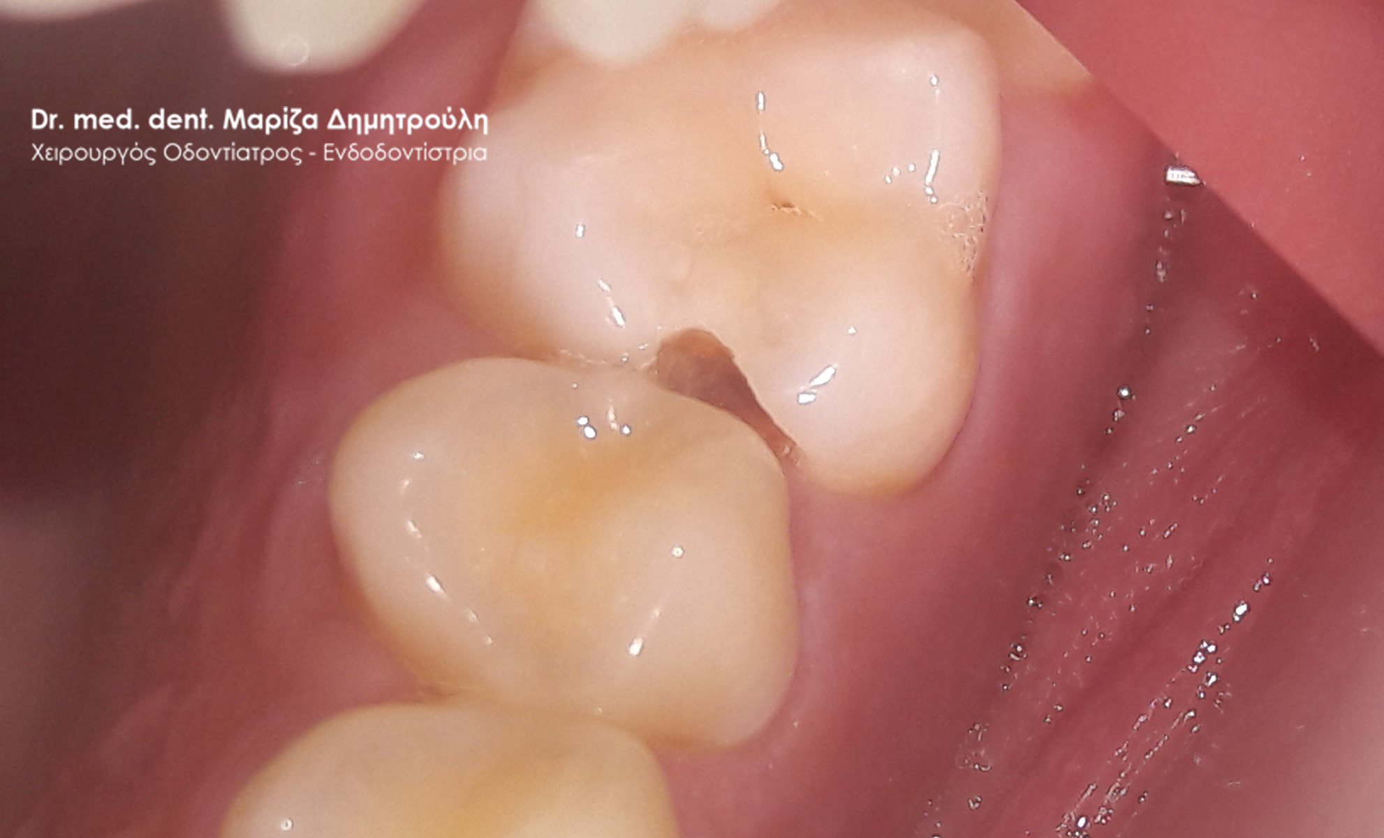
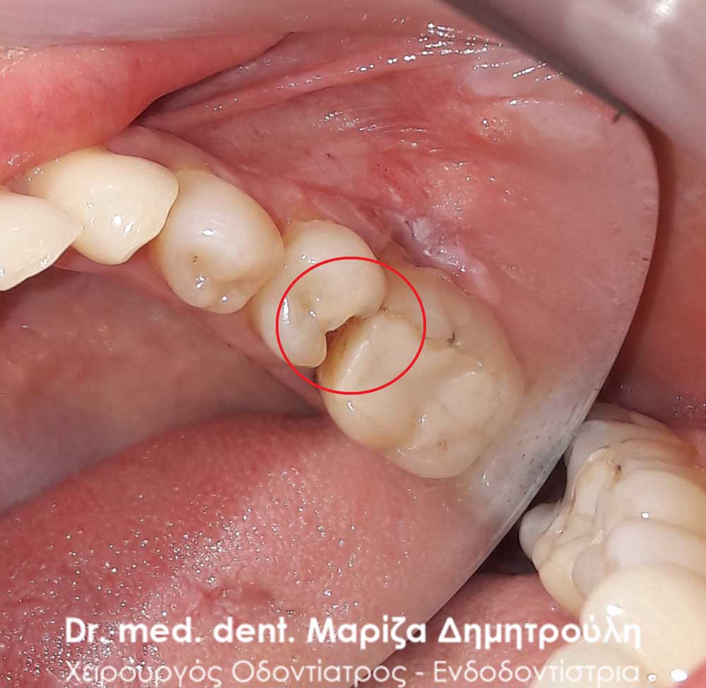
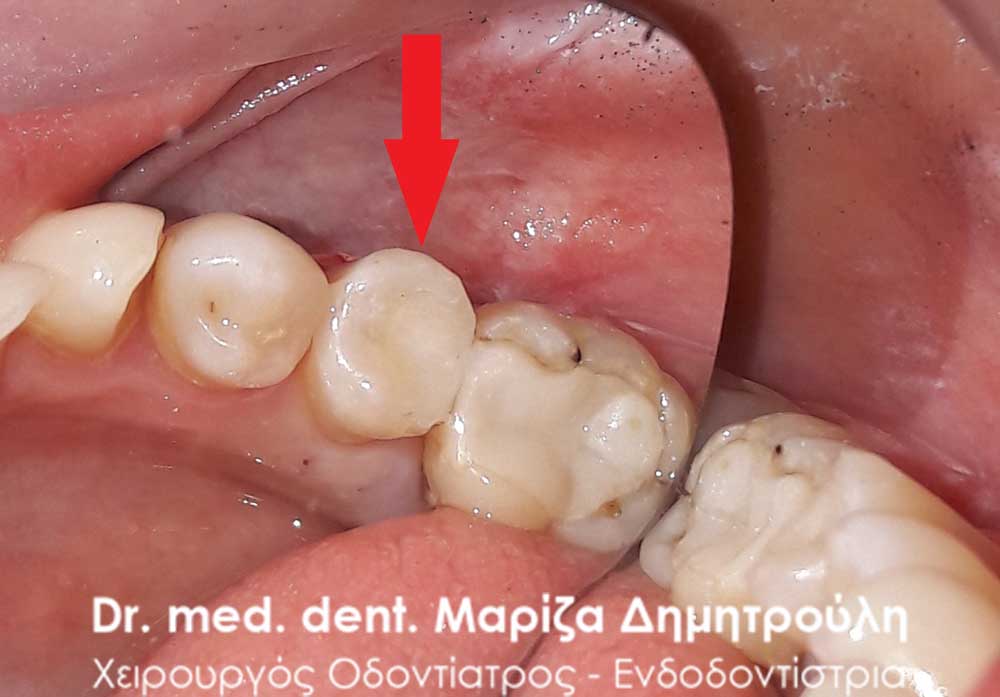
Incident – White tooth filling
The patient noticed a hole in the second premolar on the right side of the mandible. For this reason, he visited the doctor’s office with the aim of restoring the carious cavity in that particular tooth. After opening the tooth and removing the caries, the restoration of the damage was carried out with a white resin seal.
BEFORE
META
Incident – White tooth filling
The patient visited the office as he has severe pain when chewing on the left side of the upper jaw. Clinical examination of the area revealed a deep carious cavity in the left maxillary second molar, as the old amalgam filling on the tooth was re-carious. The dental work began by numbing the area, removing the old black filling and removing the decay. Because the caries extended deep, at the level of the nerve of the tooth, a special pulp protection material was placed. Finally, the tooth was reconstructed with a white resin seal.
BEFORE
META
Incident – White tooth filling
The patient reports that she has a mild pain on the left side of the upper jaw. He also adds that a piece of the old black filling was missing from the upper left first molar, which the dentist closed with a temporary filling. Her desire was the final restoration of the tooth, as she periodically has discomfort while chewing. First the first molar area was anesthetized and then both the temporary and the black filling were removed. The caries was then removed and the tooth was restored with a white resin sealer, which provided a good contact point with the adjacent tooth, so that no food debris accumulated between the teeth.
BEFORE
META
META
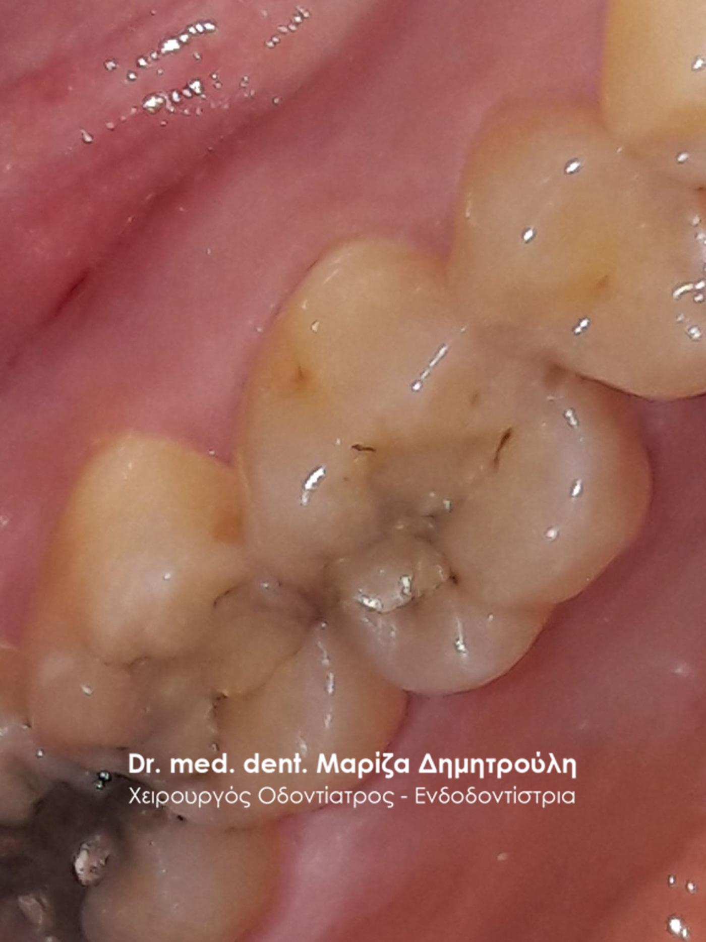
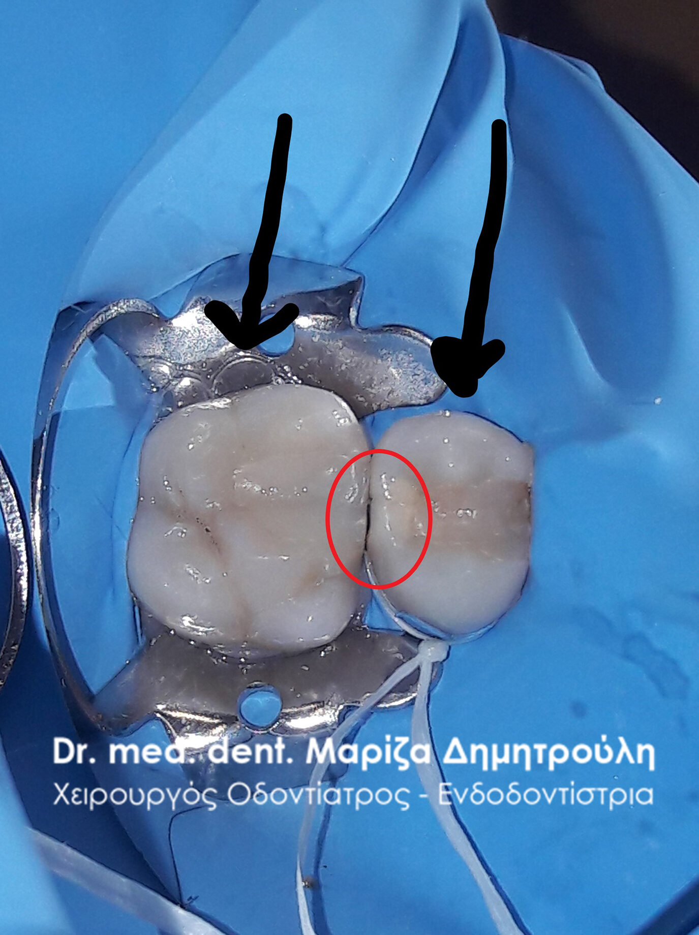
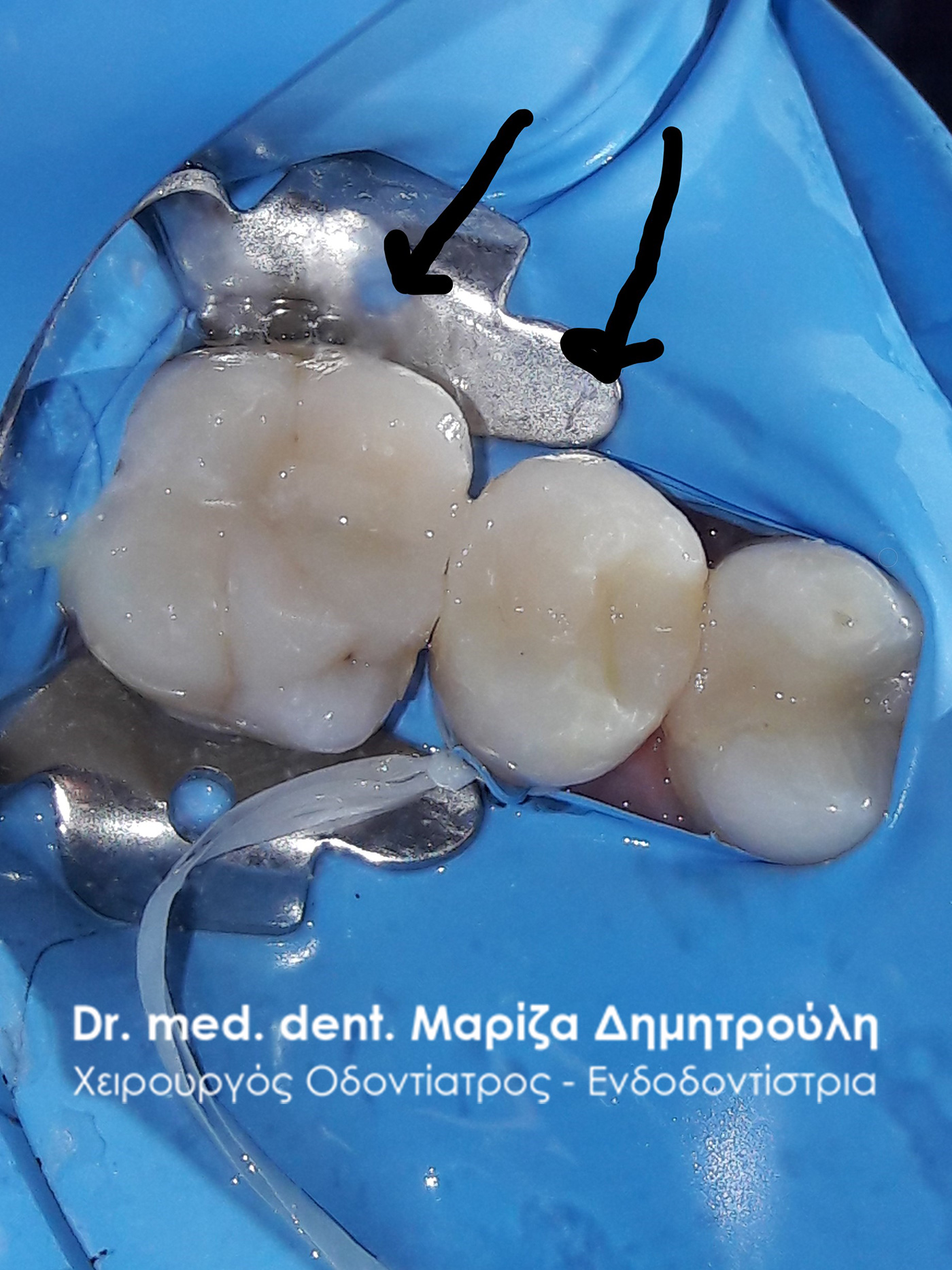
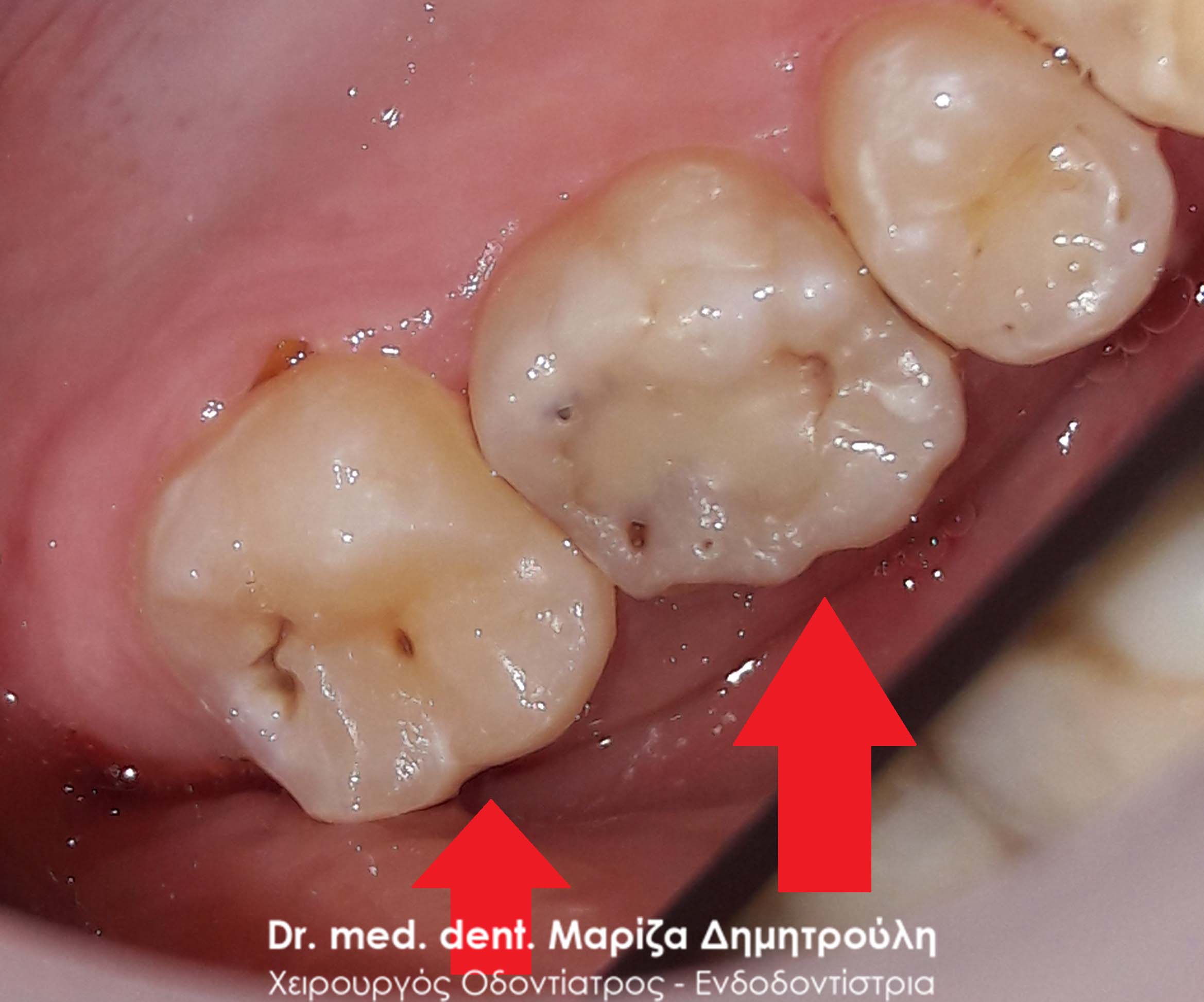
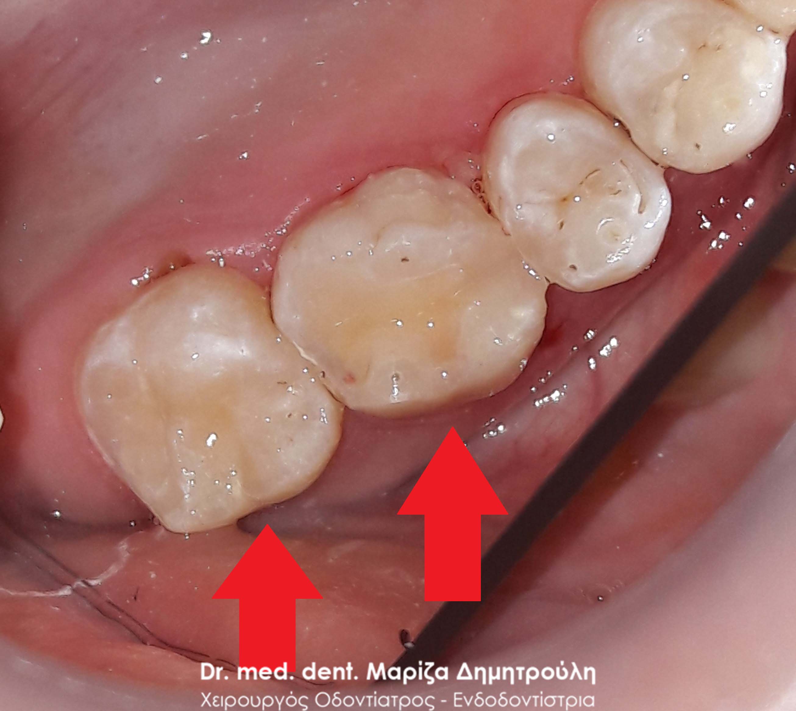
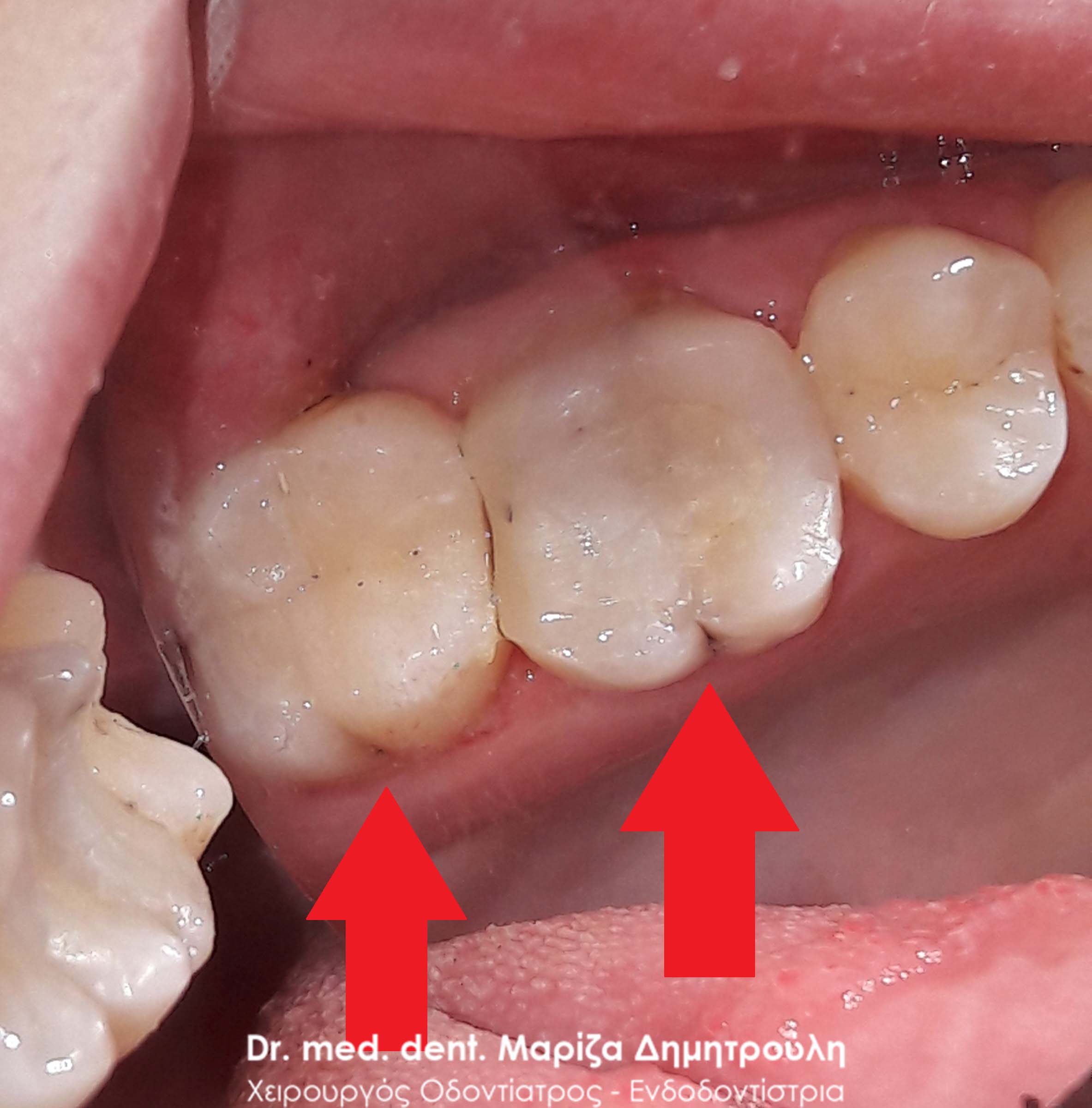
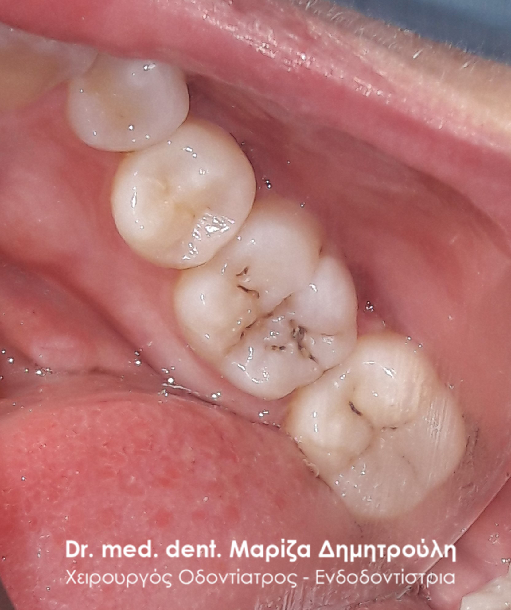
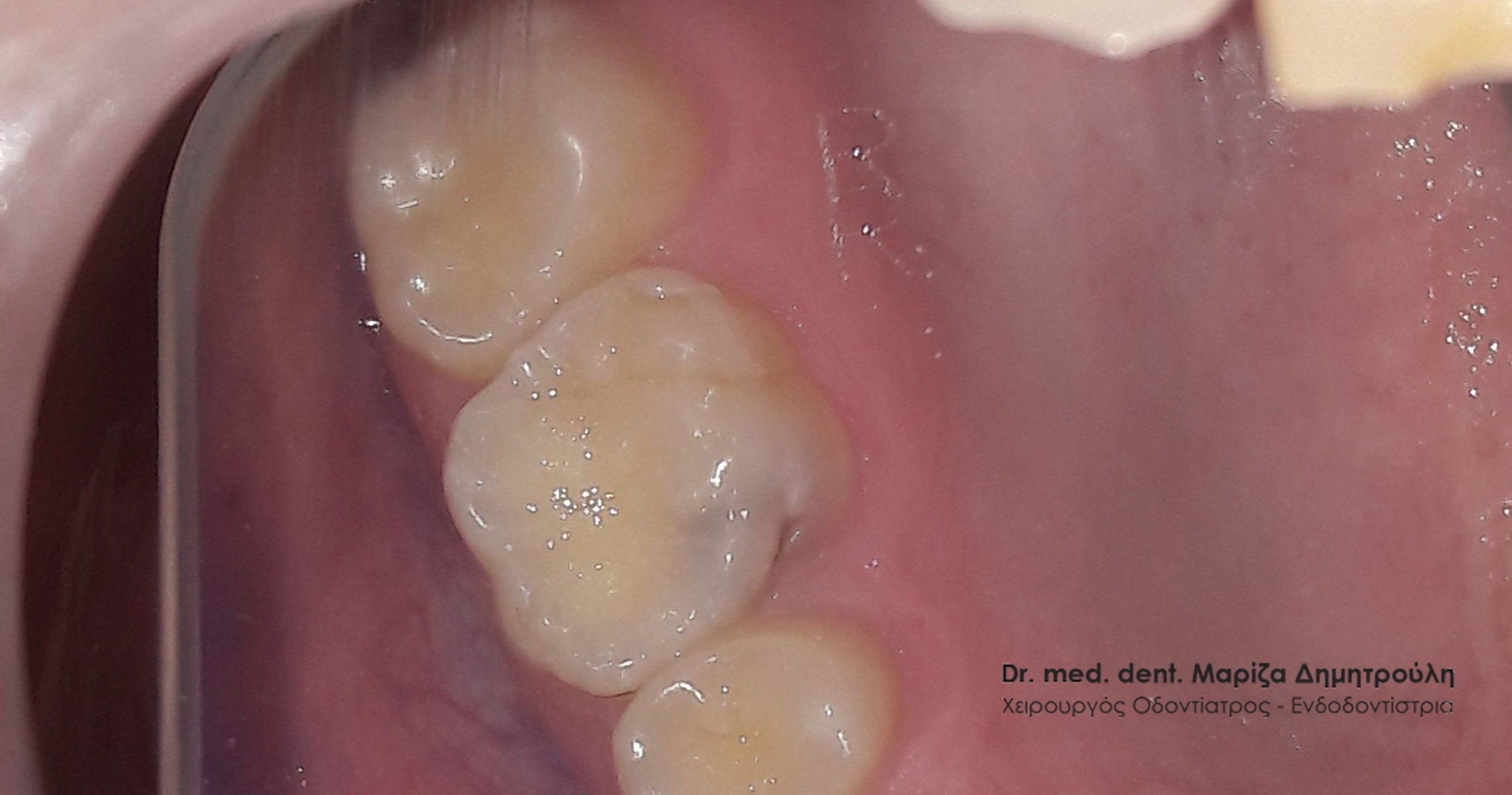
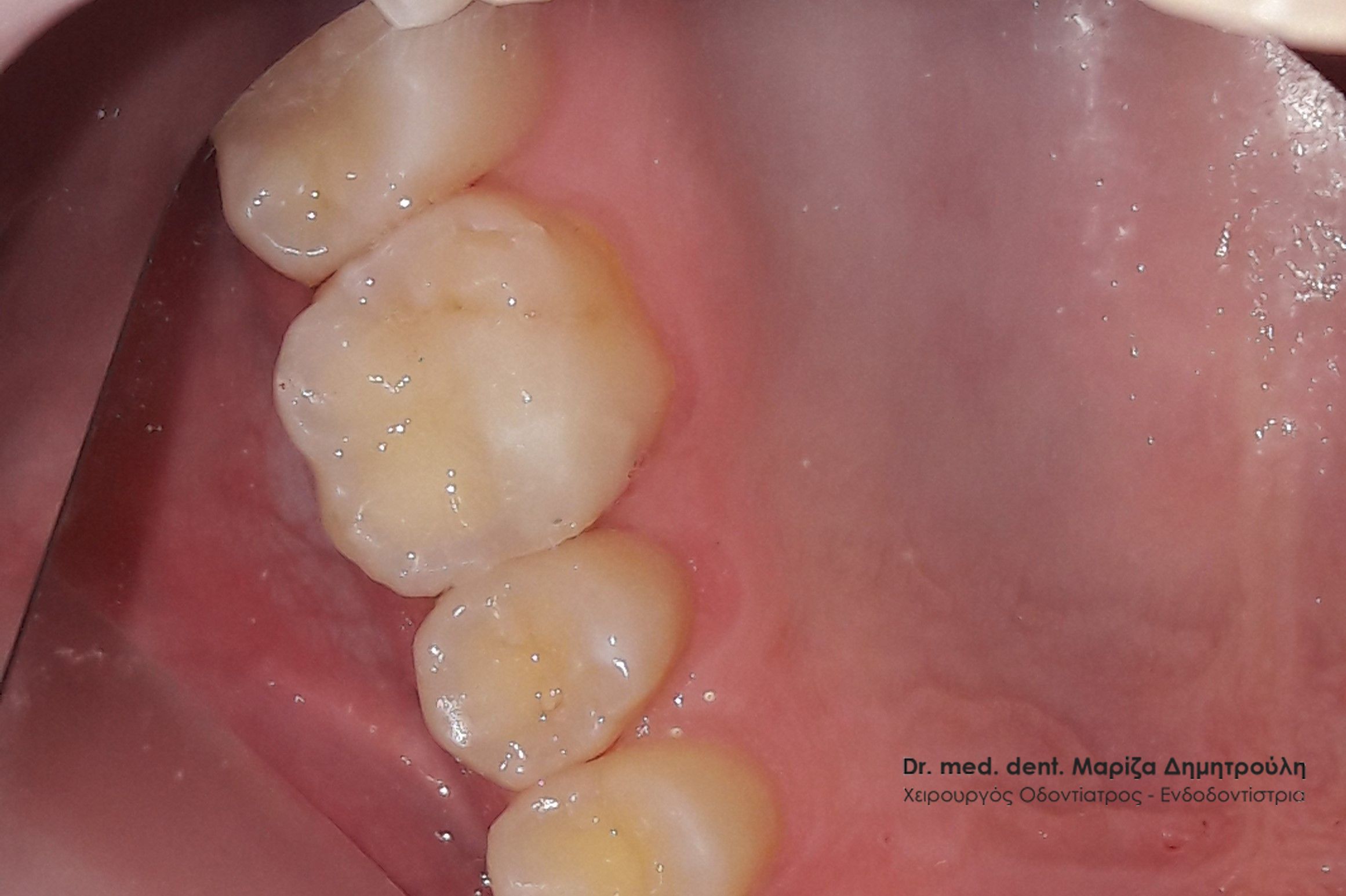
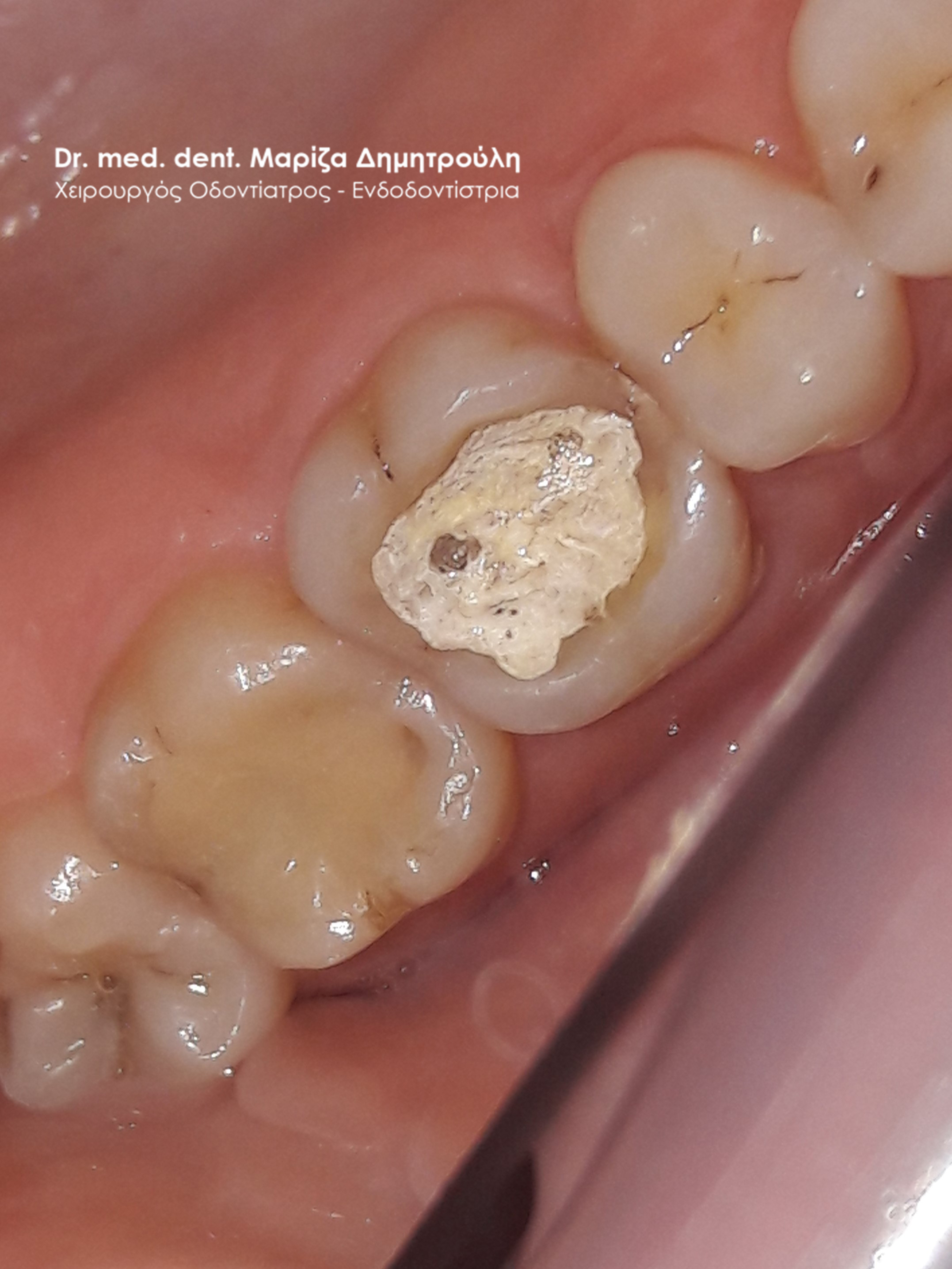
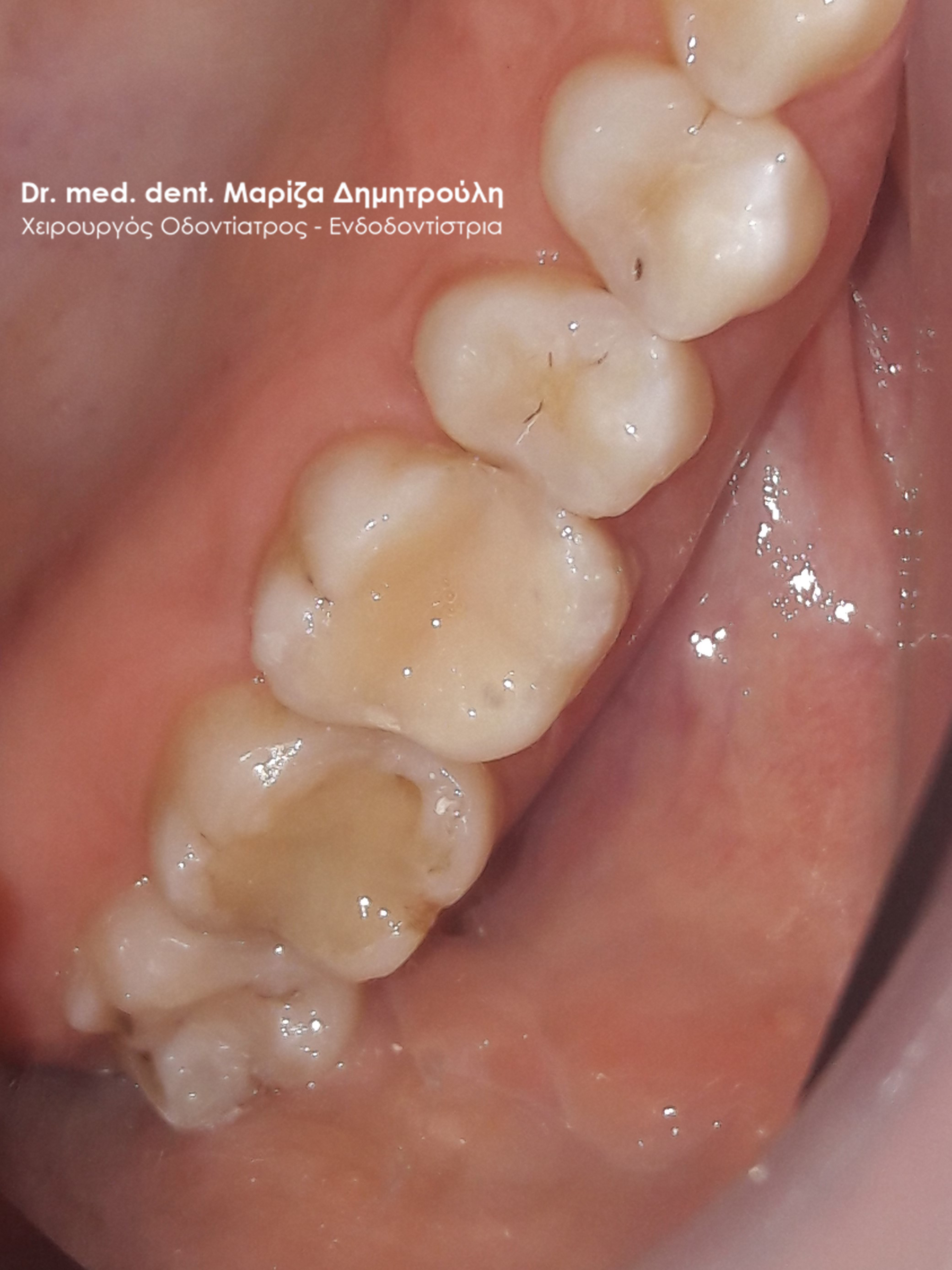
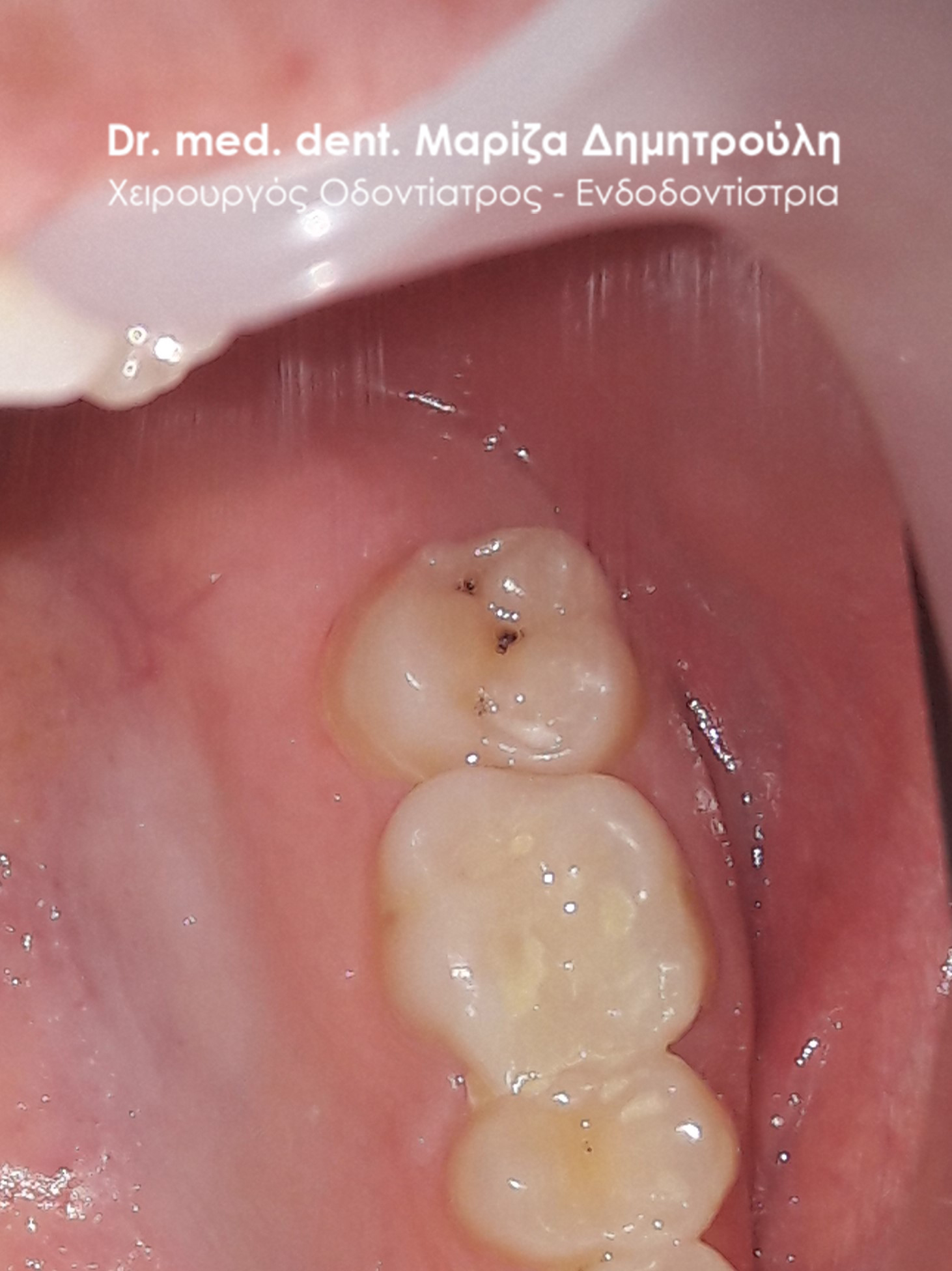
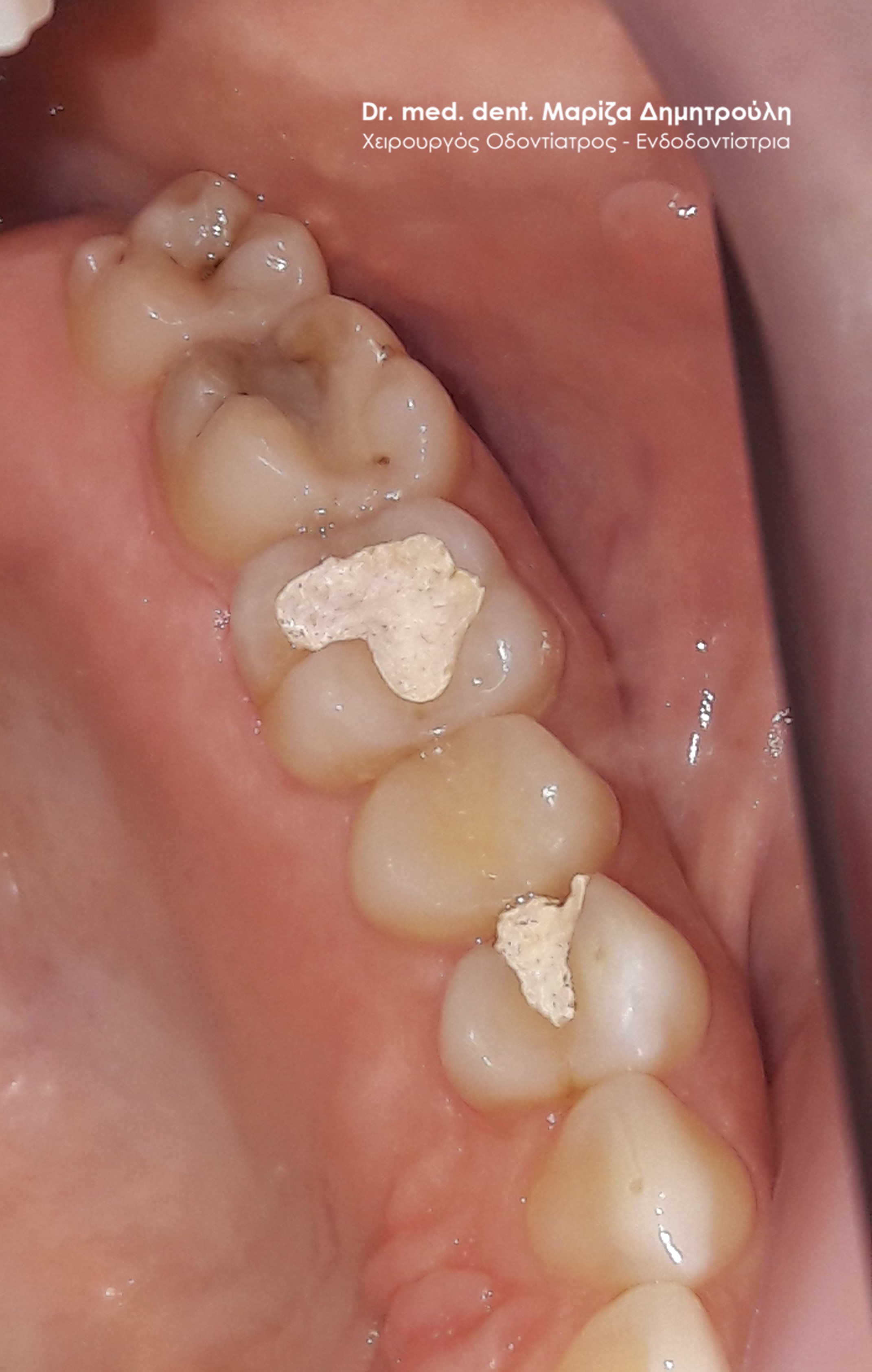
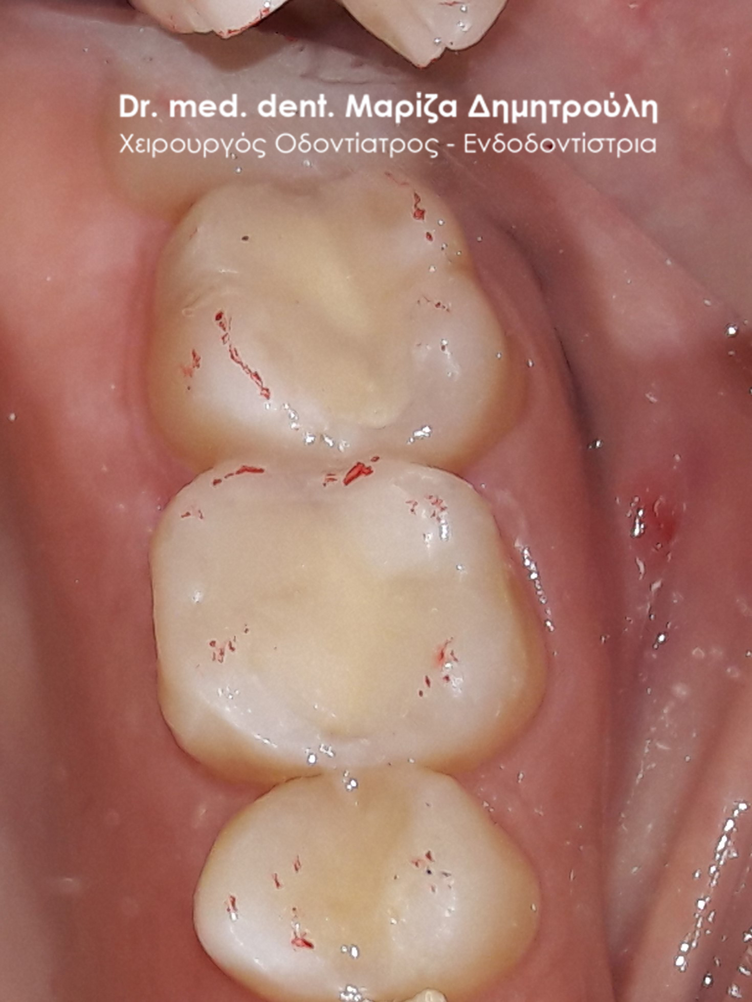
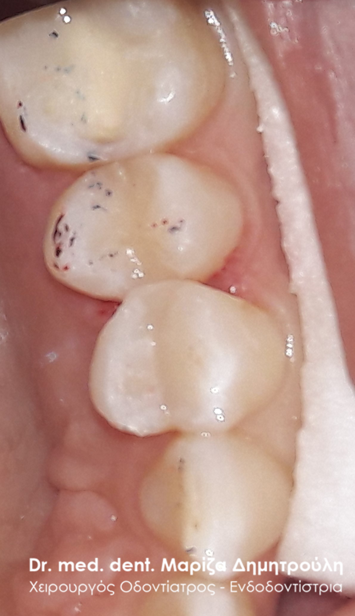
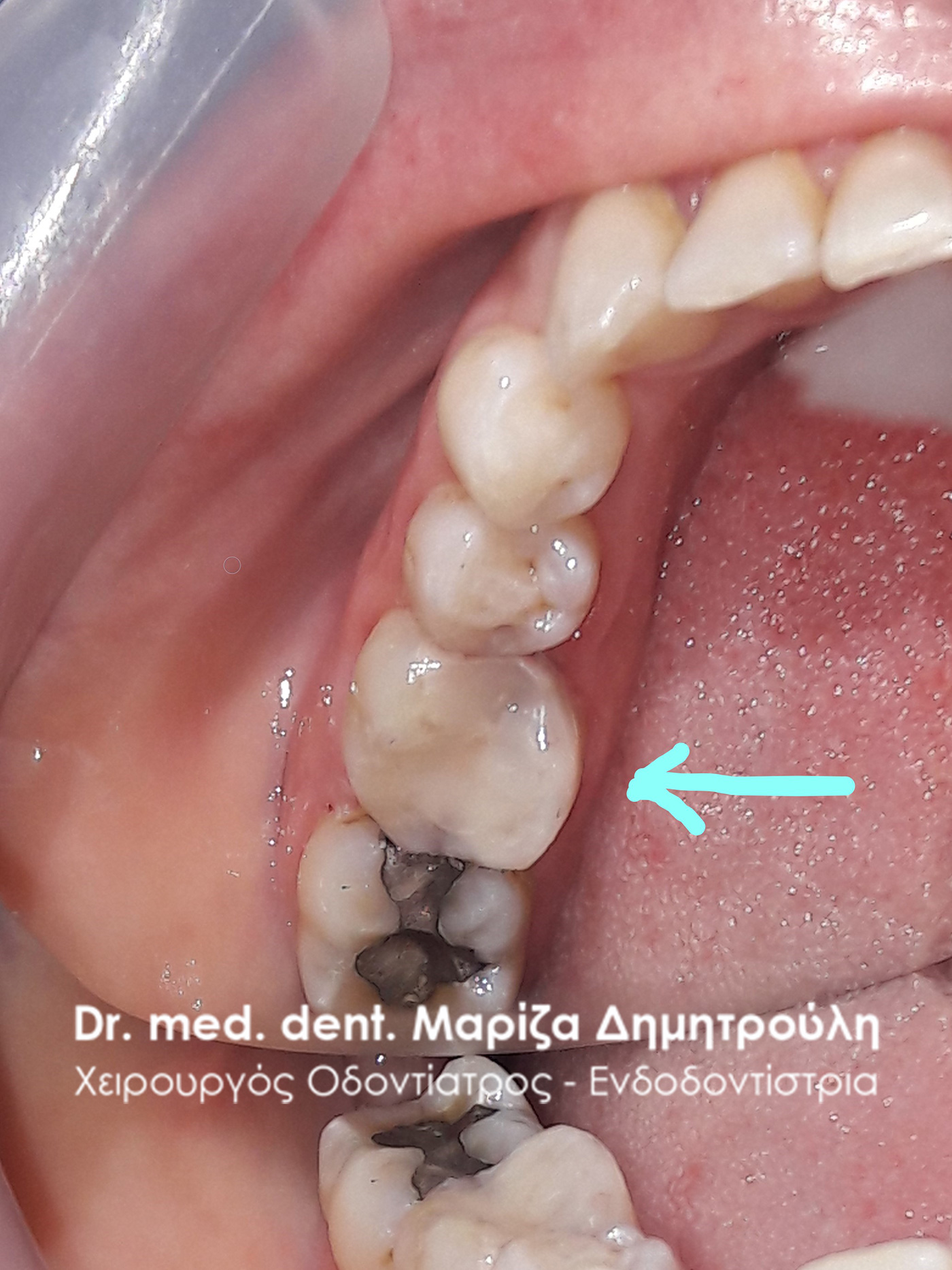
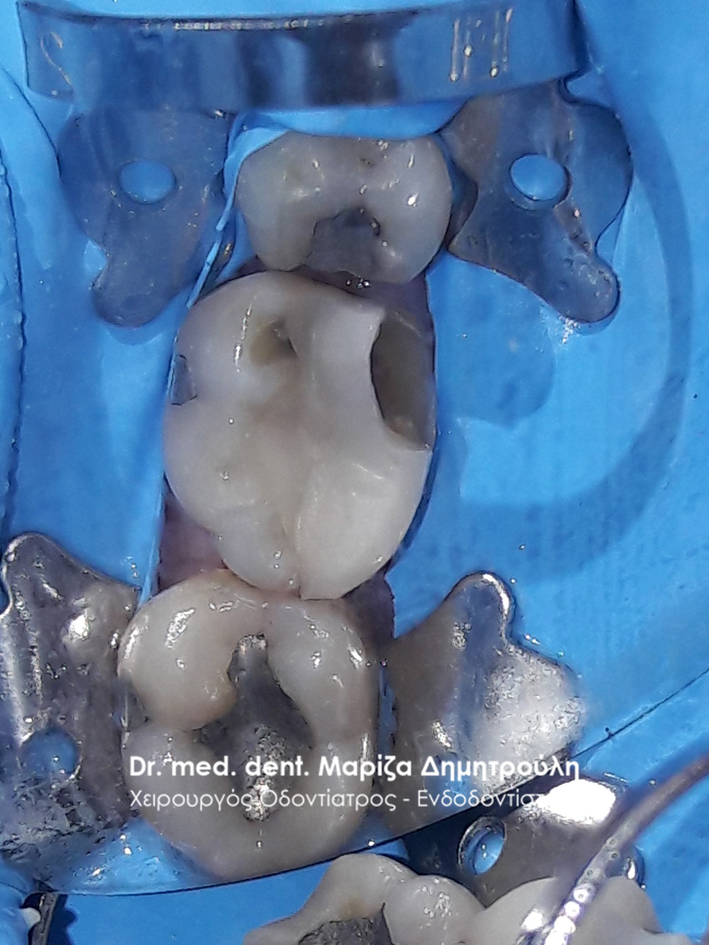
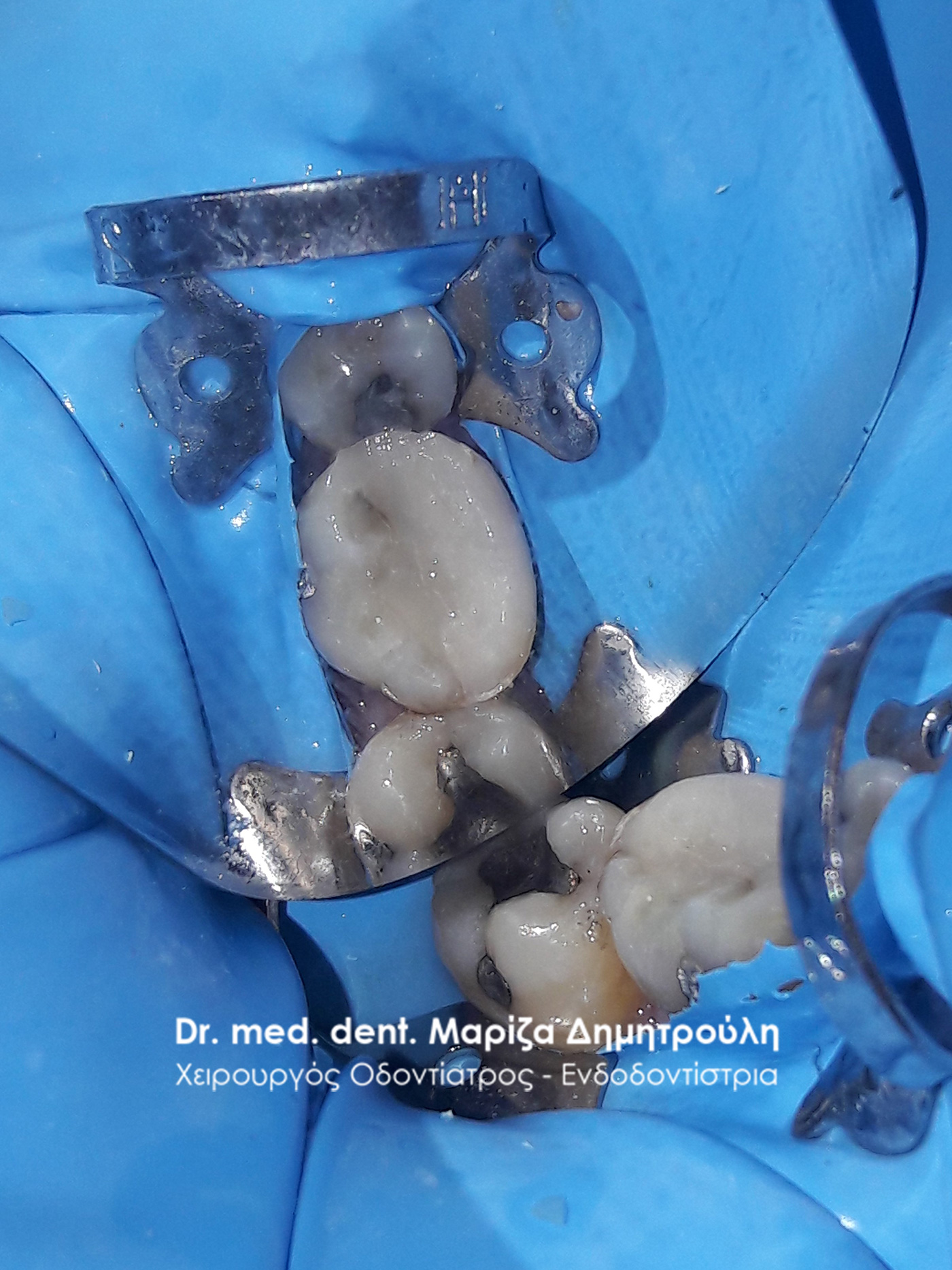
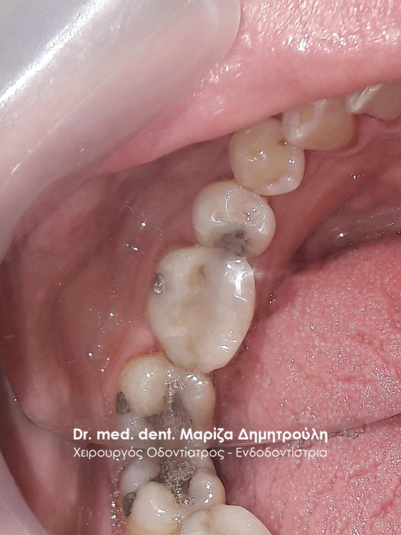
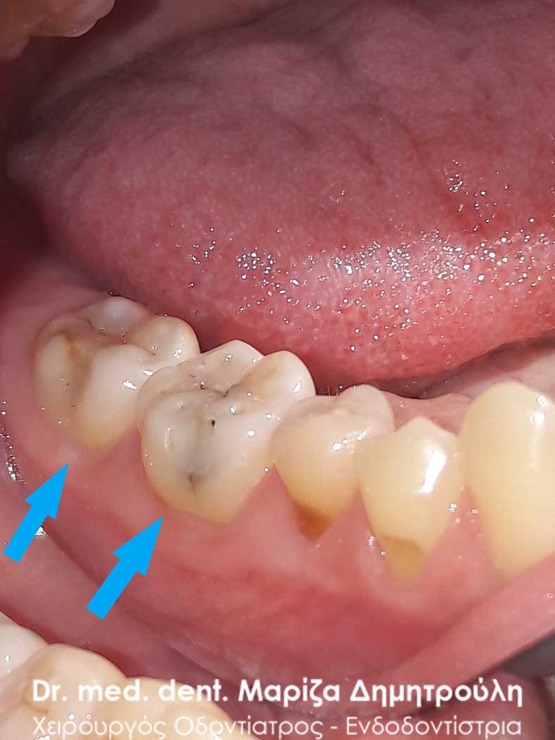
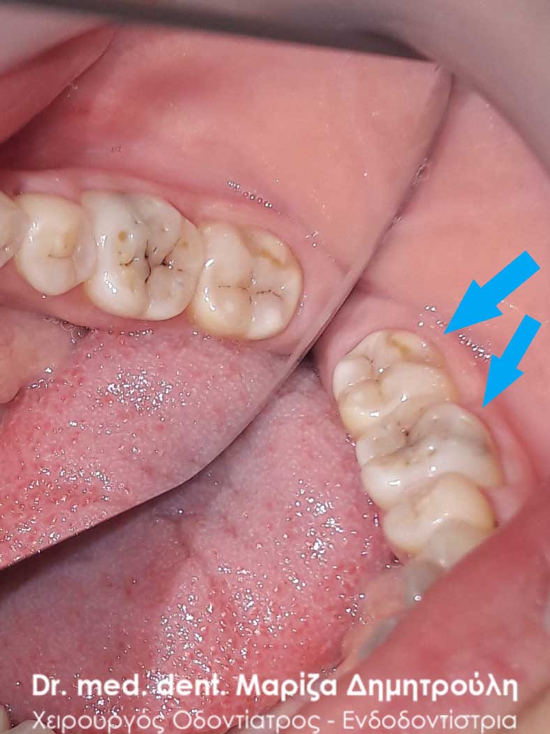
Incident – White tooth fillings
The patient had lately felt a mild discomfort when chewing on the left side of the upper jaw, without being able to determine whether the pain was coming from a specific tooth. Clinical examination revealed three old fillings, which needed immediate restoration as they had re-caried. Discoloration of the borders on an old white filling is an indication of re-caries of a tooth. After the removal of the old white fillings, under the use of local anesthesia in the area of the teeth, followed the removal of the carious dental tissues and the restoration of the teeth with new white resin fillings.
BEFORE
The deficit of dental tissues after cavity opening
BEFORE
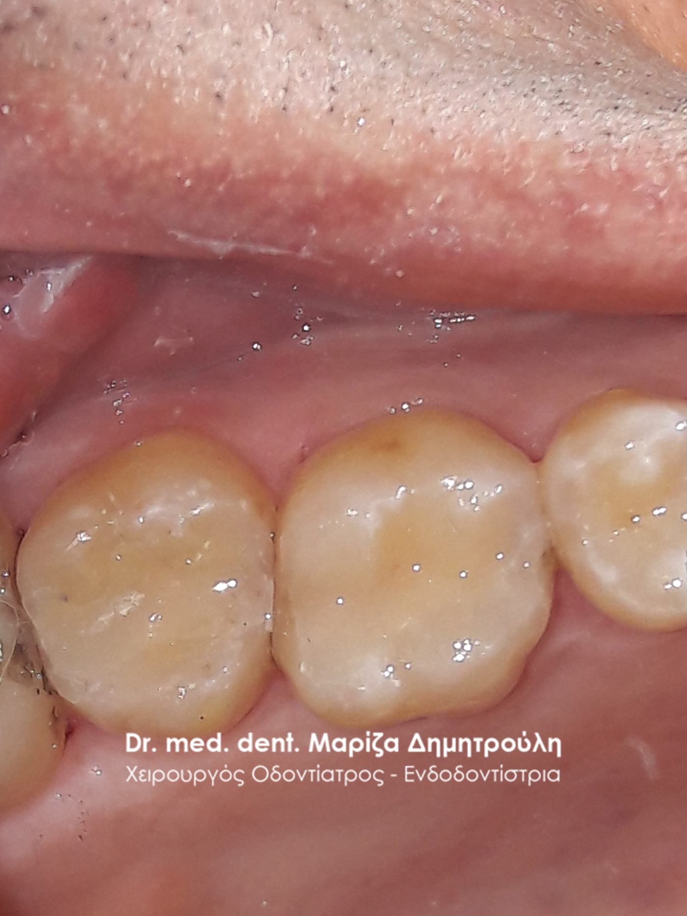
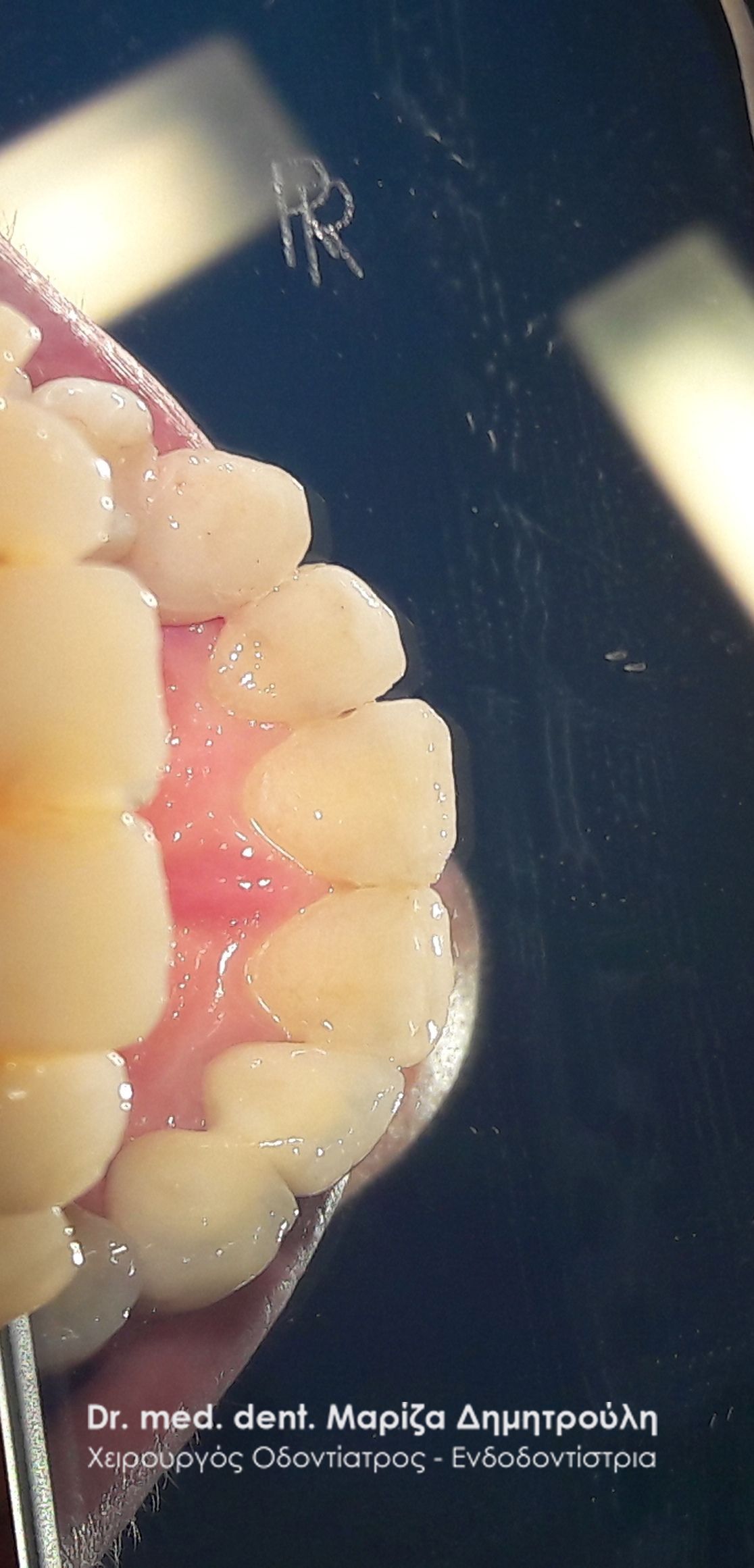
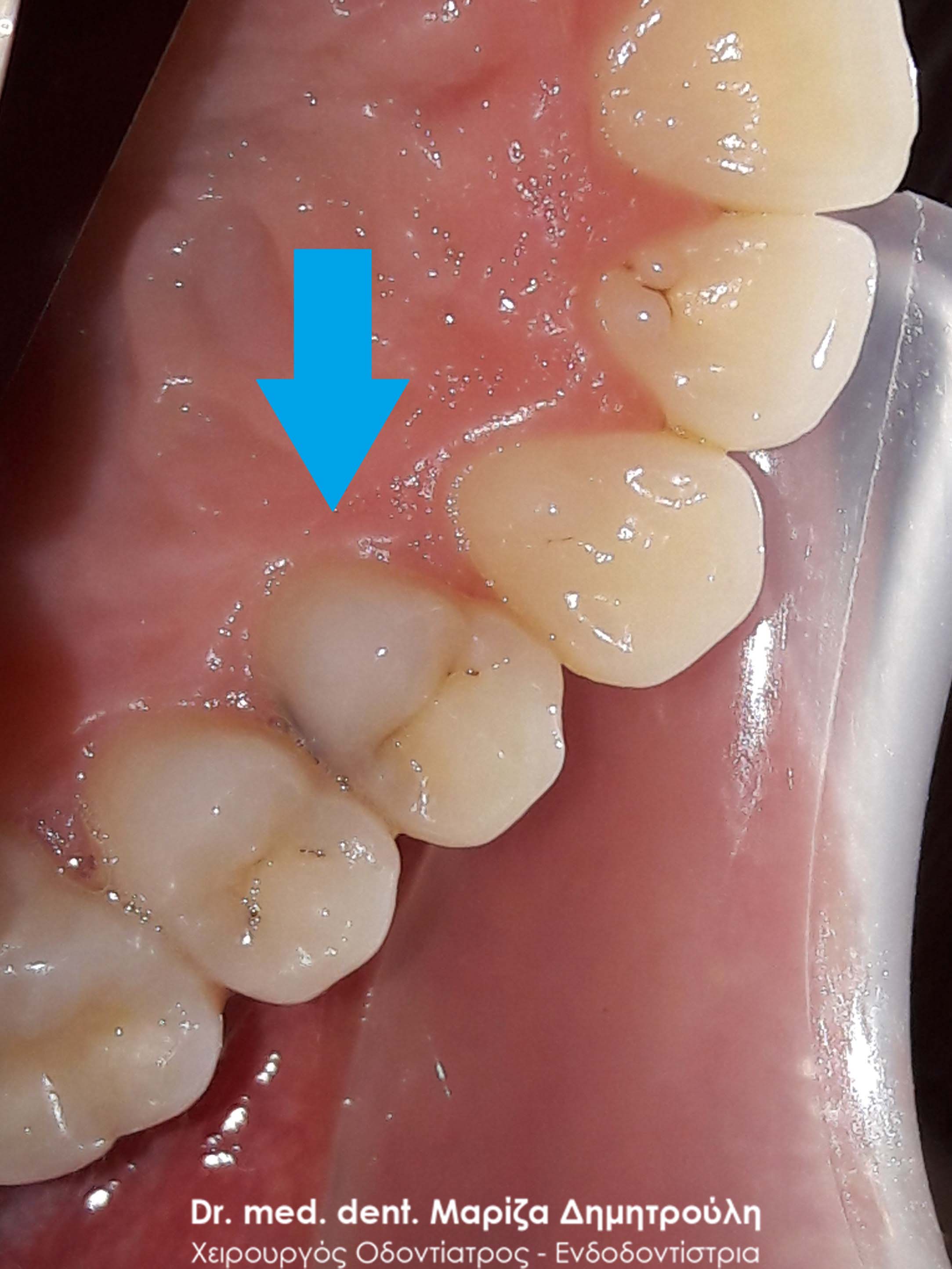
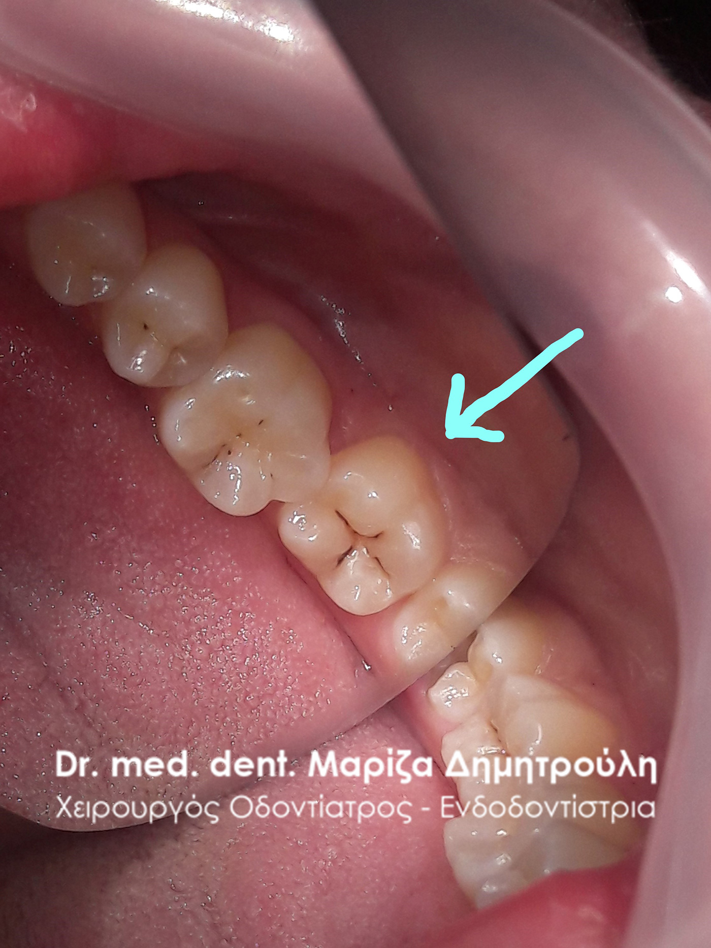
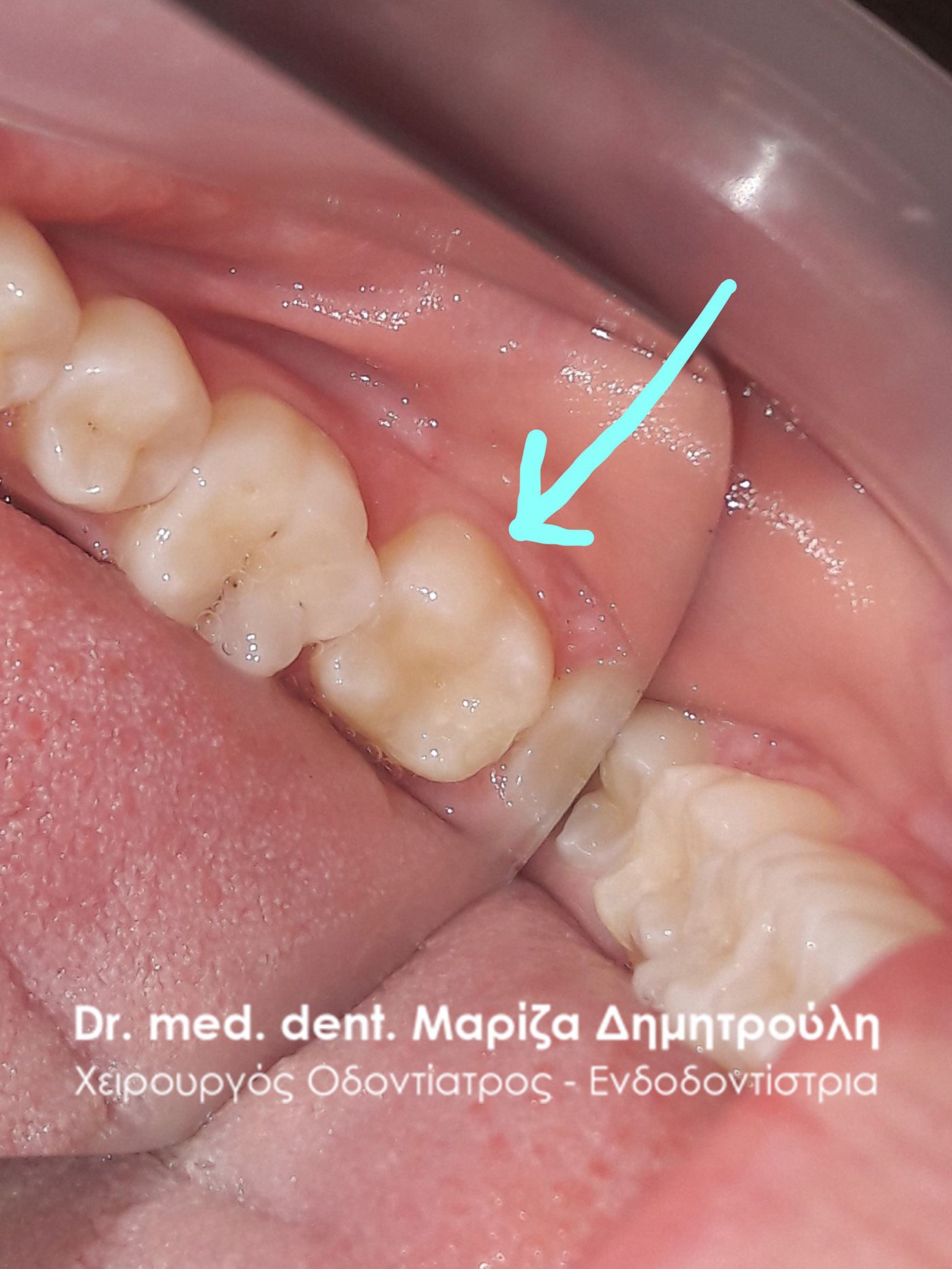
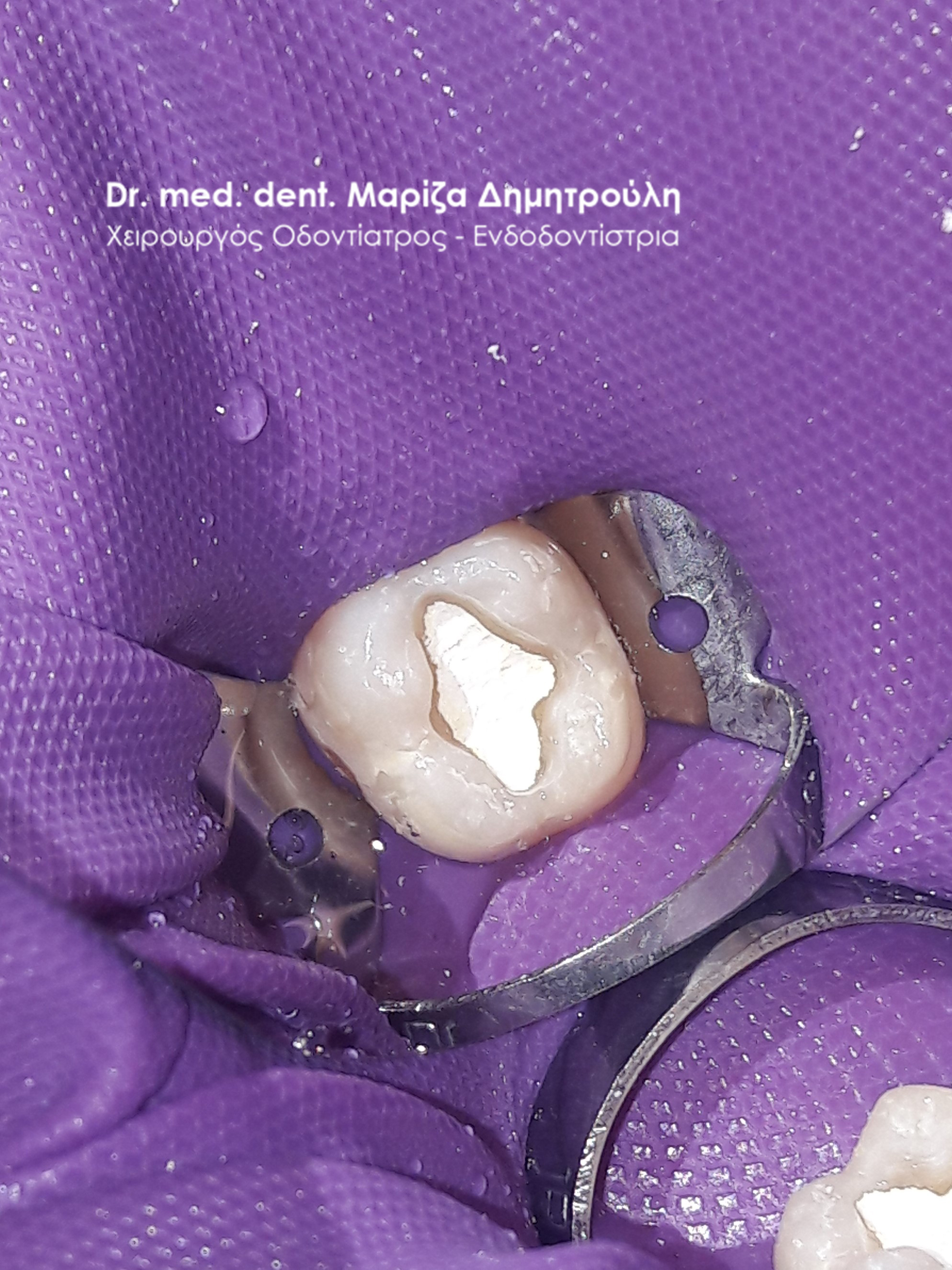
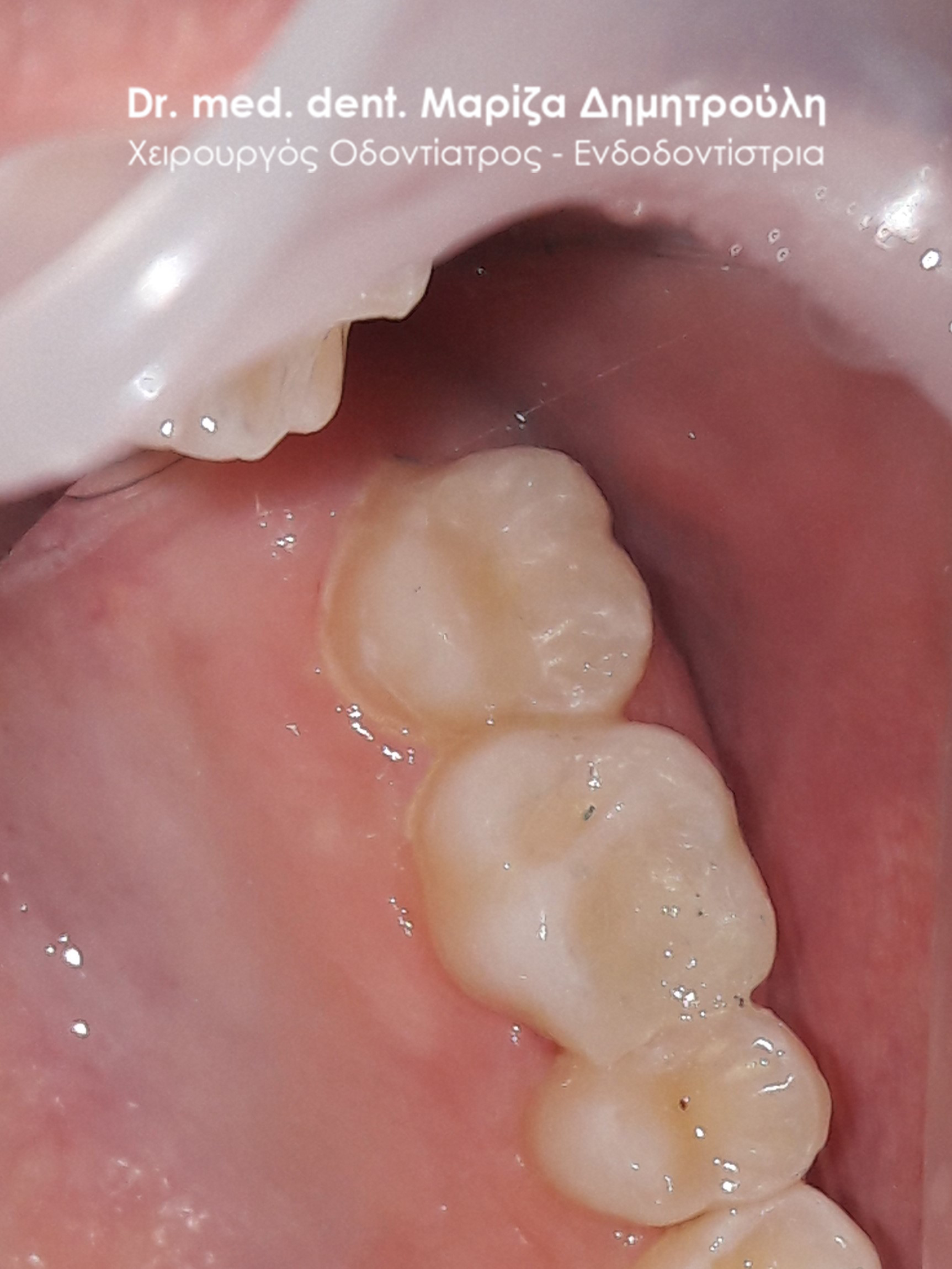
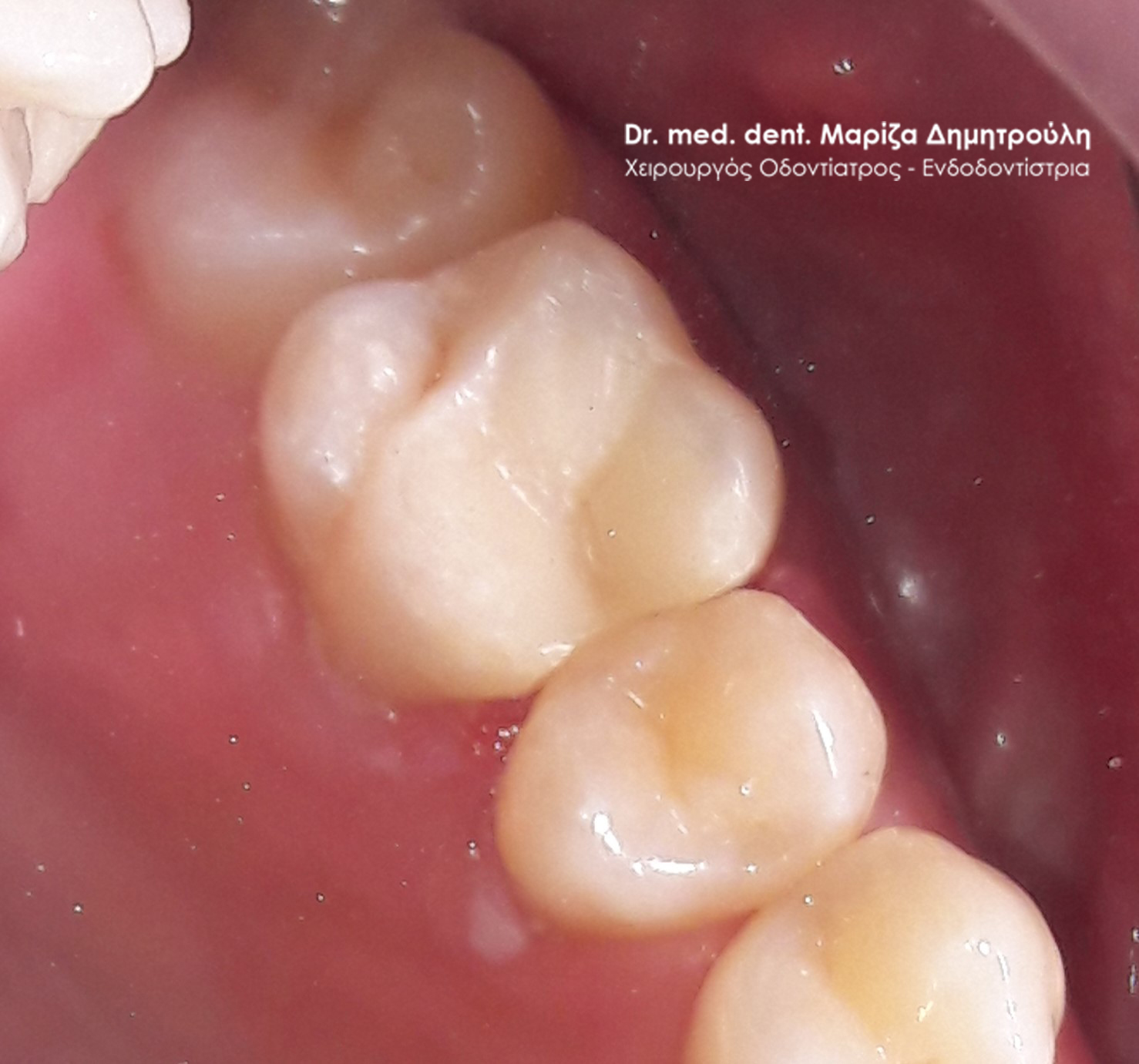
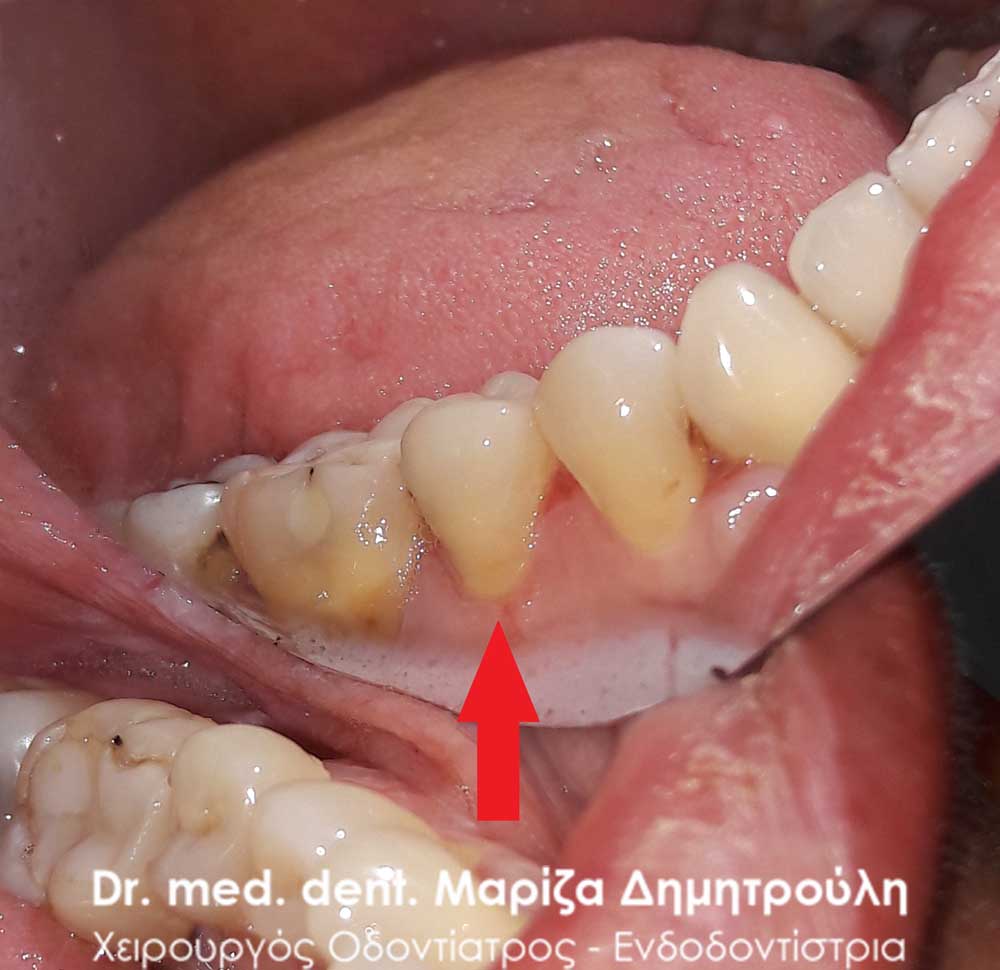
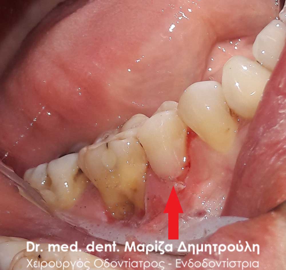
Case – Cervical white tooth filling
The patient wanted the restoration of the dental deficit in the neck of the second left lower premolar. The lack of dental tissue at that point caused the patient intense sensitivity when receiving cold stimuli (drinks and food). After the restoration of the tooth the symptoms subsided immediately.
BEFORE
BEFORE
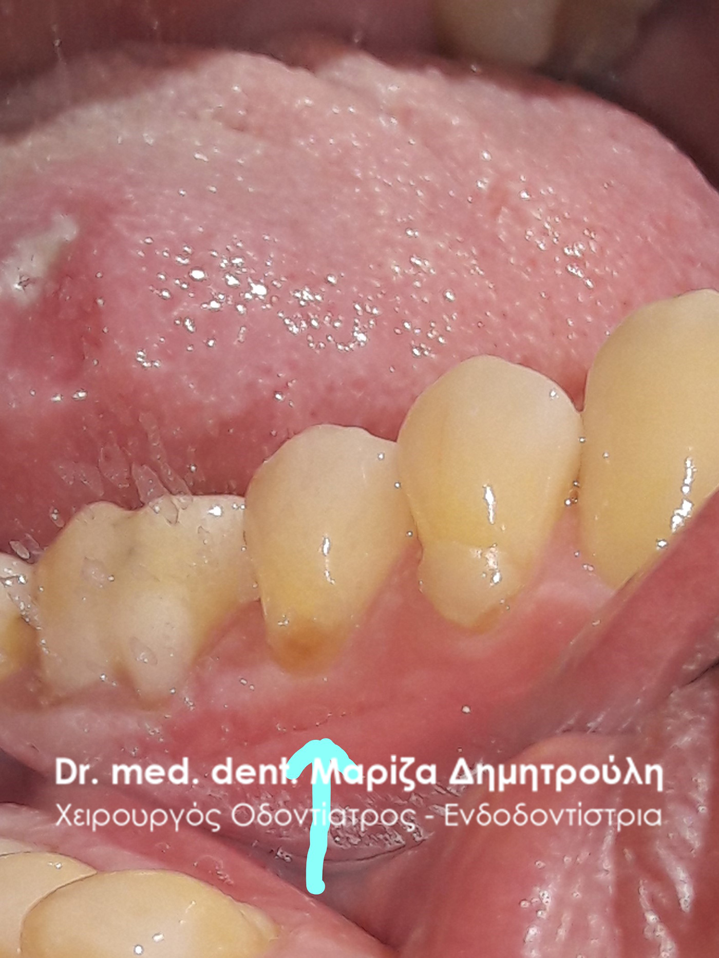
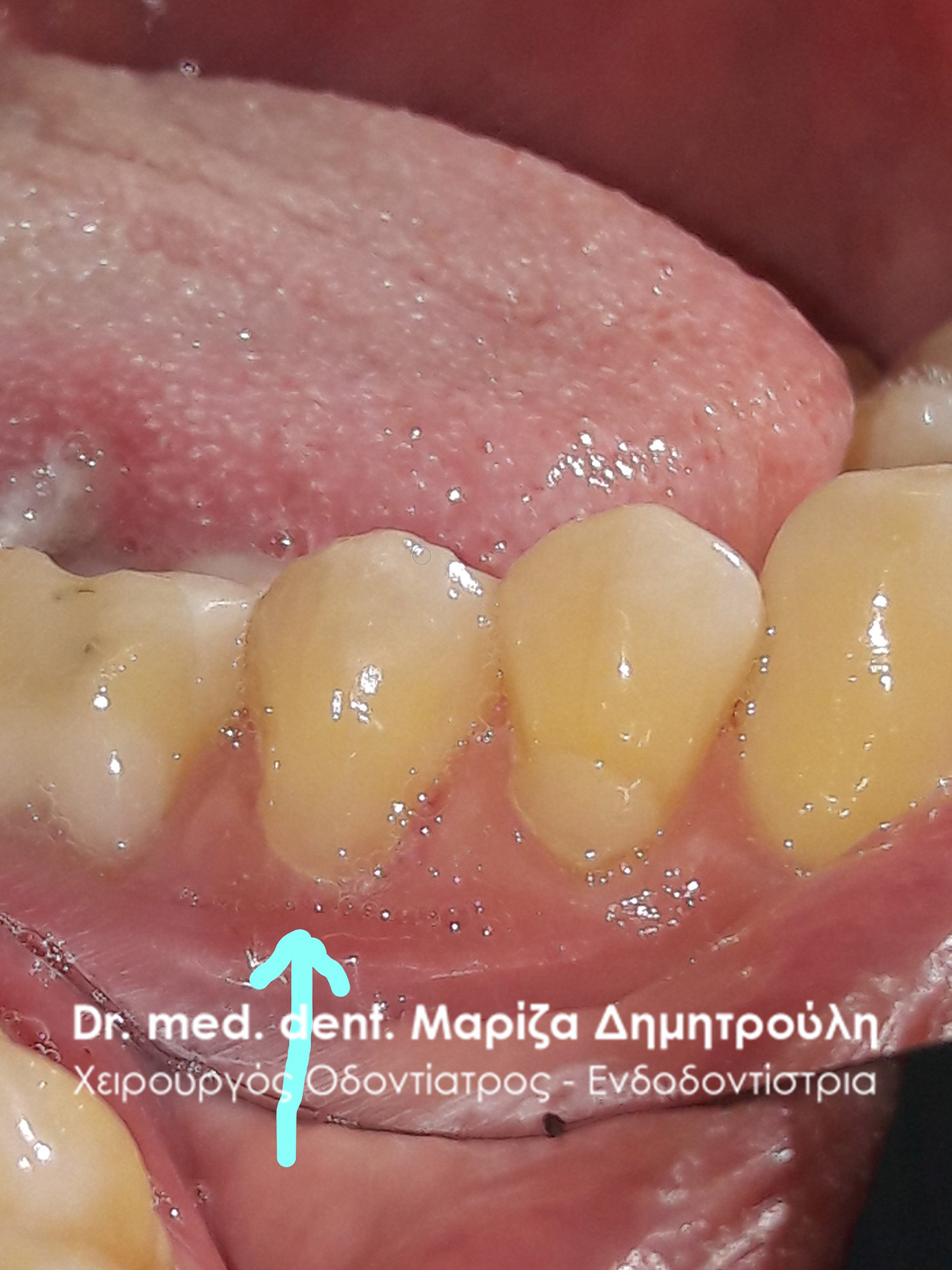
Incident – Replacement of old white tooth filling
BEFORE
BEFORE
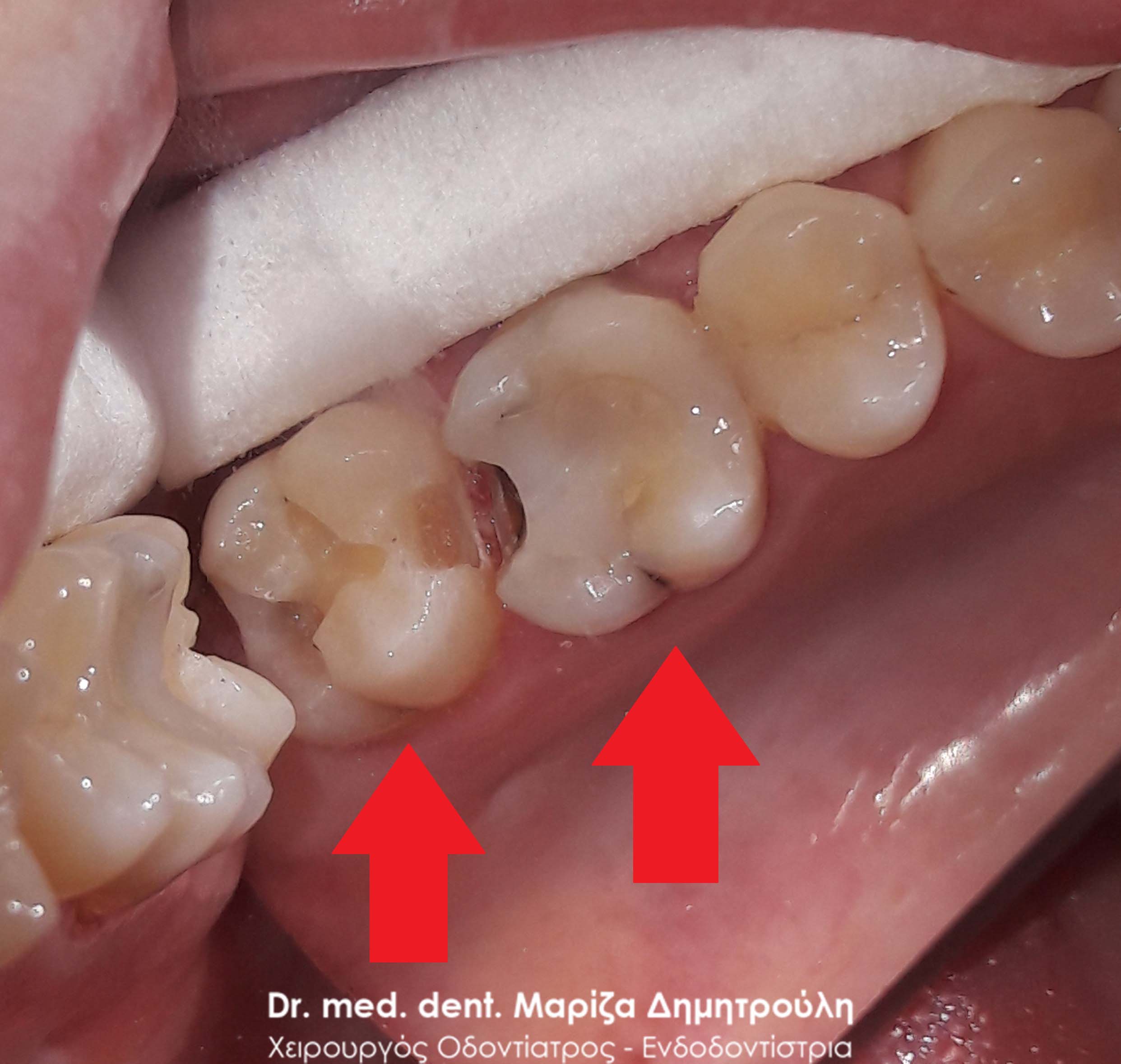
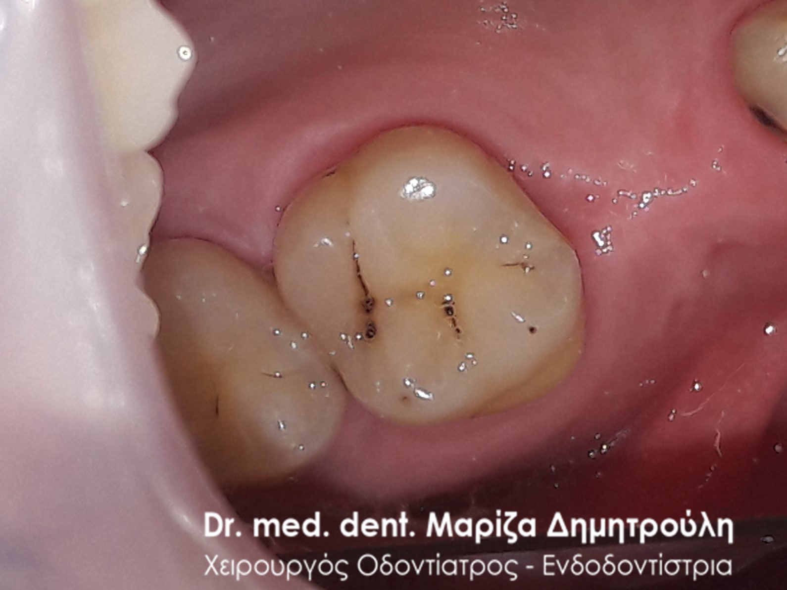
Incident – White tooth filling
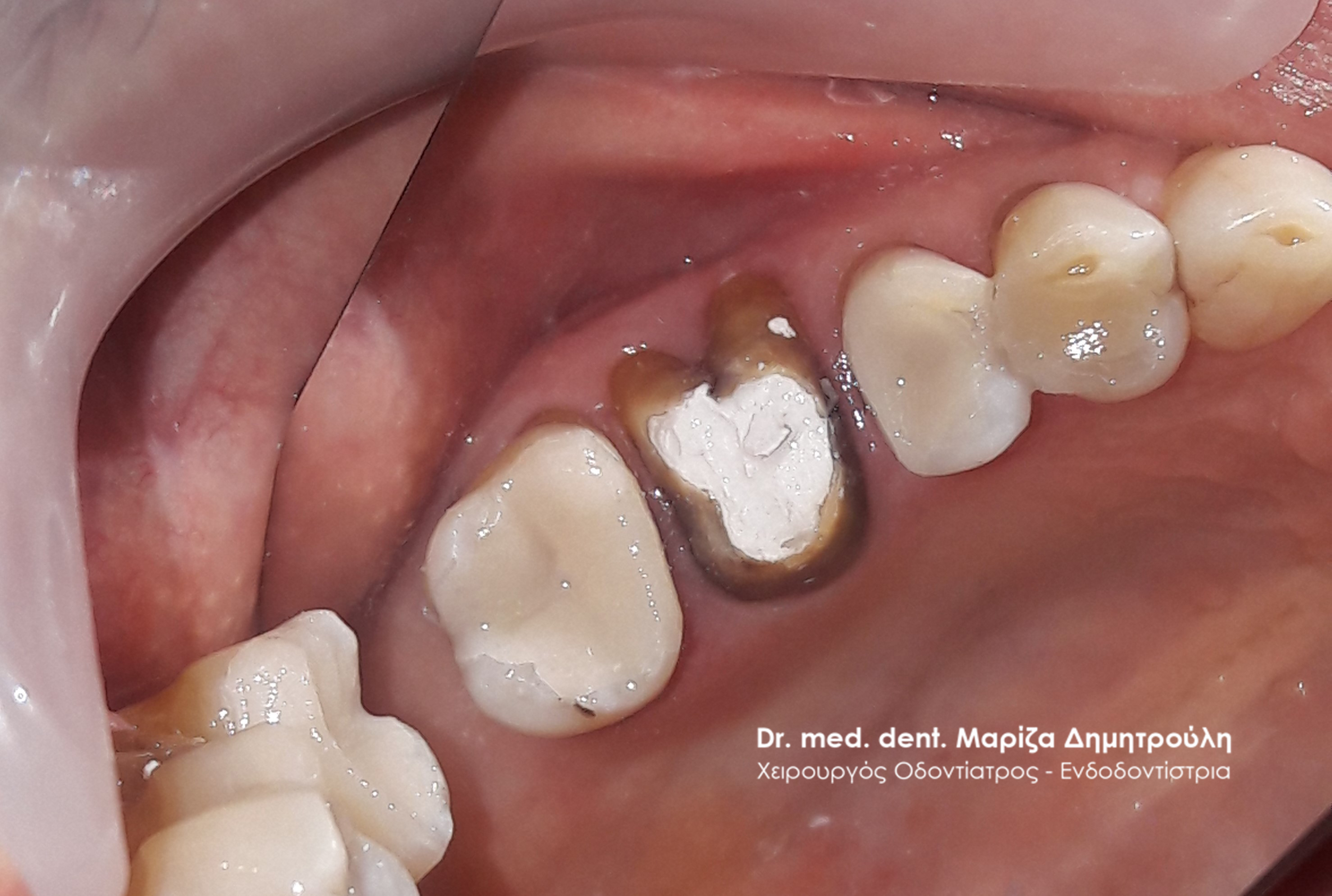
The patient visited the doctor’s office as she has considerable pain when chewing on the right side of her upper jaw. After the clinical and radiographic examination of the area, the presence of a deep “hidden” caries in the first upper right molar was established. In consultation with the patient, it was decided to open the tooth and restore it with a white composite resin filling. P
Initially, local anesthesia was administered in the area of the painful tooth and its extraction began. Opening the tooth did indeed reveal the presence of deep decay, as the photos show. This was followed by the removal of caries, the placement of special material to protect the nerve of the tooth, and finally the dental deficit was restored with a white filling.
The tooth is now asymptomatic and the patient can chew without discomfort on the right side of the upper jaw.
BEFORE
The extent of caries after opening the cavity
AFTER
Incident – White tooth filling
BEFORE
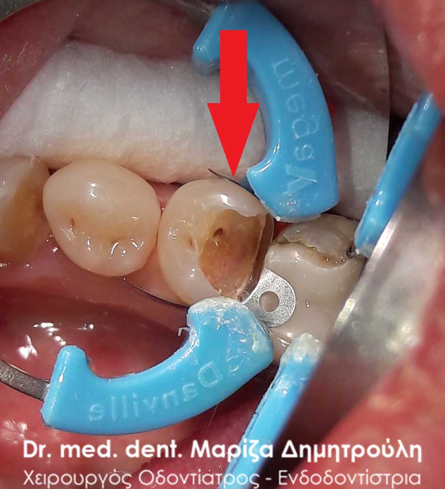
Image of deep extensive caries after tooth extraction
AFTER
BEFORE
AFTER
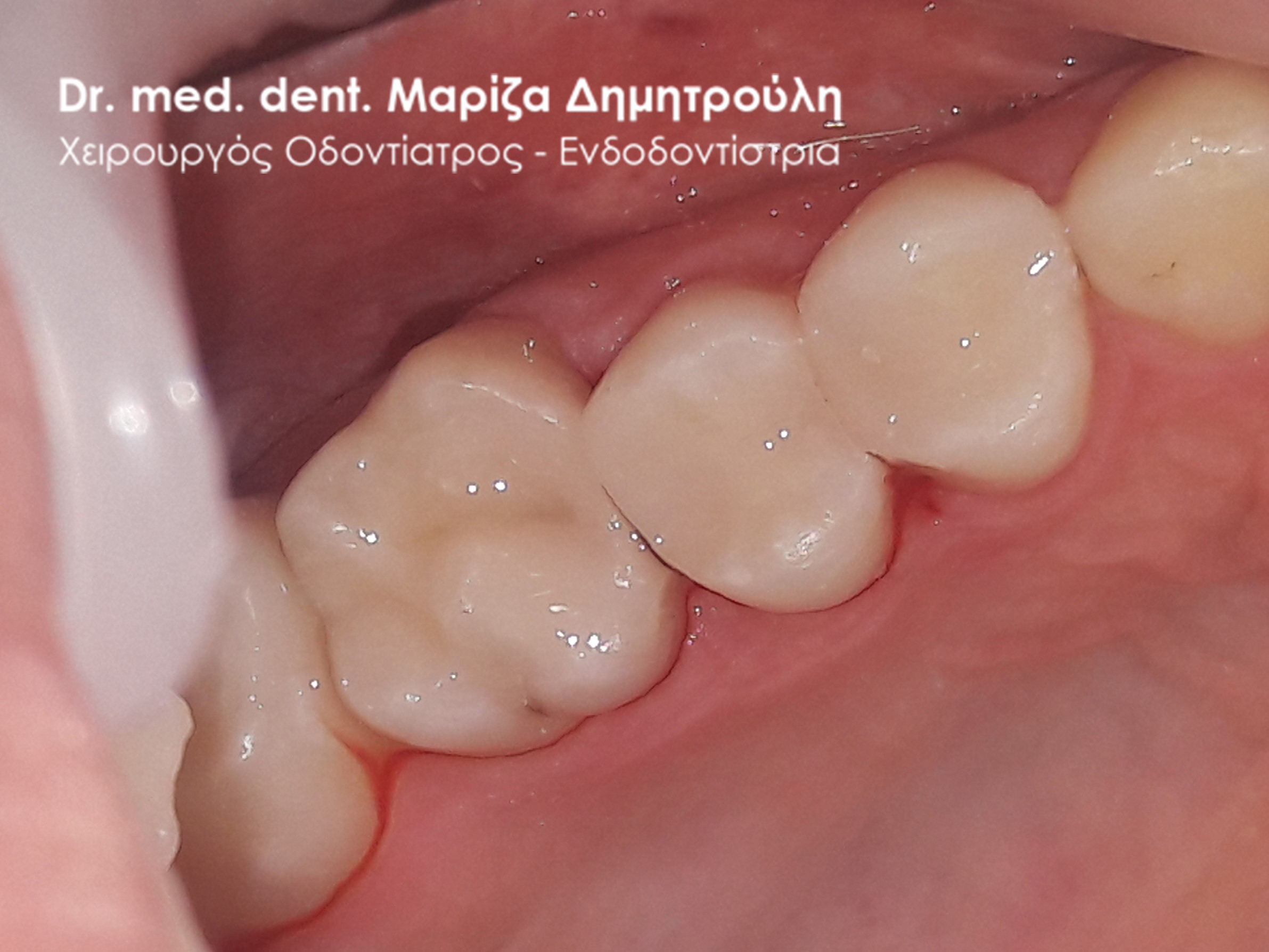
Incident – White tooth filling
BEFORE
BEFORE
Deep caries during teething
AFTER
AFTER
AFTER
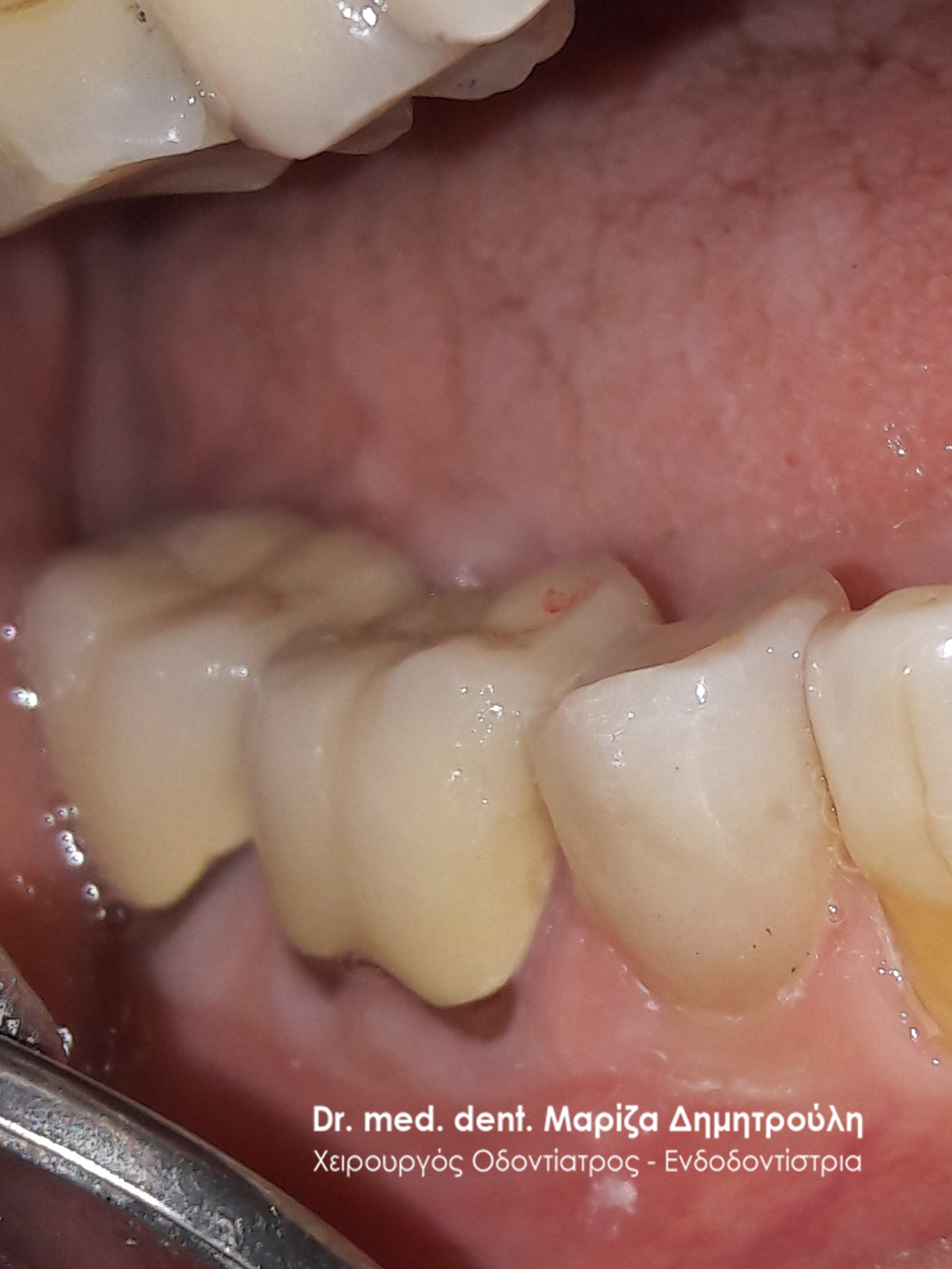
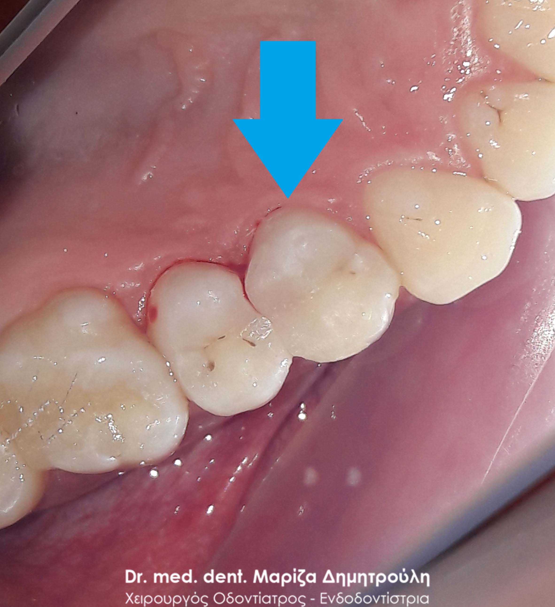
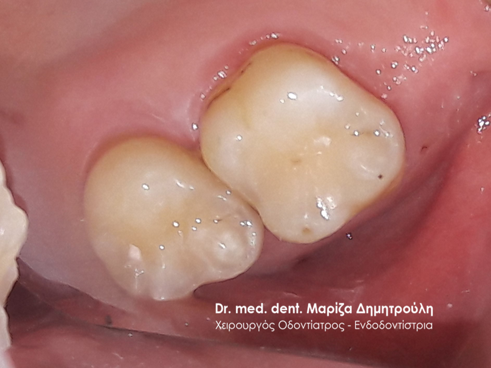
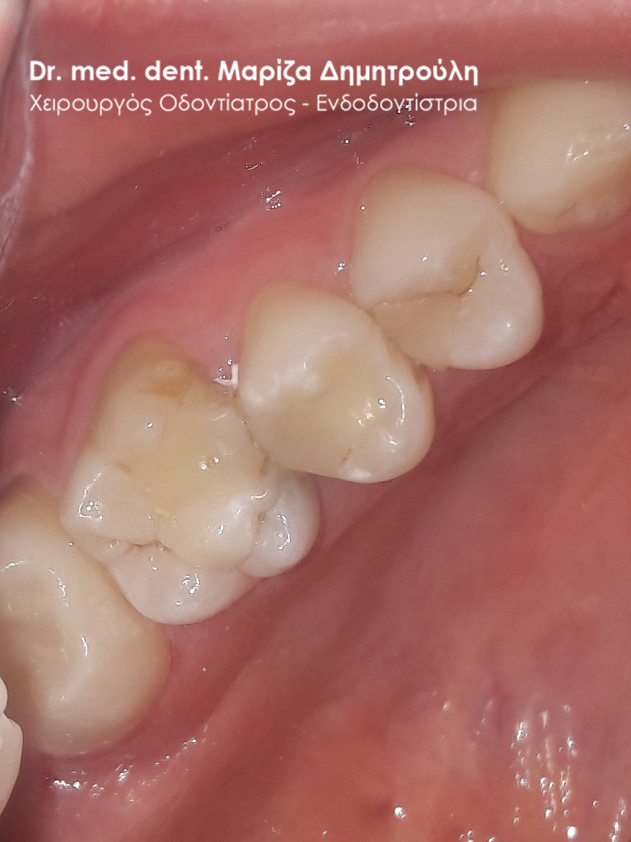
Incident – White tooth filling
BEFORE
AFTER
Widget After Content

Dr. med. dent. Mariza Dimitrouli
Dr. med. dent. Mariza Dimitrouli is a specialist at the University of Hannover, Germany (MHI) and also holds a PhD from the same university. She also worked in a dental clinic in Berlin. The title of Endodontist held by Dr. M. Dimitrouli is renewed every six years by the German Endodontics Society. She has a private practice in Thessaloniki, Thermi.